Professor Ji Donghua's First Clinical Use of DKutting® LL Peripheral Scoring Balloon in Below-the-Knee CTO Lesion

DK Medtech
Vascular Interventional Balloon Product Developer

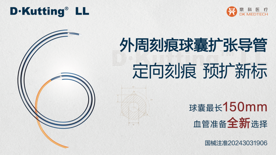
DKutting®LL continues to use DK Medtech's unique patented triangular nitinol coil technology, combined with the actual clinical needs of lower limb arteries, significantly increasing the length of the scoring element (up to 150mm). It has also developed a completely coaxial balloon delivery system compatible with 0.014"/0.018" guidewires and enriched the selection of balloon diameters with 0.5mm increments.
DK Medtech's exclusive directional scoring balloon boasts comprehensive performance with robust radial expansion and flexible axial bending. Its clinical performance has been widely recognized in both the coronary and hemodialysis access fields. The latest development is the DKutting.®LL Scoring Balloon is specifically designed for lower limb arterial intervention, demonstrating significant clinical advantages in directional expansion mechanisms and setting a new standard for peripheral vascular pre-dilation.
DK Medtech Special Release[Professor Ji Donghua: DKutting®Debut of LL Peripheral Scoring Balloon in BTK Artery CTO Lesions: Case Presentation, demonstrating the meticulous operation of each case and the clinical application of advanced equipment and instruments. From the formulation of treatment strategies for different cases, standardized intraoperative procedures and technical applications, complication prevention, to perioperative management, etc., the aim is to promote the standardization of diagnosis and treatment for vascular stenosis and occlusive diseases, enhance technical exchange and experience sharing among doctors, with the hope of providing new ideas and methods for future diagnosis and treatment, benefiting more clinical patients.
DKutting®LL Peripheral Scoring Balloon
Debut of Below-the-Knee Artery CTO Lesion
Dalian Medical University Affiliated First Hospital, Ji Donghua
Patient Information
Basic Information:Female, 89 years old.
Chief Complaint:Cyanosis of the left heel and sole with resting pain for over a month.
History of Present Illness:More than one month ago, the patient developed cyanosis in the left heel and sole without obvious cause, accompanied by pain during nighttime rest, which required painkillers for relief; the foot pain worsened in the past week, prompting a visit to the hospital for diagnosis and treatment.
Past Medical History:A history of diabetes for over 30 years, hypertension and hyperlipidemia for over 30 years, and left knee replacement surgery 5 years ago.
Physical Examination:The color of the left foot sole is cyanotic, with low skin temperature and poor local perfusion. The pulse of the left femoral popliteal artery can be felt, the dorsalis pedis artery pulse is palpable, but the distal posterior tibial artery is not detectable.
Admission Diagnosis:Lower Extremity Arteriosclerosis Obliterans (Chronic Severe Threatening Limb Ischemia, Rutherford Grade 4).
Preoperative Analysis
Preoperative Analysis:The patient has Rutherford grade 4 ischemia, a CLTI case, with the ischemic area located on the sole. The responsible vessel should be the posterior tibial artery.
Surgical Objective:
Main Objectives:Reconstruction of the posterior tibial artery and plantar arch;
Secondary Objective:Reconstruction of the plantar arch using PPL technology via the anterior tibial artery.
Surgical Strategy/Plan:
Through the anterior tibial artery to the dorsal pedal arch, the PPL technique was applied to open the plantar arch;
Retrograde opening of the posterior tibial artery through the anterior tibial artery;
If retrograde access fails, antegrade + retrograde can be applied to complete SAFARI;
Finally, the antegrade reconstruction of the posterior tibial and plantar arch was successfully completed.
The posterior tibial artery is a CTO lesion. If the lumen gain from POBA is unsatisfactory, further application can be considered.DKutting®LL Peripheral Scoring BalloonCompletion of PTA
Main Treatment Process
Time | Main Treatment Process |
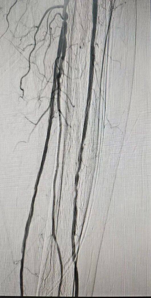
2024.12.25 | Antegrade puncture of the left femoral artery, angiography after insertion of a 6F sheath showed that the femoral-popliteal artery was patent without significant stenosis or occlusion. The posterior tibial artery below the knee was occluded almost throughout its entire course; the plantar arch was not visible, while the anterior tibial artery and peroneal artery were patent, and the dorsalis pedis arterial arch was visible. |
Using the CXI support catheter in conjunction with the Regalia guidewire, access was gained to the anterior tibial artery, advancing to the dorsalis pedis arterial arch. Through the arcuate artery, retrograde entry was achieved into the lateral plantar arch and extending distally into the posterior tibial artery. A Command ES guidewire was then exchanged and positioned within the posterior tibial artery. Subsequently, the CXI catheter was advanced over a V18 guidewire through the sheath in an antegrade fashion to the origin of the posterior tibial artery, reaching the intima down to the distal posterior tibial artery. The SAFARI technique was employed to complete the antegrade and retrograde guidewire rendezvous within the posterior tibial artery. The catheter was advanced antegradely across the plantar arch to the dorsalis pedis artery, where a Command ST guidewire was exchanged. | |
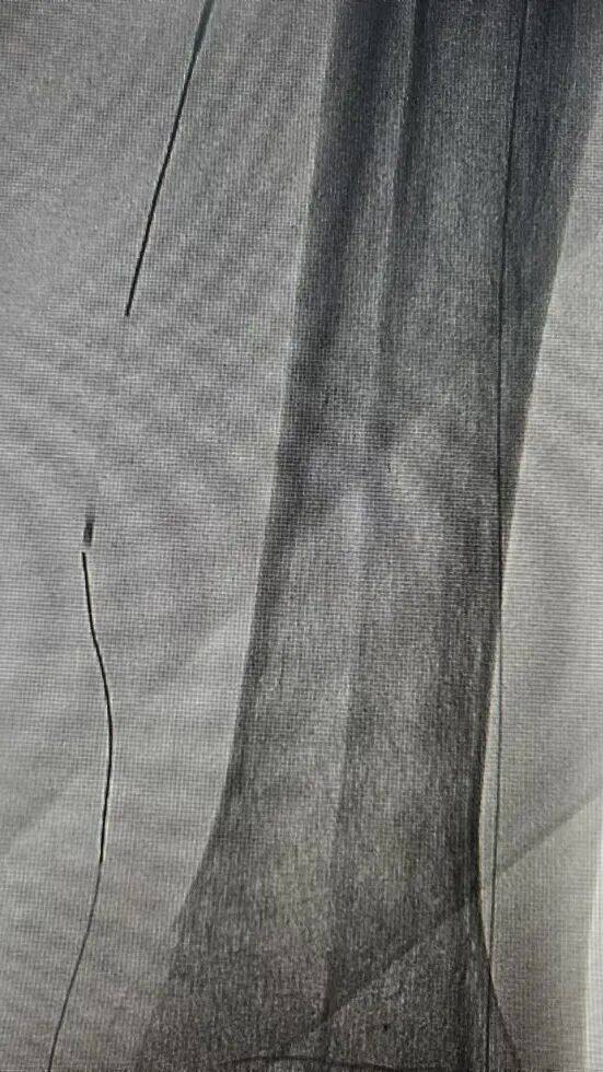
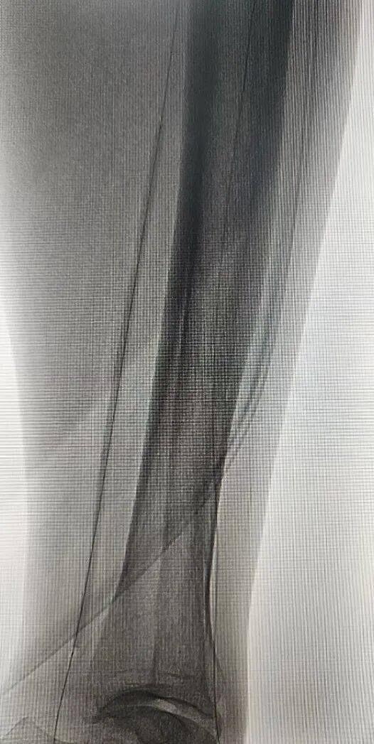
The posterior tibial artery was first treated with a 2.5*150mm balloon dilation. Follow-up angiography showed multiple small dissections and residual stenosis in the posterior tibial artery. | |
Then advance along the guidewire for exchangeDKutting®LL 3*150mm BalloonFull-length dilation of the posterior tibial artery; | |
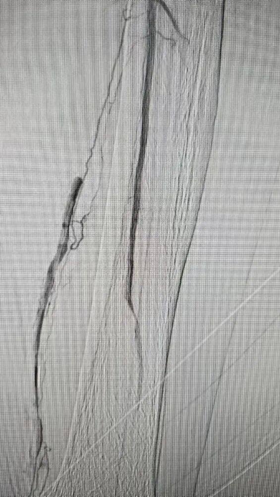
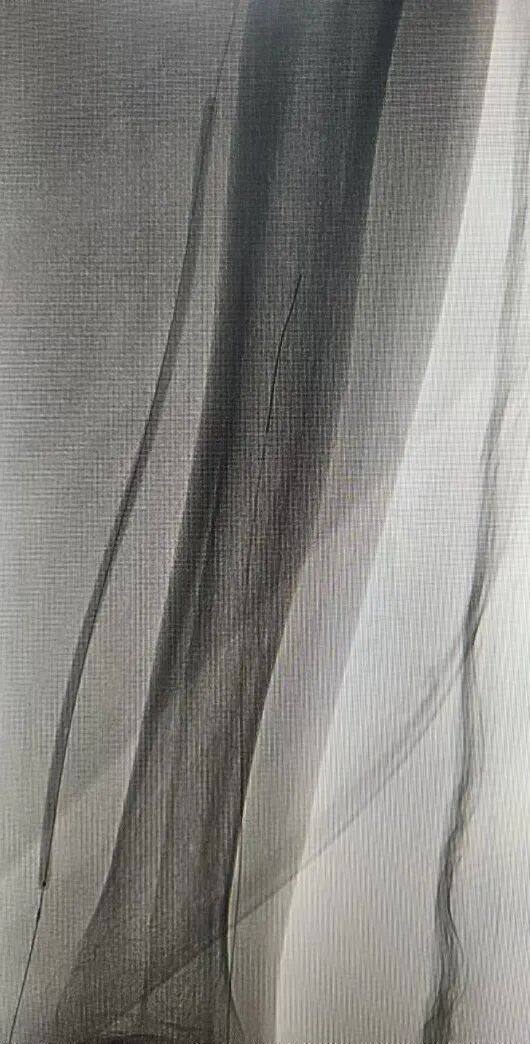
Final angiography showed satisfactory luminal gain in the posterior tibial artery, with no significant dissection or residual stenosis. |
Surgical Procedure
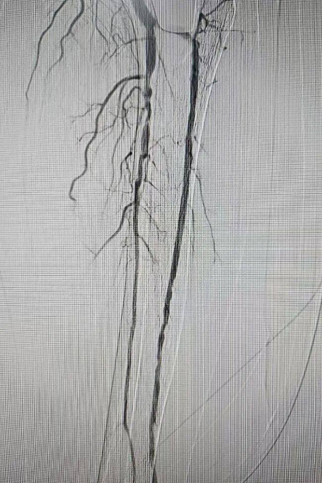
Angiography of the arteries below the knee showed that the posterior tibial artery was not visualized, while the anterior tibial artery and peroneal artery were patent with multiple stenoses.
Preoperative angiography showed the anterior tibial and dorsalis pedis arteries, but the posterior tibial and plantar arch were not visible.
During the surgery, the PPL technique was applied retrograde via the anterior tibial artery and the dorsal pedal arterial arch to access the distal posterior tibial artery, which was confirmed by angiography through a CXI catheter.
Intraoperatively, a Command ES guidewire was placed and advanced retrograde through the anterior tibial artery into the distal posterior tibial artery, followed by antegrade advancement of a CXI catheter and V18 guidewire to achieve a guidewire and catheter rendezvous in the distal posterior tibial artery.
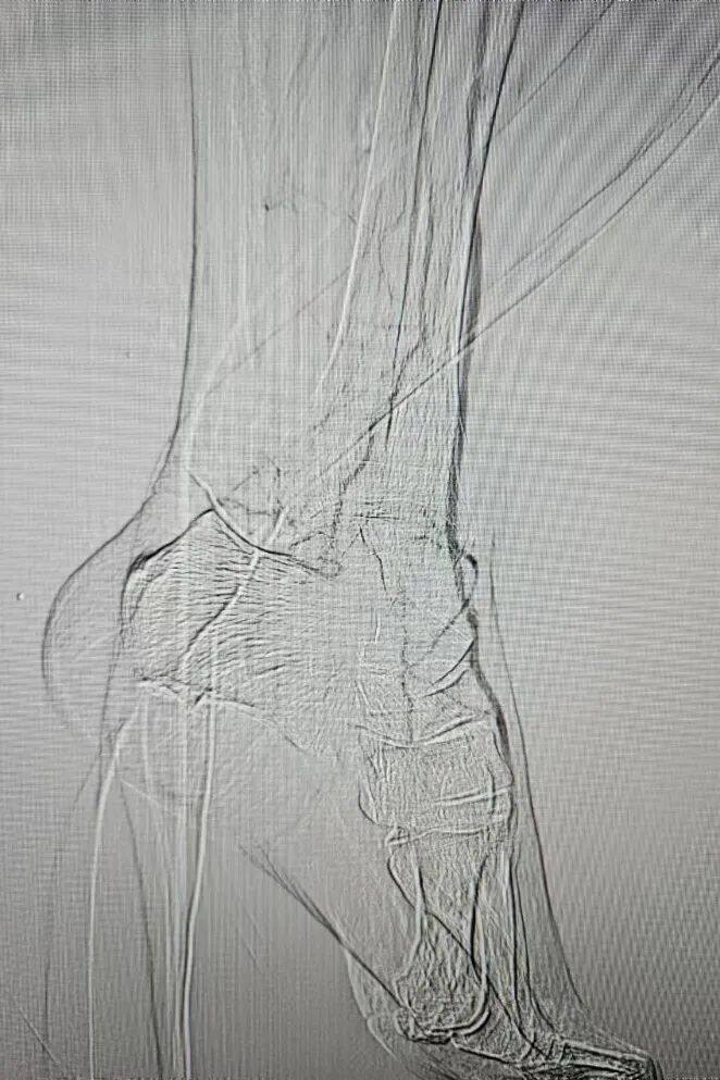
After the catheter and guidewire of the posterior tibial artery meet, the CXI catheter retrogradely advances along the guidewire into the dorsalis pedis artery and reaches the distal end of the anterior tibial artery, confirmed by angiography.
A Command ST guidewire was advanced retrograde along the posterior tibial artery into the dorsalis pedis artery, and a 2mm diameter balloon was used to dilate the dorsalis pedis artery and plantar arch.
Posterior Tibial Artery Application of 2.5*150mm Balloon Dilation
Angiography after the application of a 2.5*150mm balloon dilation on the posterior tibial artery revealed multiple small dissections and residual stenosis, with unsatisfactory lumen gain.
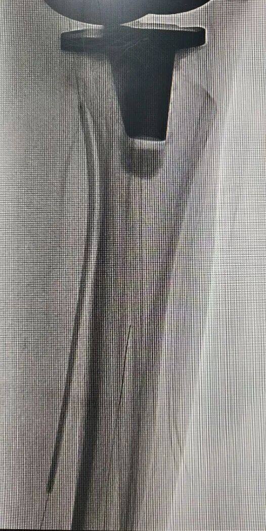
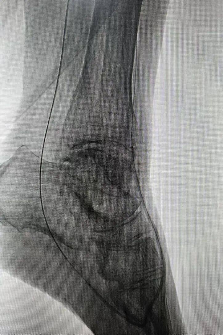
The posterior tibial artery used a 3*150mmDKutting®LL Peripheral Scoring BalloonFull-length dilation of the posterior tibial artery
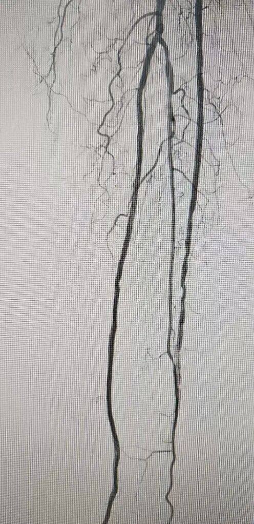
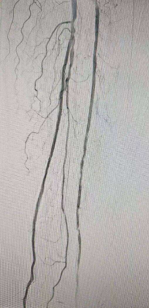
The posterior tibial artery used a 3*150mmDKutting®LL Peripheral Scoring BalloonDilation of the Posterior Tibial ArteryAngiography results from two angles showed no significant residual stenosis or dissection, with satisfactory lumen gain.
Case Summary
Case Characteristics:CTO lesion of the posterior tibial artery, dissection and residual stenosis occurred after plain balloon dilation,DKutting®LL Peripheral Scoring BalloonThe lumen was satisfactorily expanded, and the stenosis disappeared.
Characteristics/Usage Tips of the Device:DKutting®LL Peripheral Scoring BalloonExcellent performance in achieving lumen gain in infrapopliteal arteries, with good pushability, trackability, and recoil of the entire device (It is recommended to pre-dilate with a small balloon for occlusive lesions before application).

Expert Introduction


Professor Ji Donghua
M.D., Professor/Chief Physician, Master's Graduate Supervisor.
Currently serving as the Deputy Director of the Interventional Therapy Department at the First Affiliated Hospital of Dalian Medical University, he is a renowned mid-career expert in the field of endovascular treatment for diabetic foot in China. He concurrently holds positions as a member of the Expert Committee of the National Quality Control Center for Peripheral Vascular Interventional Technology, a member of the Lower Extremity Arterial Disease Group under the Vascular Surgery Professional Committee of the National Cardiovascular Disease Expert Committee, the Director of the Lower Extremity Arterial Occlusive Disease Training Center of the Chinese Vascular Surgery Training Project Expert Committee, a member of the Vascular Surgery and Tissue Engineering Group of the Medical Engineering Branch of the Chinese Medical Association, the Deputy Chairman of the World Young Committee of the International Union of Angiology, the Deputy Chairman of the Lower Extremity Artery Professional Committee of the Chinese Chapter of the International Union of Angiology, a member of the International Society for Vascular Surgery, a member of the European Society of Interventional Radiology, a member of the European Radiological Society, a member of the International Union of Endovascular Specialists, a national youth member of the Interventional Physicians Branch of the Chinese Medical Doctor Association, a member of the Complications Group of the Vascular Surgery Branch of the Chinese Medical Doctor Association, the Deputy Chairman of the Carotid Artery Group of the Peripheral Vascular Disease Professional Committee of the Chinese Integrative Medicine Association, a member of the Diabetic Foot Group of the Peripheral Vascular Disease Professional Committee of the Chinese Integrative Medicine Association, the Deputy Chairman of the Vascular Surgery Professional Committee of the Chinese Health Science and Technology Promotion Association, the Deputy Chairman of the Diabetic Foot Committee of the Chinese Health Science and Technology Promotion Association, and a national member of the Vascular Medicine Professional Committee of the Chinese Research Hospital Association. He also serves as an editorial board member for journals such as the "Chinese Journal of Clinical Medical Imaging," "Journal of Interventional Radiology," "Chinese Journal of Interventional Imaging and Therapy," "Trauma and Critical Care Medicine," "Clinical Misdiagnosis & Mistreatment," "Chinese Journal of Interventional Radiology (Electronic Edition)," "Chinese Journal of Vascular Surgery (Electronic Edition)," "Journal of Interventional Medicine," and "Translational Medicine." Additionally, he acts as a peer reviewer for journals including "Pulmonary Circulation," "The Surgeon," "CVIR Endovascular," "Chinese Journal of Clinical Physicians" (Electronic Edition), "Chinese Medical Imaging Technology," and the "Journal of Dalian Medical University."
Participated in and completed multiple scientific research projects, including the National Natural Science Foundation of China and the National "Tenth Five-Year Plan" Science and Technology Research Projects. Awarded the Dalian Science and Technology Progress Award, and in 2023, received the "Clinical Medical Research Award" at the First Clinical Medicine Awards. Authored the "Chinese Expert Consensus on Clinical Practice for Infrapopliteal Artery Revascularization" on behalf of the Lower Extremity Arterial Disease Group of the Vascular Surgery Committee of the National Cardiovascular Disease Expert Committee. Contributed to the compilation of the "Chinese Guidelines for Diagnosis and Treatment of Diabetic Foot" and the "Chinese Expert Consensus on Endovascular Treatment for Ischemic Diabetic Foot." Acted as the chief editor for one textbook published by People's Health Publishing House and co-edited six other books, including those from the same publisher. Published over 100 articles in domestic and international core journals, including 10 SCI articles.
He has successively visited and studied endovascular interventional techniques in Sweden, Germany, and South Korea. He was the first in China to clinically apply the below-the-ankle reverse puncture-assisted opening technique for total occlusion lesions below the knee. A case completed by his team, the first successful reverse puncture of the plantar arch to open three branches below the knee in China, was included in the book "Diagnosis and Treatment Strategies and Handling Techniques for Vascular Surgery Difficult Cases." He was the first in China to propose a new concept for infrapopliteal artery revascularization: the Angiographosome concept. Guided by this novel "Angiographosome" concept, he was also the first in China to perform infrapopliteal artery revascularization. The clinical research results of his team were the first to be published in a Chinese core journal, marking the first clinical paper in China on infrapopliteal artery revascularization under the guidance of the "Angiographosome" concept.

Department Introduction

The First Affiliated Hospital of Dalian Medical University has been conducting interventional treatments for nearly three decades, with independent interventional wards, interventional operating rooms, and interventional outpatient services. Currently, the Interventional Treatment Center has 57 beds, performs over 2,000 surgeries annually, and has handled a total of more than ten thousand clinical cases.
The Interventional Treatment Center of the First Affiliated Hospital of Dalian Medical University comprises three clinical subspecialties: interventional treatment for neurological diseases, interventional treatment for peripheral vascular diseases, and interventional treatment for tumors and non-vascular system diseases. The Stroke Center of the First Affiliated Hospital of Dalian Medical University, led by the Department of Interventional Therapy, was awarded the titles of "China Stroke Center Training Base" and "Five-Star Advanced Stroke Center" by the National Health Commission's Stroke Prevention and Treatment Engineering Committee in 2019. The Peripheral Vascular Specialty Group is a leader in China with advanced techniques such as 3D printing external fenestration for treating complex thoracic aortic dissections and thoracic aortic aneurysms; targeted vascular recanalization for diabetic foot, completing the first three cases of retrograde puncture via the plantar arch to assist in infrapopliteal angioplasty domestically; participating in multiple domestic clinical trials of new devices for lower limb arteries; and contributing to the writing of guidelines for interventional treatment of diabetic foot. It was also among the earliest in China to conduct interventional treatments for acute massive pulmonary embolism and contributed to the writing of guidelines for diagnosis and treatment of pulmonary artery embolism. Other projects include interventional treatment for abdominal aortic aneurysms, renal artery stenting, arteriosclerosis obliterans of the lower extremities, venous thromboembolism, visceral artery aneurysm intervention, vascular malformation intervention, and interventional treatment for venous system diseases. The Tumor and Non-Vascular Diseases Specialty Group routinely performs cutting-edge oncology treatments such as arterial chemoembolization, ablation, targeted therapy, and immunotherapy for primary liver cancer, metastatic liver cancer, and various other tumors. For many years, it has also been conducting multiple interventional diagnostic and therapeutic procedures, including stent placement for esophageal and tracheal stenosis, percutaneous biliary biopsy, biliary drainage and stent placement, TIPS treatment for portal hypertension, as well as Budd-Chiari syndrome, uterine fibroids, and hepatic hemangioma.
From 1993 to the present, the Interventional Therapy Center of Dalian Medical University First Affiliated Hospital has hosted many influential national academic conferences and national-level continuing medical education programs. In 2000, the Interventional Therapy Department was awarded the "Chinese Medical Association Practical Interventional Technology Promotion and Training Center." In 2013, it was designated as the "Peripheral Vascular Interventional Diagnosis and Treatment Training Base" by the National Health and Family Planning Commission, and in 2014, it was recognized as both a Comprehensive Interventional Diagnosis and Treatment Training Base and a Neurointerventional Diagnosis and Treatment Training Base. In 2020, the Interventional Department was approved as the "Liaoning Province Radiation Therapy Clinical Medicine Research Center."
The Interventional Treatment Center of Dalian Medical University First Affiliated Hospital boasts a professional team of medical researchers and nursing staff. It has developed into a comprehensive setup where various interventional treatment technologies for "neurological, vascular, oncological, and non-vascular diseases" are advancing in parallel. Under the leadership of Professor Wang Feng, the center continues to forge ahead, and its overall strength and influence have reached a leading position in China.
Copyright Statement: This platform aims to help healthcare professionals better understand the latest developments in relevant disease areas. The information published on this platform does not imply agreement with its descriptions or viewpoints, but is solely for providing more information. If there are any copyright issues, we kindly request the rights holders to contact us, and we will address them as soon as possible. This information is intended for healthcare professionals to stay informed and should not replace professional medical guidance in any way, nor should it be considered as diagnostic or treatment advice. If such information is used for purposes other than staying informed, this platform and its authors shall not bear any related responsibilities.Contact Email for Cooperation:vascular@edoctor.work。