
H&H Healthcare
R&D and Producer of Interventional Medical Devices for Heart Disease
Recently,The First Affiliated Hospital of Xi'an Jiaotong UniversityProfessor Yuan Zuyi's Team, Renji Hospital Affiliated to Shanghai Jiao Tong University School of MedicineProfessor Bu Jun's Team, Ningxia Medical University General Hospital Cardiovascular and Cerebrovascular Diseases HospitalProfessor Liu Zhijun's Team, People's Hospital of Xinjiang Uygur Autonomous RegionProfessor Yang Yi'ning's Team,The Second Affiliated Hospital of Xi'an Jiaotong UniversityProfessor Deng Jie's Team,People's Hospital of Ningxia Hui Autonomous RegionProfessor Li Youjin's Team(The ranking is不分先后)Six Well-Known Cardiac Centers in China Successfully Apply HuiHe Healthcare's K-Clip®Transcatheter Tricuspid Annuloplasty System Completes First Implants. Pioneering a New Paradigm in Tricuspid Valve Intervention with Annular Repair.
K-Clip®It is a transcatheter tricuspid annuloplasty system independently developed by HuiHe Healthcare, and also the first approved interventional tricuspid device in China. Using minimally invasive interventional methods, it mimics the principles of classic surgical valve annuloplasty by folding and cinching the dilated tricuspid annulus to reduce its circumference, thereby increasing the coaptation of the leaflets and improving tricuspid regurgitation.
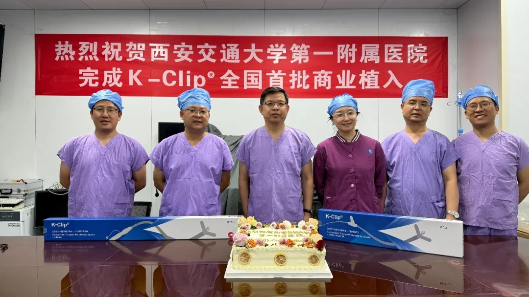
✧ The First Affiliated Hospital of Xi'an Jiaotong University ✧
Professor Zuyi Yuan, Professor Ke Han Team
Patient Baseline
Patient, female, 65 years old, was found to have tricuspid regurgitation one year ago during examination. One month ago, a follow-up examination showed progression to severe regurgitation, seeking further treatment.
Preoperative Imaging:Normal blood flow velocity below the tricuspid valve, severe regurgitation (4+) above the valve; CTA indicates a tricuspid annulus area of 1370mm².
Surgical Strategy:Plan to implant 2 K-Clips
Surgical Procedure and Outcomes
Intraoperatively, under the guidance of transesophageal echocardiography, one 12T K-Clip was implanted at the posterior annulus and another at the anterior-posterior annulus. Immediately postoperatively, reflux decreased from baseline severe (4+) to mild (1+).
'%20fill='%23FFFFFF'%3E%3Crect%20x='249'%20y='126'%20width='1'%20height='1'%3E%3C/rect%3E%3C/g%3E%3C/g%3E%3C/svg%3E)
Preoperative Ultrasound
Postoperative Ultrasound
✧ Renji Hospital, School of Medicine, Shanghai Jiao Tong University ✧
Professor Bu Jun's Team
Patient Baseline
Patient, female, 68 years old, has repeatedly experienced lower limb edema, abdominal distension, and dyspnea over the years.
Preoperative Imaging:The blood flow velocity below the tricuspid valve is normal, with severe regurgitation (5+) above the valve; CTA indicates a tricuspid annulus area of 1632 mm².
Surgical Strategy:Plan to implant 2 K-Clips
Surgical Procedure and Outcomes
During the surgery, under the guidance of intraoperative echocardiography, one 14T K-Clip was implanted at the posterior annulus and another at the anterior-posterior annulus. Immediately after the operation, the regurgitation decreased from baseline severe-to-profound (5+) to moderate-to-severe (3+).
Preoperative Ultrasound
Postoperative Ultrasound
✧ Ningxia Medical University General Hospital Cardiovascular and Cerebrovascular Diseases Hospital ✧
Professor Liu Zhijun's Team & Professor Song Guangyuan
Patient Baseline
Patient, male, 86 years old, presented with poor appetite accompanied by chest tightness and shortness of breath for 2 months, worsening over the past day. Admitted with a diagnosis of tricuspid regurgitation.
Preoperative Imaging:The blood flow velocity under the tricuspid valve is normal, with severe regurgitation (5+) above the valve; CTA indicates a tricuspid annulus area of 2650 mm².
Surgical Strategy:Plan to implant 2 K-Clips
Surgical Procedure and Outcomes
During the operation, under the guidance of esophageal ultrasound, one 14T K-Clip was implanted at the posterior annulus and another at the anteroposterior annulus. Immediately after the operation, reflux decreased from baseline severe (5+) to moderate (2+).
Preoperative Ultrasound
Postoperative Ultrasound
✧ People's Hospital of Xinjiang Uygur Autonomous Region ✧
Professor Yang Yi'ning's Team & Professor Liu Xianbao
Patient Baseline
Patient, female, 84 years old, with chest tightness and shortness of breath for 40 years, worsened in the past week. History of atrial fibrillation for over a decade. Cardiac ultrasound indicates tricuspid regurgitation.
Preoperative Imaging:The blood flow velocity below the tricuspid valve is normal, with severe regurgitation above the valve (6+); CTA indicates a tricuspid annulus area of 1314mm².
Surgical Strategy:Plan to implant 2 K-Clips
Surgical Procedure and Outcomes
During the operation, under the guidance of transesophageal echocardiography, one 12T K-Clip was implanted at the posterior annulus and one 14T K-Clip at the anterior-posterior annulus. Immediately after the operation, the regurgitation decreased from a baseline torrential (6+) to moderate-to-severe (3+).
Preoperative Ultrasound
Postoperative Ultrasound
✧ The Second Affiliated Hospital of Xi'an Jiaotong University ✧
Professor Deng Jie's Team
Patient Baseline
Patient, female, 66 years old, with intermittent chest tightness and chest pain for more than 1 year. Further examination confirmed persistent atrial fibrillation, high-degree atrioventricular block, and severe tricuspid regurgitation.
Preoperative Imaging:The blood flow velocity below the tricuspid valve is normal, with severe regurgitation (5+) above the valve; CTA indicates a tricuspid annular area of 1289mm².
Surgical Strategy:Plan to implant 2 K-Clips
Surgical Procedure and Outcomes
Intraoperatively, under the guidance of transesophageal echocardiography, one 14T K-Clip and one 12T K-Clip were implanted at the posterior annulus and the anterior-posterior annulus respectively. Immediately postoperatively, the regurgitation decreased from baseline severe (5+) to moderate (2+).
Preoperative Ultrasound
Postoperative Ultrasound
✧ People's Hospital of Ningxia Hui Autonomous Region ✧
Professor Youjin Li's Team & Professor Weidong Li
Patient Baseline
Patient, female, 77 years old, with chest tightness, shortness of breath accompanied by dizziness for more than 2 months, worsened in 1 day, post mechanical mitral valve replacement surgery. Cardiac ultrasound shows: severe tricuspid regurgitation.
Preoperative Imaging:The blood flow velocity below the tricuspid valve is normal, with severe regurgitation (4+) above the valve; CTA indicates a tricuspid annulus area of 1587 mm².
Surgical Strategy:Plan to implant 2 K-Clips
Surgical Procedure and Outcomes
Intraoperatively, under the guidance of transesophageal echocardiography, one 12T K-Clip and one 14T K-Clip were implanted at the posterior annulus and the anterior-posterior annulus respectively. Immediately postoperatively, the regurgitation decreased from baseline severe (4+) to moderate (2+).
Preoperative Ultrasound
Postoperative Ultrasound
Tricuspid regurgitation was once called "the forgotten valvular disease" because of its complex anatomy and limited treatment options, which have long led to high mortality and low intervention rates. As an increasing number of clinical centers adopt K-Clip®Transcatheter Tricuspid Annuloplasty Surgery: The treatment landscape for tricuspid regurgitation is also set to undergo a revolutionary change. K-Clip®Through a simple, safe approach and the concept of physiological repair, it takes a solid first step in the lifelong management of patients with tricuspid regurgitation.
K-Clip®Transcatheter Tricuspid Annuloplasty System:
1. Through vascular puncture approach, minimal damage, with a sheath outer diameter of 18Fr.
2. Simple operation, all completed on the atrial surface, with a short learning curve.
3. All operational steps before dissociation are reversible, and the surgical outcome is controllable.
4. Physiological annuloplasty technique, no damage to native valve leaflets.