
Percutek Therapeutics
Developer of Minimally Invasive Cardiovascular Treatment Devices

Today, I will introduce to youProfessor Zhou Bin's Team from the Department of Vascular Surgery, Oriental Hospital, ShanghaiTEVAR Surgery Sharing for Patients with Thoracic Aortic Hematoma and Penetrating Ulcer. In this patient, the proximal end of the hematoma lesion involves the root of the left subclavian artery (LSA), and the distal end extends to the bifurcation of the abdominal aorta; the ulcer lesion is located at the beginning of the descending aorta, adjacent to the root of the LSA, with a wide range of lesions and insufficient proximal anchoring area.
Facing the complex condition of the patient, a pressing challenge stood before the medical team: how to devise a surgical plan that could not only safely and effectively cure the patient but also simultaneously complete arch reconstruction while isolating the lesion, ensuring unobstructed blood flow in the arch branch vessels? After careful consideration and repeated deliberation, Professor Zhou Bin’s team successfully performed an in-situ fenestration on the Percutek Therapeutics thoracic aortic stent graft using a 0.014" soft-tip guidewire, efficiently and precisely reconstructing the LSA and successfully isolating the lesion. Post-surgery, the patient recovered well.

Gender:Female
Age:71 years old
Chief Complaint:Sudden abdominal pain accompanied by back pain for 2 days.
Past Medical History:History of hypertension for over 2 years, usually taking Valsartan orally with good blood pressure control; history of cholecystectomy.
Physical Examination:The patient is conscious, breathing steadily, with coarse breath sounds in both lungs, no dry or wet rales heard. The abdomen is soft, no fluid thrill or succussion splash detected. Mild tenderness is present in the upper-middle abdomen, without rebound tenderness or muscle rigidity. Bilateral femoral artery pulses are normal.
Detailed Explanation of CTA:Hematoma of the thoracic aorta combined with penetrating ulcer; the proximal end of the hematoma involves the root of the left subclavian artery (LSA), and the distal end involves the bifurcation of the abdominal aorta. The ulcer lesion is located at the beginning of the descending aorta, close to the root of the LSA, approximately 14.8 mm from the opening of the root of the left common carotid artery (LCCA), with a size of about 17.1*7.3 mm. The distance between the LSA and the LCCA is approximately 6.9 mm, and the diameter of the aorta at the posterior edge of the LCCA is 31.5 mm. The left and right vertebral arteries are equipotential, and there are extensive calcified plaques in the abdominal aortic segment.

Preoperative Three-dimensional Reconstruction

Preoperative CTA Cross-Section

The lesion involves the root of the LSA, with a large range of involvement.

The lesion range is extensive, with the proximal end involving the root of the LSA and the distal end reaching the bifurcation of the abdominal aorta, necessitating a stent graft with excellent sealing properties.
The lesion is adjacent to the LSA, with an obvious insufficient proximal anchoring zone, requiring extension of the anchoring zone proximally and reconstruction of the LSA.
The patient is an elderly woman with a thin brachial artery, making it difficult to introduce a steerable sheath. Additionally, the left subclavian artery (LSA) forms an acute angle with the aortic arch, posing challenges for in-situ fenestration.

Surgical Plan Strategy
Endovascular Repair of the Thoracic Aorta with Direct Coverage of the LSA Using a Stent-Graft: The procedure is simple and effective, but covering the LSA may lead to clinical manifestations of posterior circulation ischemia and upper limb ischemia in patients.
Thoracic Aortic Stent-Graft Endovascular Repair + LSA Chimney Technique: The surgical procedure is relatively simple, but there is a higher risk of endoleaks and occlusions, with suboptimal mid- to long-term outcomes.
Endovascular Repair of Thoracic Aortic Stent Graft with In Vitro Fenestration: Excellent sealing of the lesion, preservation of original hemodynamic characteristics, but complex operation. Preoperative stent modification is required based on measurement results, which is time-consuming; intraoperative precise alignment and super-selective window positioning are required, posing higher risks.
Endovascular Repair of Thoracic Aortic Stent Graft + In-situ Fenestration: Excellent sealing of the lesion, no need for preoperative stent modification. However, traditional in-situ fenestration of aortic stent grafts has high requirements for interventional devices, necessitating specialized membrane-puncturing instruments such as in-situ fenestration needles, lasers, and biopsy needles.
The Percutek Therapeutics Thoracic Aortic Stent Graft can penetrate the graft for in-situ fenestration using only a 0.014" CTO guidewire with a soft tip. Considering the mid- to long-term treatment outcomes and the simplicity of intraoperative manipulation, Professor Zhou Bin's team comprehensively evaluated and selected the Percutek Therapeutics Thoracic Aortic Stent Graft for endovascular repair, reconstructing the LSA through in-situ fenestration technology.

01.The patient was placed in a supine position, with routine disinfection and draping. Bilateral radial arteries and the right femoral artery were punctured, and 5F and 8F sheaths were inserted respectively. Two suturing devices were pre-placed in the right femoral artery. A gold-marked pigtail catheter was introduced into the ascending aorta through the right femoral artery; a 6F crossover sheath was introduced through the left brachial artery, with its tip positioned in the aorta.

Preoperative Angiography
02.The super hard guidewire was inserted through the femoral artery into the ascending aorta to form a loop, and Huamao Tianyi was introduced along the guidewire.®Thoracic Aortic Stent Graft System PTBS3430210, Angiographic Positioning.

Implant Percutek
03.After positioning is completed, release Percutek Therapeutics.®Thoracic aortic stent graft; after the complete release of the stent graft, the proximal bare stent was released. Angiography after release showed accurate and ideal stent positioning.

Release Coated Stent

Release Bare Stent

Post-release Contrast
04.Due to the thin left brachial artery, the adjustable bend sheath was difficult to introduce and lacked sufficient support. Therefore, a 6F-45cm crossover sheath was used for support, allowing the successful use of a 0.014" ASAHI Conquest Pro guidewire with a soft tip in conjunction with the support sheath for membrane puncture and fenestration. The success of the fenestration was confirmed from multiple angles.

LSA Placement Through Contralateral Sheath

Successful Membrane Penetration
05.The guidewire of the left brachial artery was pulled through the snare to the right femoral artery sheath to create tension, followed by the sequential introduction and dilation of a 4-80mm balloon and an 8-80mm balloon to open the fenestration.

Grasping Guide Wire

4-80mm Balloon Dilation Window Opening

8-80mm Balloon Dilation Window Opening
06.The left subclavian artery (LSA) was identified by angiography via the LSA sheath, followed by the introduction of a 10-39mm balloon-expandable covered stent through the femoral artery and its deployment to reconstruct the LSA.

Contrast Localization of Left Vertebra

Placement of 10-39mm Balloon-Expandable Covered Stent

Release Balloon-Expandable Covered Stent
07.Postoperative angiography showed: the aortic stent graft and LSA branch stent were in good shape, blood flow in the aorta and its branches was unobstructed, no imaging of the aortic arch ulcer was observed, and there was no significant endoleak. All guidewires, catheters, and sheaths were withdrawn, the puncture site was closed, pressure dressing was applied for hemostasis, and the incision was closed layer by layer. The operation was completed.

Postoperative Angiography

Preoperative and Postoperative Comparison

Preoperative/Postoperative 3D Reconstruction Comparison

Preoperative/Postoperative CTA Axial Comparison
The patient recovered well after the surgery. A follow-up CTA examination at 5 months showed that the aortic stent graft and the LSA branch stent were in good condition, with no displacement, smooth blood flow, and no endoleak.
Summary of Case Experience
This shared case involves a patient with a thoracic aortic hematoma combined with a penetrating ulcer. The hematoma lesion extends proximally to the root of the LSA and distally to the bifurcation of the abdominal aorta; the ulcer lesion is located at the beginning of the descending aorta, adjacent to the root of the LSA. The lesion range is extensive, with insufficient proximal anchoring area, necessitating supra-arch branch reconstruction of the LSA. The surgery is highly challenging, with high demands on the sealing performance of the stent graft, the accuracy of positioning, and the passage of the delivery system.
Professor Zhou Bin's team ultimately chose the Percutek Therapeutics Thoracic Aortic Stent Graft for endovascular repair. This stent graft is currently the only product on the market that can achieve in-situ reconstruction solely through guidewire penetration. It offers precise positioning, no endoleaks, and ease of operation. During the procedure, a 0.014" CTO guidewire with a soft tip can easily penetrate the membrane. The fenestration area of the graft is easy to penetrate and expand, and after implantation of the branch stent, it demonstrates excellent morphology with no membrane leakage.At the 5-month postoperative follow-up, CTA showed an ideal stent morphology after implantation, no displacement, complete absorption of the patient's hematoma, good aortic remodeling, smooth blood flow, and no endoleak. The treatment outcome was clear.

Expert Introduction

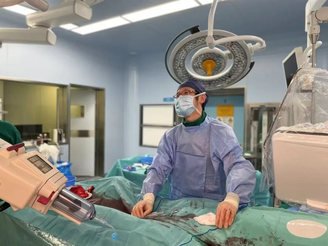
Professor Zhou Bin
Chief of the Vascular Surgery Department at the Lujiazui Campus of Shanghai East Hospital, Deputy Chief Physician, Associate Professor.
Proficient in total endovascular repair of aortic aneurysms and aortic dissections, diagnosis and treatment of peripheral vascular diseases such as lower extremity arterial sclerosis occlusion, visceral artery aneurysms, and venous thrombosis; minimally invasive treatment of lower extremity varicose veins.
Currently serving as a Youth Committee Member of the International Vascular Alliance, a committee member of the Peripheral Vascular Medicine Branch of the China International Exchange and Promotive Association for Medical and Healthcare, and a member of the Research and Transformation Group of the Vascular Surgery Specialty Branch of the Shanghai Medical Association.


Department Introduction
Department of Vascular Surgery, Oriental Hospital of Shanghai
The Department of Vascular Surgery at Shanghai East Hospital was established in 2001, led by Director Liu Jie. The department has eight doctors, including one chief physician, three deputy chief physicians, and four attending physicians, with three holding doctoral degrees and four holding master’s degrees. The hospital is equipped with advanced hardware facilities, and the department is distinguished by its specialized medical techniques, particularly in the rapid diagnosis and minimally invasive treatment of vascular diseases. It has extensive clinical experience in the diagnosis and treatment of critical and complex vascular conditions. The department regularly engages in academic exchanges with renowned experts and research institutions both domestically and internationally, ensuring that its diagnostic and therapeutic technologies, as well as research standards, remain aligned with international advancements. This fosters a world-class vascular surgery platform integrating medical care, teaching, research, and prevention, providing professional diagnosis, treatment, and care for patients with vascular diseases from China and abroad.
The main diagnosis and treatment scope of vascular surgery includes all vascular diseases except cardiovascular and cerebrovascular diseases, including arterial, venous, vascular malformation, and lymphatic system diseases.
1.Arterial Disease:Arterial stenosis/occlusion caused by atherosclerotic plaques, arterial embolism, and thrombosis, such as arteriosclerosis obliterans (peripheral arteries, superior mesenteric artery, renal artery, carotid artery, etc.), diabetic peripheral vascular disease; arterial inflammation, such as Takayasu arteritis, thromboangiitis obliterans; arterial dilatation diseases, such as aneurysm, pseudoaneurysm, and aortic dissection; arterial dysfunction, such as Raynaud's disease (phenomenon), erythromelalgia.
2. Venous Diseases:Varicose veins of the lower extremities, primary deep venous valve insufficiency, deep vein thrombosis of the lower extremities, sequelae of deep vein thrombosis of the lower extremities, Cockett syndrome, Budd-Chiari syndrome, and thrombophlebitis, etc.
3. Vascular Malformations:Arterial malformations, venous malformations, arteriovenous fistulas, and K-T syndrome, etc.
4. Lymphatic Diseases:Lymphatic malformation, lymphangitis, lymphedema, etc.
The main treatment methods in vascular surgery are divided into physical therapy, medication, interventional therapy, and surgical treatment.
1.Interventional Treatment:Endovascular repair of the aorta, stent implantation in the inferior vena cava and iliac veins, stent implantation for renal artery stenosis (for treating renovascular hypertension), balloon angioplasty and stent implantation for limb artery stenosis/occlusion, balloon angioplasty and stent implantation for carotid artery stenosis, stent implantation for nutcracker syndrome, inferior vena cava filter implantation, embolization and sclerotherapy for vascular malformations, etc. Interventional treatments have the advantages of minimal damage, fewer complications, and rapid recovery.
2. Surgical Treatment:Surgical treatment for aortic aneurysm resection and artificial blood vessel reconstruction, bypass surgery for lower limb arteriosclerosis obliterans, varicose vein surgery of the lower limbs (stripping, laser, and radiofrequency), carotid endarterectomy, vascular injury, arterial embolism, carotid body tumor, Budd-Chiari syndrome, etc.


END
Copyright Statement: This platform aims to help medical and health professionals better understand the latest developments in related disease areas. The information content published on this platform does not imply agreement with its descriptions or viewpoints, but is merely for providing more information. If there are any copyright issues, we kindly request the rights holders to contact us, and we will handle it as soon as possible. This information is intended solely for medical and health professionals to stay informed. Such information cannot replace professional medical guidance in any way and should not be considered as diagnostic or therapeutic advice. If such information is used for purposes other than staying informed, this platform and the author assume no responsibility.Contact Email for Cooperation:vascular@edoctor.work。