Prof. Li Guoliang's Team: Management of Recurrent Type I Stenosis Using DKutting™ High-Pressure Scoring Balloon – A Clinical Case Series

DK Medtech
Vascular Interventional Balloon Product Developer
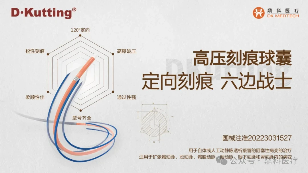
With the vigorous development of China's peripheral vascular intervention market, ordinary peripheral balloons have entered a stage of intense competition. However, for the increasing number of highly resistant stenotic lesions, ordinary balloons can easily cause complications such as excessive vascular injury, flow-limiting dissections, and hematomas. On the other hand, pressure-focused balloons utilize cutting/notching elements between the vessel wall and the balloon’s outer diameter during expansion to enhance localized pressure and achieve efficient directional dilation. This reduces vascular elastic recoil and represents a new direction in the development of vascular intervention balloons.
DK Medtech's independently developed DKutting™ High-Pressure Scoring Balloon, featuring an exclusive patented design (CN201810478242.X), boasts numerous advantages such as excellent deliverability, uniform expansion, and high burst pressure. In terms of overall product performance, it can be called a "hexagonal warrior" with virtually no weaknesses. This represents a significant breakthrough for local companies in innovating to catch up with and surpass top imported products.
DK Medtech Special Release[Professor Li Guoliang's Team: Management of Recurrent Type I Stenotic Lesions] Case Presentation, demonstrating the meticulous operation of each case and the clinical application of advanced equipment and instruments. From the formulation of treatment strategies for different cases, standardized intraoperative procedures and technical applications, complication prevention, perioperative management, and other aspects, the aim is to promote the standardization of diagnosis and treatment for vascular stenosis and occlusive diseases, strengthen technical exchanges and experience sharing among doctors, with the hope of providing new ideas and methods for future diagnosis and treatment, benefiting more clinical patients.

Management of Recurrent Type I Stenotic Lesions
People's Hospital of Tibet Autonomous Region, Li Guoliang, Suolang Quzhen

Patient Information
Basic Information:Male patient, 67 years old.
Chief Complaint:More than 2 years of maintenance hemodialysis, poor function of the internal fistula for 3 days.
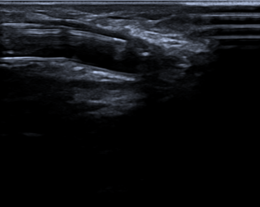
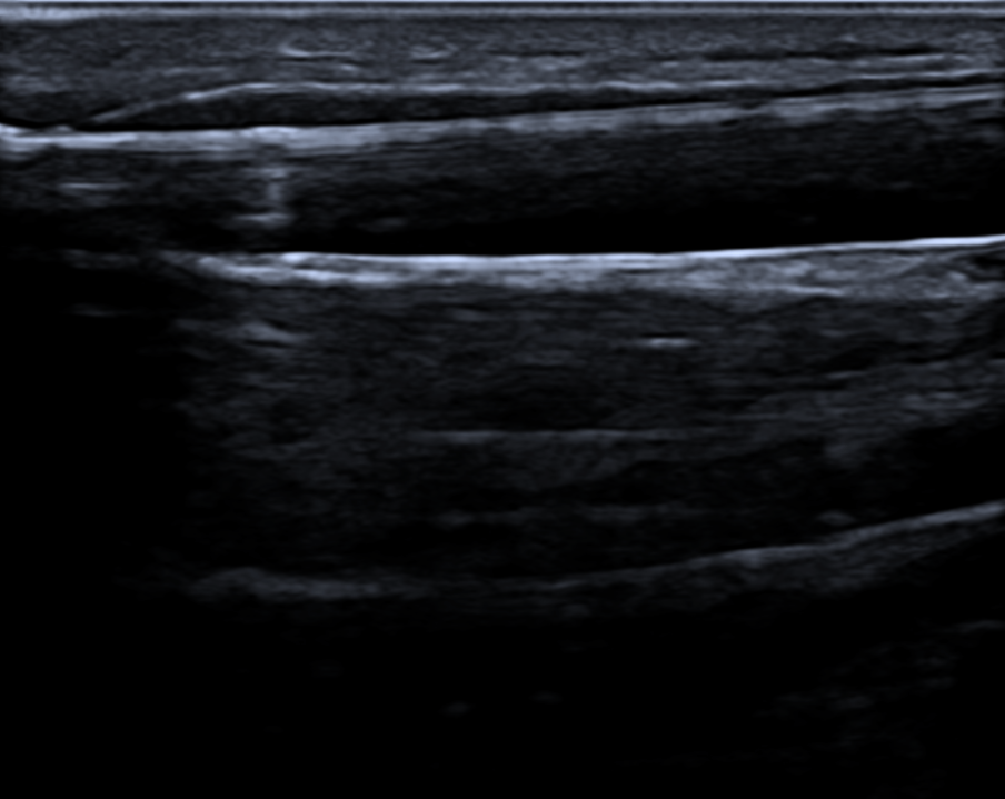
History of Present Illness:The patient was diagnosed with "chronic kidney disease stage 5" two years ago due to fatigue and edema. The primary disease was considered diabetic nephropathy. On March 17, 2022, a temporary catheter was placed in the right femoral vein, followed by hemodialysis treatment. On March 31, a semi-permanent catheter was inserted into the right internal jugular vein, and in April of the same year, an arteriovenous fistula surgery was performed on the left forearm. Two months later, hemodialysis treatment was continued using the fistula as the vascular access until now. Three days ago, during dialysis, the patient experienced insufficient flow upon initiation, suggesting stenosis of the fistula. An outpatient ultrasound indicated Type I stenosis, and the patient was admitted for treatment with a diagnosis of "dysfunction of the arteriovenous fistula."
Past Medical History:Past medical history includes "Type 2 Diabetes" for 18 years and "Hypertension" for 6 years. Denies history of chronic diseases such as "Heart Disease" and infectious diseases.。
Physical Examination:Physical Examination of the Left Upper Limb
Visual Examination: The skin of the left upper limb is rosy, and there are two surgical scars visible on the right forearm, located approximately 3cm and 5cm above the wrist, each about 3cm in length.Traces: Two blunt needle puncture scars are visible on the cephalic vein of the left forearm, without aneurysmal dilation or skin breakdown.
Palpation: The skin temperature of the left upper limb is normal. Pulsation can be felt at the anastomosis of the internal fistula, and weaker thrill can be palpated at the arterial puncture site.
Auscultation: Systolic murmur at the anastomosis site, high-pitched systolic murmur at the arterial puncture site.
Admission Diagnosis:
Stenosis of Left Forearm Internal Fistula (Type I);
Chronic Kidney Disease Stage 5, Maintenance Hemodialysis;
Diabetic Nephropathy;
Type 2 Diabetes;
Hypertension Grade 3, Very High Risk.
Previous interventional treatment
Time | Main Treatment Process |
March 17, 2022 | Temporary dialysis catheter in the right femoral vein initiated hemodialysis treatment. |
March 31, 2022 | Insertion of a semi-permanent catheter into the right internal jugular vein, removal of the temporary catheter from the right femoral vein |
April 3, 2022 | Underwent left forearm arteriovenous fistula surgery, the fistula matured two months post-operation, and the right neck semi-permanent catheter was removed. |
February 5, 2023 | Type I stenosis of left forearm arteriovenous fistula, PTA treatment |
April 16, 2023 | Type I stenosis of the left forearm arteriovenous fistula, followed by proximal reconstruction of the left forearm arteriovenous fistula after poor efficacy of PTA treatment. |
Preoperative Analysis
Preoperative Analysis:The patient had stenosis of the left forearm fistula and underwent PTA treatment twice. The stenosis recurred in a short period, and the patient also had diabetes, with significant intimal hyperplasia at the stenosis site. Afterward, proximal reconstruction was performed, but intimal hyperplasia reoccurred in less than a year. The effect of ordinary balloon dilation was poor, and it might be necessary to choose a balloon suitable for the characteristics. This time, the use of a scoring balloon for treatment is being considered.
Surgical Objective:
Main Objectives:Resolve stenosis;
Secondary Objective:Improve the patency rate of internal fistulas and reduce the recurrence rate.
Surgical Strategy/Plan:Percutaneous Transluminal Angioplasty of Left Forearm Arteriovenous Fistula Under B-Mode Ultrasound Guidance.
Surgical Procedure
Puncture the cephalic vein of the forearm against the blood flow direction. After successful puncture, insert the needle sheath, remove the needle core, and then insert the sheath guidewire along the needle sheath to establish support. Subsequently, insert the 6F catheter sheath.
A 0.035" standard hydrophilic guidewire was inserted along the catheter, passing retrogradely through the stenotic lesion → anastomosis → proximal radial artery → brachial artery, followed by an injection of 20mg of sodium heparin.
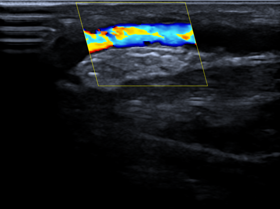
A 6mm*40mm DKutting high-pressure scoring balloon was advanced over the guidewire to dilate the stenotic lesion, with noticeable scoring observed at working pressure.
Gradually increase the pressure to 20atm, be sure to open it significantly and maintain for 3 minutes.
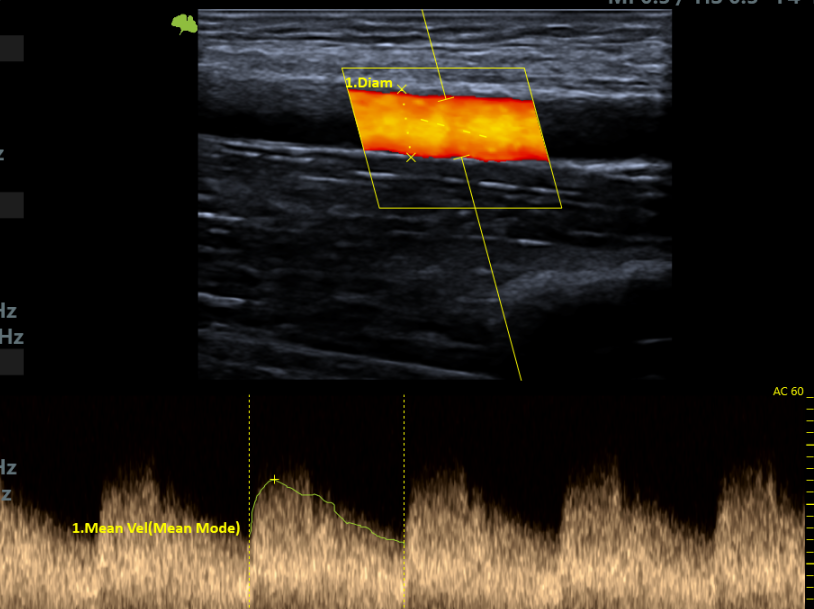
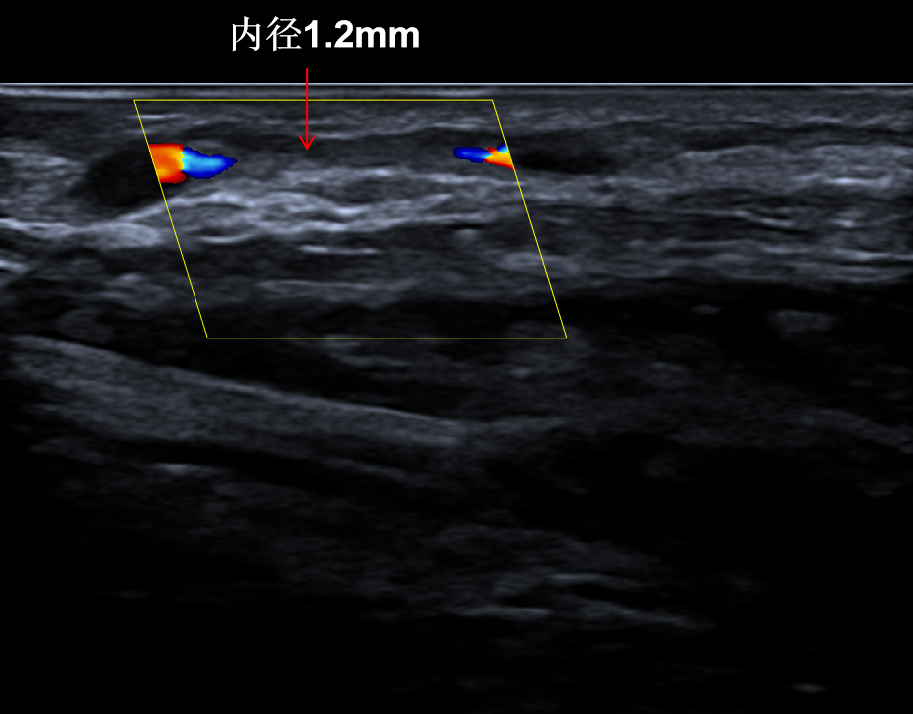
Postoperative blood flow signal is full, and postoperative VF1435.2ml/min.
Follow-up
Discharge Status:Postoperative vascular dilation was significant, and the internal fistula was used normally with a blood flow rate of 250ml/min during dialysis. On March 18, 2024, an outpatient review showed that the stenosis had been resolved, and the brachial artery flow reached 870ml/min.
Case Summary
Case Characteristics:The patient's left forearm fistula has repeatedly narrowed in a short period of time, with intimal hyperplasia being the main lesion.
Preoperative Assessment Key Points:Evaluate vascular proliferation, select balloon size, and formulate a reasonable surgical plan.
Surgical Strategy/Technical Key Points:The patient's stenotic lesion is short, so it is recommended to use a pressure-focused balloon for vascular preparation. The balloon size should be 6mm, and the dilation time should last for 3 minutes.
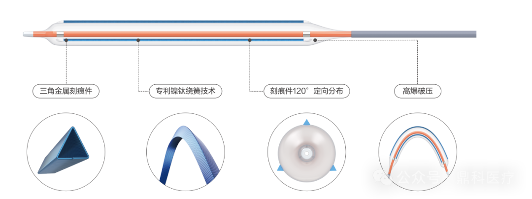
Device Features/Usage Tips:Under the same balloon pressure, the small contact area generates localized high pressure, resulting in effective dilation. The directional tearing of scoring elements can reduce injury to the vascular intima.

Expert Introduction


Dr. Guoliang Li, Associate Chief Physician
Surgeon of This Case
Group Leader of the Access Team, Department of Nephrology, People's Hospital of Tibet Autonomous Region;Member of the Vascular Access Intervention Group of the Ultrasound Intervention Committee of the Interventional Physicians Branch of the Chinese Medical Doctor Association.Hosted 1 in-hospital research project;Published multiple articles in national core journals.Secretary of the Tibet Blood Purification Quality Control Center.Proficient in the establishment, maintenance, and interventional treatment of vascular access.

Dr. Solang Quzhen, Attending Physician
The Surgeon of This Case
Attending Physician, Department of Nephrology, People's Hospital of Tibet Autonomous Region;Secretary of the Tibet Medical Association Nephrology Society;Member of the Vascular Access Group, Blood Purification Branch, Tibet Medical Association.Hosted 1 in-hospital research project.In March 2022, I attended an advanced training course on "Endovascular Techniques for Vascular Access and Interventional Nephrology" at Haidian Hospital, Beijing.Expertise: Hemodialysis technology, dialysis vascular access, renal biopsy.

Department Introduction

Our department was established in May 2014, with a current capacity of 32 beds, 54 dialysis machines of various types, and a total of 46 medical staff members.
Our department was the first in the province to establish and maintain arteriovenous fistulas. In 2017, we introduced renal biopsy techniques in collaboration with Peking University People's Hospital. In 2021, we began performing ultrasound-guided endovascular treatments and artificial blood vessel implantation in the same year. In 2023, we implemented DSA-guided central venography and PTA techniques for central venous lesions, currently at a leading level in the province. Additionally, our department provides vascular access surgeries for various dialysis centers in the Tibet region.