Professor Qiu Mingsheng: Application of Scoring Drug-Coated Balloon in Recurrently Stenotic Hemodialysis Fistula Lesions

DK Medtech
Vascular Interventional Balloon Product Developer
Stenosis of autologous/artificial arteriovenous fistula (AVF/AVG) is the most common complication in hemodialysis patients. Percutaneous transluminal angioplasty (PTA) has become the primary method for maintaining dialysis access. However, the blunt and irregular tearing of the intima and part of the media by traditional balloons during PTA can cause excessive damage to the endothelial vessels, leading to intense proliferation of vascular smooth muscle cells and macrophages, which quickly results in restenosis.
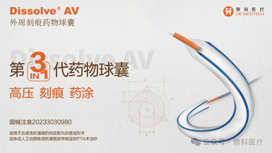
In recent years, there have been continuous explorations and clinical studies internationally regarding the use of drug-coated balloons for PTA treatment in dialysis access. DK Medtech has developed the Dissolve™ AV Scoring Drug-Coated Balloon, which integrates three features—"scoring," "high-pressure," and "drug-coating"—and is the world's first third-generation drug-coated balloon:
Directional Scoring: Uniform and regular tearing of the intima to reduce damage from blunt rupture.
High Burst Pressure: For high-resistance lesions, improving technical success rate.
Paclitaxel Coating: Effectively Inhibits Excessive Proliferation of Smooth Muscle Cells.
DK Medtech Special Release[Professor Ming-Sheng Qiu: Application of Scored Drug-Coated Balloons in Repeatedly Stenotic Dialysis Fistula Lesions] Case Presentation, demonstrating the fine operations of each case and the clinical application of advanced equipment and instruments. From the formulation of treatment strategies for different cases, standardized intraoperative procedures and technical applications, complication prevention, perioperative management, and other aspects, the aim is to promote the standardization of diagnosis and treatment for vascular stenosis and occlusive diseases, strengthen technical exchanges and experience sharing among doctors, with the hope of providing new ideas and methods for future diagnosis and treatment, benefiting more clinical patients.

Scored Drug Balloons Applied to Repeated Stenosis
Dialysis Fistula Lesion
Qiu Mingsheng, the First People's Hospital of Zhumadian City

Patient Information
Basic Information:The patient is a 47-year-old female.
Chief Complaint:Decreased thrill of the internal fistula was noted 1 week ago.
History of Present Illness:One week ago, the patient was found to have weakened thrill in the left forearm fistula during dialysis initiation. The fistula opening was mainly pulsating, with no limb pain and no local redness or swelling. The pump blood flow was 180ml/min.
Past Medical History:Suffering from "systemic lupus erythematosus" for 10 years, renal failure for 3 years, and started hemodialysis treatment 1 year ago, with no history of "diabetes."
Physical Examination:
Visual Examination:The skin of the left upper limb is normal, with no swelling in the arm. A longitudinal surgical scar approximately 3 cm in length is visible 2 cm above the transverse wrist crease. Rope ladder puncture scars are visible on the cephalic vein, with no aneurysmal dilation observed and no signs of skin ulceration.
Palpation:The skin temperature of the left upper limb is normal. Pulsation and weak thrill can be palpated at the anastomosis of the internal fistula. The venous segment about 4 cm in length above the anastomosis is cord-like and stiff, with a thrill palpable in the proximal end, and the thrill transmits towards the proximal direction.
Auscultation:A fine, weak, blowing murmur can be heard at the anastomosis site of the internal fistula, and a high-pitched, short blowing murmur can be heard at the arterial puncture site.
Color Doppler Ultrasound Examination:Venous intimal hyperplastic stenosis near the anastomosis, vascular diameter 1.9mm, length 50mm, preoperative brachial artery flow 265ml/min。
Admission Diagnosis:
Chronic Kidney Disease Stage 5, Maintenance Hemodialysis;
Stenosis of Arteriovenous Fistula in Left Upper Limb (Type I).
Previous interventional treatment
Time | Main Treatment Process |
May 2022 | Renal Function Failure Detected, Left Forearm Native Arteriovenous Fistula Established |
October 2022 | Application of Left Forearm Arteriovenous Fistula for Hemodialysis Treatment |
January 2023 | Type I Stenosis and Occlusion of Left Forearm Arteriovenous Fistula Treated with PTA |
April 2023 | Type I stenosis and occlusion of arteriovenous fistula in the left forearm treated with PTA |
September 2023 | Left forearm arteriovenous fistula type I stenosis with insufficient blood flow underwent PTA treatment |
January 2024 | Type I Stenosis of Left Antebrachial Arteriovenous Fistula with Insufficient Blood Flow |
Preoperative Analysis
Preoperative Analysis:The patient has stenosis of the arteriovenous fistula in the left forearm, and has undergone multiple PTA treatments. Two months after the last treatment, restenosis occurred with significant intimal hyperplasia at the stenotic site; the effect of repeated dilation treatment cannot be guaranteed.
Surgical Objective:
Main Objectives:Use a scored drug balloon to dilate the stenotic lesion, reducing vascular intimal injury, and after dilation, the stenosis is less than 15%.
Secondary Objectives:No bleeding or intimal tearing.
Surgical Strategy/Plan:
Under B-ultrasound guidance, enter the sheath;
Under DSA guidance, perform angiography, insert guidewire and catheter, and complete endovascular balloon angioplasty (Dissolve AV Peripheral Scoring Drug-Coated Balloon Dilatation Catheter ).
Surgical Procedure
Through the retrograde approach of the cephalic vein along the transverse elbow line, a 6F sheath was inserted. The proximal end of the cephalic vein was pressurized for angiography to confirm the distal path of the cephalic vein.
Along the 6F sheath guidewire catheter, enter the proximal end of the radial artery anastomosis, and confirm with catheter angiography.
Due to insufficient imaging of the stenosis at the anastomotic floating segment, contrast was re-administered by compressing the proximal cephalic vein, revealing a narrowed lumen diameter (approximately 1.6 mm) and length (approximately 50 mm) near the anastomosis.
A 6mm*60mm Dissolve AV high-pressure scoring drug balloon was advanced along the sheath and inflated to 20atm to achieve full dilation of the stenosis.
After the balloon is fully expanded, perform two inflations at 20atm+12atm, each maintaining pressure for 2 minutes, to ensure the drug is fully adhered to the vessel wall.
After dilation, balloon catheter anastomosis angiography showed good expansion of the stenotic segment, with an inner diameter reaching 5.8mm and no significant rebound (residual stenosis less than 15%).
Follow-up
Discharge Status:Good postoperative thrill of the internal fistula, no abnormal pulsation was palpated, and postoperative color Doppler ultrasound examination showed a narrowed segment.Inner Diameter5.5mm, brachial artery flow 884ml/min, dialysis proceeded smoothly on the same day (pump-controlled blood flow rate 220ml/min), outpatient follow-up scheduled for 3 months later.
Case Summary
Case Characteristics:The patient underwent repeated angioplasty for arteriovenous fistula stenosis in the left upper limb more than three times within a year, with the last dilation lasting less than three months.
Preoperative Assessment Key Points:It is necessary to understand the characteristics of scored drug-coated balloons and select a balloon of the appropriate size based on the lesion location, the inner diameter, and length of the blood vessels around the narrowed area.
Surgical Strategy/Technical Key Points:Based on the degree of intimal hyperplasia at the stenotic site and the pressure previously used for balloon dilation, choose the appropriate scoring drug. The scoring drug balloon Dissolve AV offers the combined advantages of high pressure, scoring, and drug coating.
Characteristics/Usage Tips of the Device:It is recommended to use a larger sheath for easy insertion and removal of the scoring drug-coated balloon. After the balloon is fully expanded, maintain pressure for at least 3 minutes to ensure adequate drug adherence to the vessel wall.

Expert Introduction


Dr. Ming-Sheng Qiu, Chief Physician
Surgeon of This Case
Chief Physician, Master of Medicine, Director of the Nephrology Department at Zhumadian First People's Hospital; Vice Chairman of the Zhumadian Kidney Disease Association; Vice Chairman of the Zhumadian Integrated Traditional Chinese and Western Medicine Association; Member of the Henan Kidney Disease Association; Standing Committee Member of the Henan Science Popularization Society Kidney Disease Association; Expert in Medical Accident Technical Appraisal for the Henan Medical Association; Health Science Popularization Expert in Yicheng District, Zhumadian City;Zhumadian Young Science and Technology Talent; Zhumadian Top Talent; Zhumadian Academic Leader; Zhumadian Tianzhong Science and Technology Innovation Youth.

Department Introduction

The Nephrology Department of the First People's Hospital of Zhumadian City was established in 2012. The department currently has 1 chief physician, 3 deputy chief physicians, 6 attending physicians, and 2 resident physicians, including 4 master's degree holders. There is also 1 chief nurse, 3 associate chief nurses, 7 nurses, and 4 nursing staff.
The nephrology department has 72 open beds, 23 hemodialysis machines, and 5 CRRT machines. The main services conducted in the ward include diagnosis, treatment, teaching, and clinical research of nephrology diseases. The department has rich clinical experience in the diagnosis and treatment of primary and secondary glomerular diseases such as acute and chronic glomerulonephritis, nephrotic syndrome, IgA nephropathy, Henoch-Schönlein purpura nephritis, lupus nephritis, diabetic nephropathy, hypertensive renal damage, small vessel vasculitis-induced kidney injury, tubulointerstitial diseases, urinary tract infections, and acute and chronic renal failure. The department performs procedures including hemodialysis, peritoneal dialysis, colon dialysis, renal biopsy puncture, renal cyst puncture sclerotherapy, arteriovenous vascular anastomosis, medium- and long-term dialysis catheter implantation, peritoneal dialysis catheter implantation, ultrasound (DSA)-guided balloon angioplasty for arteriovenous fistula stenosis or occlusion (PTA), central venous stenosis and occlusion recanalization with covered stent implantation, and other related techniques.