Case Report by Prof. Yu Hua's Team: Application of DKutting™ Scoring Balloon in Vascular Access

DK Medtech
Vascular Interventional Balloon Product Developer
With the vigorous development of China's peripheral vascular intervention market, common balloons tend to cause complications such as excessive vascular injury, flow-limiting dissections, and hematomas when dealing with the increasing number of highly resistant stenotic lesions. In contrast, pressure-focused balloons utilize cutting/notching elements between the vessel wall and the balloon’s outer diameter during expansion to enhance localized pressure and achieve efficient directional dilation. This reduces vascular elastic recoil and represents a new direction in the evolution of vascular intervention balloons.
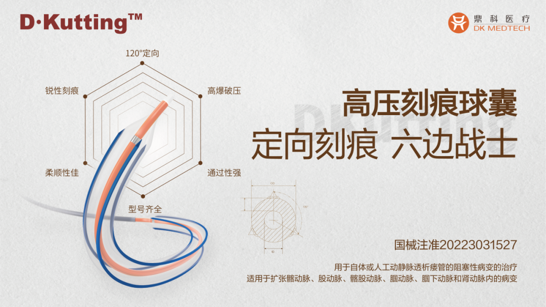
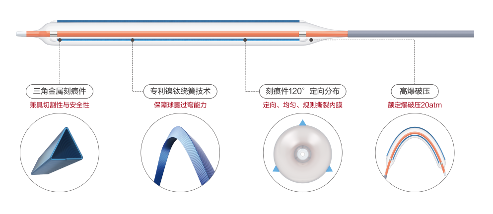
DK Medtech's independently developed DKutting™ High-Pressure Scoring Balloon, featuring an exclusive patented design (CN201810478242.X), boasts numerous advantages such as excellent deliverability, uniform expansion, and high burst pressure. In terms of overall product performance, it can be described as a "hexagonal warrior," marking a significant breakthrough for local enterprises in innovating to surpass top-tier imported products.
DK Medtech Special Release[Professor Yu Hua's Team: Experience with the Use of a Scored Balloon in Vascular Access] Case Presentation, demonstrating the detailed operations of each case and the clinical application of advanced equipment and instruments. From the formulation of treatment strategies for different cases, standardized intraoperative procedures and technical applications, complication prevention, perioperative management, and other aspects, the aim is to promote the standardization of diagnosis and treatment for vascular stenosis and occlusive diseases, strengthen technical exchanges and experience sharing among doctors, with the hope of providing new ideas and methods for future diagnosis and treatment, benefiting more clinical patients.

Experience with the Use of a Notched Balloon in Vascular Access

Yu Hua, Zhabei Central Hospital of Jing'an District, Shanghai
Patient Information
Basic Information:The patient is a 63-year-old male.
Chief Complaint:Maintenance hemodialysis for more than 3 months.
History of Present Illness:More than three months ago, the patient started dialysis treatment due to "Stage 5 Chronic Kidney Disease, Diabetic Nephropathy" with a temporary catheter in the right internal jugular vein, while undergoing an arteriovenous fistula formation surgery on the left forearm. The arteriovenous fistula did not mature well, and ultrasound examination suggested stenosis of the arteriovenous fistula. The patient is now admitted to our department for further treatment.
Past Medical History:Hypertension, Type 2 Diabetes, Coronary Atherosclerotic Heart Disease, Cerebral Infarction.
Physical Examination:The patient is conscious, with coarse breath sounds in both lungs, no obvious dry or wet rales heard. Heart rate is 80 beats/min, regular rhythm. The abdomen is soft, with no tenderness. No edema in the lower extremities. Vascular murmurs can be heard in the left forearm, with palpable thrill. Blood pressure is 156/80 mmHg.
Admission Diagnosis:
Stenosis of Arteriovenous Fistula;
Chronic Kidney Disease Stage 5, Diabetic Nephropathy, Hemodialysis;
Type 2 Diabetes Mellitus with Multiple Complications;
Grade 3 Hypertension (Very High Risk);
Coronary Atherosclerotic Heart Disease;
History of Cerebral Infarction。
Preoperative Analysis
Preoperative Analysis:The patient has been on hemodialysis for over three months. Currently, dialysis is being performed via a temporary right internal jugular vein catheter (Non-Tunnel Catheter, NTC). The left forearm arteriovenous fistula has not matured well. Vascular ultrasound examination revealed stenosis in the middle segment of the left forearm arteriovenous fistula due to valve calcification, along with intimal hyperplasia around the valve.
Surgical Objective:
Main Objectives:Dilate the stenosis of the arteriovenous fistula to relieve the stenosis and improve the poor maturation of the fistula.
Secondary Objectives:Perform dilation at the anastomosis of the arteriovenous fistula to improve inadequate expansion at the fistula site.
Surgical Strategy/Plan:A 6F vascular sheath was inserted through a puncture approximately 3-4 cm below the left elbow joint. After inserting the guidewire, a 4mm balloon was used to perform the initial dilation at the stenosis and anastomosis site.DK Medtech 6.0*60mm DKutting™ High-Pressure Scoring BalloonCystToRepeat expansion at the site of valve calcification and stenosis.
Surgical Procedure
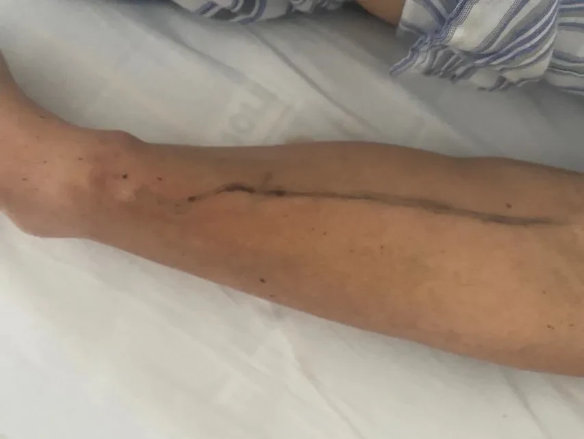
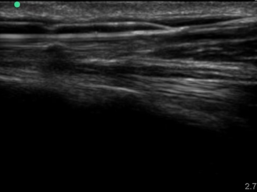
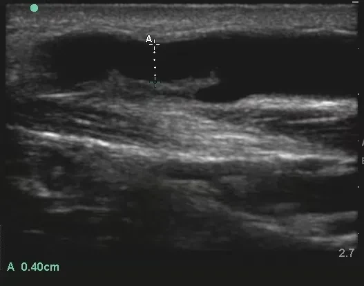
Marking of the course and stenosis location of arteriovenous fistula vessels.
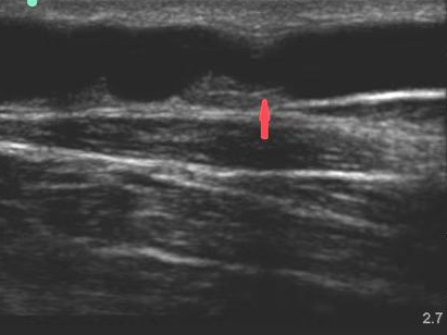
The arrow points to the stenosis of the arteriovenous fistula.
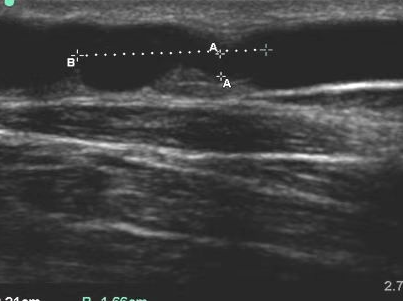
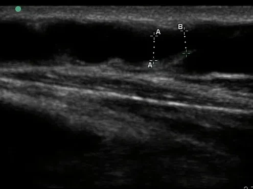
Total length of stenosis 16.6mm, diameter 2.1mm。
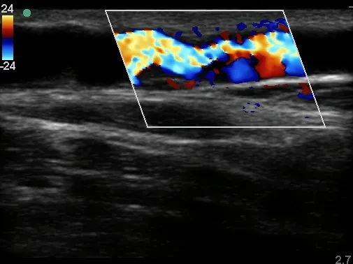
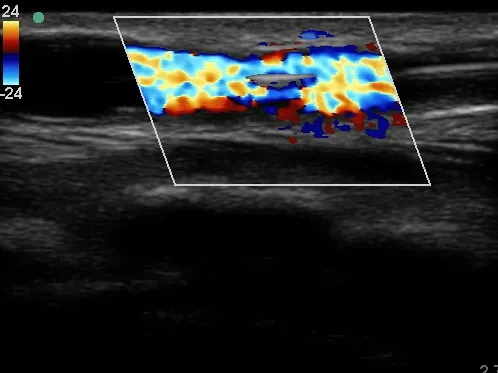
The blood flow at the stenosis is poor.
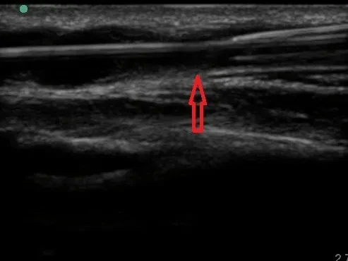
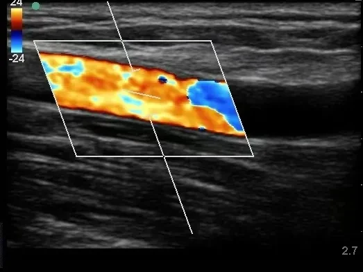
Stenosis of the internal fistula after guidewire placement.
Pre-dilation Blood Flow Diagram.
DK Medtech 6.0*60mm DKutting™ High-Pressure Scoring BalloonDilate the narrowed section.
Dilation pressure 22atm, duration 1min30s.
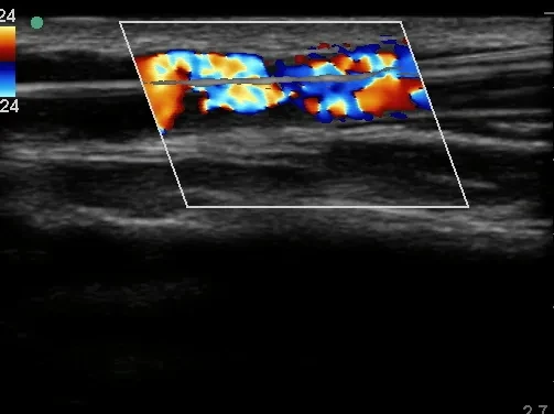
Blood Flow Diagram at the Stenosis of the Expanded Internal Fistula.
The diameter can reach up to 3.6mm immediately after the operation, with a blood flow rate of 973ml/min.
Follow-up
Discharge Status:The thrill of the patient's arteriovenous fistula significantly increased after the surgery. The blood flow through the arteriovenous fistula puncture was satisfactory, and the temporary catheter in the right internal jugular vein was removed.
The diameter of the narrowest part can reach 4mm at its widest point 2 days after the operation.
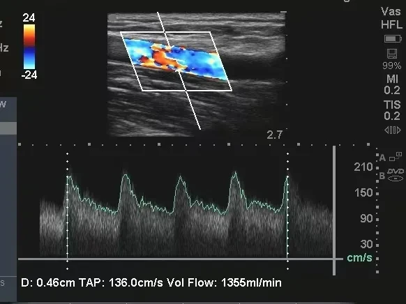
The blood flow of the arteriovenous fistula reached 1355ml/min two days after the operation.
Case Summary
Case Characteristics:The patient's arteriovenous fistula has poor maturation due to valve calcification and thickening, leading to stenosis.
Preoperative Assessment Key Points:Preoperative ultrasound carefully evaluates the lesion site, degree of stenosis, and blood flow volume of the arteriovenous fistula.
Surgical Strategy/Technical Key Points:During the operation, the patient experienced retrograde blood flow over the guidewire, making it difficult for the guidewire to pass through the stenotic area. The correct position was selected based on the direction of blood flow. After continuous random rotation control of the guidewire, it successfully passed through the lesion area. Considering that the patient's lesion was valvular with significant surrounding hyperplasia and the fistula was new, high-pressure dilation could easily cause vascular rupture. Therefore, a scoring balloon was chosen for uniform cutting of the lesion. Care was taken during the procedure to avoid vascular injury.
Features/Usage Tips of the Device:Select a scored balloon of appropriate size based on the width of the patient's fistula. Avoid excessively forceful expansion of the blood vessel, and ensure accurate positioning when using the scored balloon during the procedure.
References:Expert Consensus on Standardized Operating Procedures for Percutaneous Transluminal Angioplasty of Arteriovenous Fistula Under Ultrasound Guidance (2024).

Expert Introduction


Professor Hua Yu
Surgeon of This Case
Deputy Chief Physician of Nephrology Department, Zhabei Central Hospital, Jing'an District, Shanghai.Proficient in the establishment of vascular access, including arteriovenous fistula formation, long-term tunneled catheter placement with Dacron cuff, balloon angioplasty for arteriovenous fistula, etc.Received 2 research projects from the Health Commission of Jing'an District, Shanghai in 2018 and 2023, and participated in 4 projects.Published more than 10 papers in China and 1 SCI paper.

Department Introduction

Department of Nephrology, Zhabei Central Hospital, Jing'an District, Shanghai
In 1992, the Hemodialysis Room was established.
In 1994, the Nephrology Professional Group was established.
In 2003, the Department of Nephrology was officially established.
Key Medical Discipline of Zhabei District, 2004
2009 Zhabei District Medical Demonstration Discipline
Group Leader Unit of Nephrology Professional Quality Control Group, Zhabei District, 2010
2012 Renal Disease Prevention and Treatment Master Studio, Zhabei District
Key Medical Specialty of Shanghai in 2012
2016 Jing'an District Nephrology Professional Quality Control Group Leader Unit
Group Leader Unit of the Nephrology Group, Jing'an District Medical Association, 2017
2017 National Drug Clinical Trial Base GCP
Key Medical Specialty of Shanghai in 2019