
Percutek Therapeutics
Developer of Minimally Invasive Cardiovascular Treatment Devices

Today's sharing is byProfessor Xu Liang's Team from the Second People's Hospital of Nanyang CityA Case of Highly Complex TEVAR Surgery Successfully Completed. This surgery achieved in-situ reconstruction of the arch branch using only a V-18 guidewire, showcasing the team's exceptional skill and innovative spirit. The patient’s condition was complex, with the dissection entry located on the anterior wall of the lesser curvature side, retrograde dissection, the proximal lesion involving the root of the left subclavian artery (LSA), and the distal lesion extending to the left common iliac artery. High demands were placed on the stent’s sealing ability as well as the delivery system’s trackability and flexibility.
In response to these challenges, Professor Xu Liang's team skillfully and innovatively aligned the proximal end of Percutek Therapeutics' thoracic aortic stent graft with the posterior edge of the left common carotid artery (LCCA) ostium before deployment. They successfully performed in-situ fenestration via the V-18 guidewire penetrating the graft membrane, completing the in-situ reconstruction TEVAR procedure for the left subclavian artery (LSA). This approach precisely overcame technical difficulties such as controlling the angle of in-situ fenestration and establishing vascular access, ensuring complete closure of the lesion while efficiently reconstructing LSA blood supply.

Gender:Male
Age:78 years old
Chief Complaint:Chest and back pain for 5 hours.
History of Present Illness:Chest and back pain occurred 5 hours ago without obvious cause, presenting as persistent tearing pain. Self-administered pain medication (details unknown) had little effect. The patient came to our hospital for treatment and was admitted by the emergency department with a diagnosis of "aortic dissection formation."
Past Medical History:History of hypertension for over 1 year, with a maximum of 160/110 mmHg. Irregular medication use, and blood pressure is generally controlled.
CTA Details:Stanford Type B Aortic Dissection, the tear is located on the anterior wall of the lesser curvature side. The proximal lesion involves the root of the left subclavian artery (LSA). The distance between the root of the left common carotid artery (LCCA) and the root of the LSA is approximately 14mm, with an aortic diameter of about 29mm at the posterior edge of the LCCA root opening. The lesion has a wide range, with the distal end involving the left common iliac artery. The left vertebral artery is dominant, and the true lumen supplies blood to the celiac trunk, superior mesenteric artery, and bilateral renal arteries. Calcified plaques are present in the aortic arch, the distal abdominal aorta, and the bilateral common iliac arteries.
Preoperative Three-dimensional Reconstruction

Preoperative CTA Cross-Section

Condition of Dissection Lesions

Stanford Type B Aortic Dissection, with the proximal lesion involving the root of the LSA, showing a significant lack of proximal anchoring zone. It is necessary to extend the anchoring zone proximally and reconstruct the LSA.
The lesion range of aortic dissection is extensive, with calcification present on the lesser curvature side of the aorta corresponding to the root of the left subclavian artery (LSA), and the anatomical conditions of the anchoring zone are poor. High demands are placed on the sealing, flexibility, conformability, and anchoring stability of thoracic aortic stent grafts.
Calcified plaques are present in the aortic arch, the distal abdominal aorta, and the bilateral common iliac arteries; gentle manipulation is required.

Surgical Plan Strategy
Endovascular Repair of Thoracic Aortic Stent Graft Directly Covering the Left Subclavian Artery: The procedure is simple and effective, but covering the left subclavian artery may lead to clinical manifestations of posterior circulation ischemia and upper limb ischemia in patients.
Endovascular Repair of Thoracic Aortic Stent Graft + Left Subclavian Artery Chimney Technique: The surgical procedure is relatively simple, but the risks of endoleak and occlusion are significant, with suboptimal mid- to long-term outcomes.
Endovascular Repair of Thoracic Aortic Stent Grafts with In Vitro Fenestration: Excellent sealing of lesions, preservation of original hemodynamic characteristics, but complex operation. Preoperative stent modification is required based on measurement results, which is time-consuming; intraoperative precise alignment and ultra-selective window positioning are required, posing higher risks.
Endovascular Repair of Thoracic Aortic Stent Graft + In-situ Fenestration: Excellent sealing of the lesion, no preoperative stent modification required. However, traditional in-situ fenestration of aortic stent grafts demands advanced interventional equipment, such as in-situ fenestration needles, lasers, biopsy needles, and other specialized membrane-piercing devices.
The Hua Mai thoracic aortic stent graft can perform in-situ fenestration using the soft tip of a CTO guidewire. Considering the mid-to-long term treatment outcomes and the simplicity of intraoperative procedures, after comprehensive evaluation, Professor Xu Liang's team chose Hua Mai • Tian Yi.®Endovascular repair of the thoracic aorta with a covered stent graft and reconstruction of the left subclavian artery using the in-situ fenestration technique.

01.The patient was placed in the supine position, routinely disinfected and draped. After local anesthesia, the right femoral artery was punctured; the left brachial artery was punctured, a 6F arterial sheath was placed, and a guidewire was inserted along with a pigtail catheter which was advanced into the ascending aorta and connected to a high-pressure injector.
02.Angiography of the ascending aorta showed: A dissection shadow visible in the descending aorta, with the entry tear located approximately 7mm distal to the LSA. A localized dissection is visible in the left common iliac artery, with slow blood flow.

Preoperative Thoracic Angiography 1

Preoperative Thoracic Angiography 2

Preoperative Abdominal Aortic Angiography
03.Through the right femoral artery, the true lumen was selectively accessed, and the device was advanced into the ascending aorta. After exchanging for a stiff guidewire, a Percutek Therapeutics PTBS2828150 thoracic aortic stent graft was placed. The restrictive stent was positioned 10 cm distal to the ostium of the left subclavian artery (LSA) and deployed.

Release Restrictive Stent

Post-release of bare stent
04.The Percutek Therapeutics thoracic aortic stent graft PTBS3434150 was implanted proximally and precisely deployed distal to the LCCA ostium. The overlap between the distal end and the restrictive stent was approximately 3 cm. Angiography after deployment showed: accurate positioning of the proximal end of the stent without displacement, good sealing of the rupture, and excellent opening of the distal true lumen.

Proximal Stent Placement Angiography

Release Proximal Stent

Post-release of bare stent

Post-release Contrast
05.A 6F long sheath was delivered through the left brachial artery sheath, and the LSA fenestration was successfully performed using a V-18 guidewire in conjunction with a single-bend catheter. Balloon dilation of the fenestration site was sequentially carried out using 4mm, 6mm, and 8mm balloons until the balloon waistline disappeared.

Contrast Localization of Left Vertebra

V-18 Guidewire Successfully Opens Window

4mm Balloon Dilation Opening

6mm Balloon Dilation Opening

8mm Balloon Dilation Opening
06.LSA implanted a 10-38mm balloon-expandable covered stent and released it. Postoperative angiography showed: satisfactory closure of the dissection, no type I endoleak at the proximal end, good position and morphology of each stent, and unobstructed blood flow in each branch of the aortic arch.

Postoperative Angiography
07.Withdraw the delivery device and guidewire. After ligating the femoral artery purse-string, there was no bleeding. The incision was sutured layer by layer and compressed with a bandage. The arterial sheath at the left brachial artery incision was removed and then compressed and fixed with a bandage. The procedure was completed.

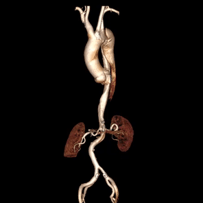
Comparison of One Week Postoperative with Preoperative

Preoperative/Postoperative 3D Reconstruction Comparison

Preoperative/Postoperative CTA Axial Contrast
Postoperative review showed good recovery. The thoracic main stent conformed to the three-dimensional torsion of the aorta, with good wall apposition. Blood flow in each branch above the arch was unobstructed, with no signs of cerebral infarction or cerebral ischemia. The aortic dissection was completely isolated, with no endoleak. The true lumen was restored, and the false lumen was thrombosed.

Summary of Case Experience
The case reported this time is a Stanford Type B aortic dissection, with the tear located only 7mm distal to the LSA orifice. The proximal retrograde dissection extends to the root of the LSA, resulting in a severe lack of proximal anchoring zone. Additionally, multiple calcified plaques are present on the concave side of the aorta, further weakening the structural condition of the anchoring area. This situation places higher demands on the anchoring stability, sealing performance, compliance, and conformability of the stent graft. Under these circumstances, ensuring effective closure of the lesion while safeguarding the patient’s long-term prognosis has become the primary challenge in treating this case.
In response to the above challenges, Professor Xu Liang's team made a precise decision to use the Percutek Therapeutics Thoracic Aortic Stent Graft System for treatment. The operation proceeded smoothly, with the puncture easily completed using only a V-18 guidewire. The balloon-expandable stent graft process at the fenestration site was simple and safe, efficiently achieving supra-arch reconstruction of the LSA. Postoperative review showed that blood flow in all arch branches was unobstructed, the stent had an excellent profile, the dissection was completely sealed without endoleaks, and the false lumen achieved complete thrombosis. The patient had a favorable prognosis, and the LSA reconstruction outcome met expectations, fully demonstrating the surgical team’s exceptional technical expertise and extensive clinical experience.

Introduction of Experts

Professor Liang Xu
Deputy Chief Physician of the Department of Cardiac and Vascular Surgery, Nanyang Second People's Hospital. Engaged in cardiac and vascular surgery for over 10 years. Completed advanced studies in vascular surgery at the General Hospital of the Chinese People's Liberation Army (Beijing 301 Hospital), Henan Provincial People’s Hospital (Fu Wai Central China Cardiovascular Hospital), and Shanghai Renji Hospital. Mainly engaged in the treatment and prevention of vascular-related diseases: aortic dissection, thoracic and abdominal aortic aneurysms, deep vein thrombosis, pulmonary embolism, lower extremity arteriosclerosis obliterans, acute limb arterial embolism, iliac vein compression syndrome, congenital heart defect occlusion, mesenteric artery, renal artery, subclavian artery stenosis, Budd-Chiari syndrome, varicose veins of the great saphenous vein, hemangioma, lymphedema, etc. Actively involved in research and application of hybrid techniques for combined surgical procedures. Published seven papers in core journals, participated in editing two monographs, and holds one invention patent.
Currently serving as a Quality Control Committee Member of the Peripheral Vascular Group at the Henan Province Cardiovascular System Disease Quality Control Center, a Quality Control Committee Member of the Great Vessels Group at the Henan Province Peripheral Vascular Intervention Technology Management Committee Quality Control Center, a Standing Committee Member of the Vascular Surgery Branch of the Henan Province Hospital Management Association, a Standing Committee Member of the Structural Heart Disease Society of the Henan Province Biomedical Engineering Association, a Committee Member of the Cardiac and Great Vessels Surgery Branch of the Henan Province Physician Association, the Vice President of the Vascular Surgery Branch of the Nanyang City Physician Association, a Committee Member of the Henan Province Interventional Therapy Society, a Committee Member of the Vascular Surgery Society, and a Committee Member of the Vascular Disease Branch of the Henan Province Respiratory and Critical Care Medicine Society.


Department Introduction
Cardiovascular and Great Vessels Surgery Ward II, Department of the Second People's Hospital of Nanyang City
The Second Ward of the Cardiac and Great Vessel Surgery Department at Nanyang Second People's Hospital is located on the 9th floor, west wing, of Building 5 in the inpatient department. The ward has 35 open beds and a medical team consisting of two chief physicians, two deputy chief physicians, one attending physician, and five resident physicians. Focusing primarily on minimally invasive interventional surgeries, the department routinely performs endovascular repair for aortic dissection, endovascular repair for thoracic and abdominal aortic aneurysms, minimally invasive transcatheter aortic valve replacement (TAVR), balloon dilation for mitral stenosis, interventional procedures for congenital heart diseases (including closure of patent foramen ovale, atrial septal defect closure, ventricular septal defect closure, patent ductus arteriosus closure, and balloon dilation for pulmonary valve stenosis), endovascular angioplasty for lower extremity arterial occlusive disease and diabetic foot, thrombectomy and endovascular treatment for deep vein thrombosis and pulmonary embolism, endovascular treatment for renal artery stenosis, endovascular treatment for mesenteric artery dissection, visceral arteriovenous diseases, as well as conventional and minimally invasive surgery for varicose veins of the great saphenous vein and lymphedema. The department’s philosophy is "Virtue, Integrity, Innovation, Excellence," and with your trust, we strive to restore your health!


END
Copyright Statement: This platform aims to help medical and health professionals better understand the latest developments in relevant disease areas. The information content published on this platform does not imply agreement with its descriptions or viewpoints; it is only for providing more information. If there are any copyright issues, we kindly request the rights holders to contact us, and we will handle them as soon as possible. This information is intended solely for medical and health professionals to stay informed, and such information cannot replace professional medical guidance in any way, nor should it be regarded as medical advice. If such information is used for purposes other than staying informed, this platform and the author shall not bear any related responsibilities.Contact email for cooperation:vascular@edoctor.work。