Home Professor Zhuang Hui's Team Reports First-in-Nation Clinical Application of DEEPQUAKE Peripheral Intravascular Lithotripsy System for Diabetic Foot Revascularization

Professor Zhuang Hui's Team Reports First-in-Nation Clinical Application of DEEPQUAKE Peripheral Intravascular Lithotripsy System for Diabetic Foot Revascularization

Oct 20, 2025 07:31 CST Updated 07:31
Trulive

Structural Heart Disease Device Developer

Image
Image


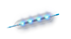
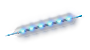
In the field of pan-vascular intervention, the treatment of calcified lesions is one of the key and difficult points in clinical practice. Produced by Trulive and promoted by Grand Pharmaceutical Group Limited,DEEPQUAKE Peripheral Vascular Intravascular Shockwave SystemOfficially launched (China Medical Device Registration No. 20243012335). This system is suitable for the pretreatment of calcified lesions and balloon dilation in the iliac artery, femoral artery, popliteal artery, renal artery, and infrapopliteal arteries, providing new treatment options for clinical use.


This issue is shared with everyoneDEEPQUAKE Peripheral Shock Wave System: First Application in China — A Case of Diabetic Foot and Lower Limb Vascular Intervention, by the Cardiovascular Hospital Affiliated to Xiamen UniversityProfessor Zhuang Hui's TeamCompleted, welcome everyone to read, study, exchange, and discuss.


Features of DEEPQUAKE Peripheral Shock Wave System
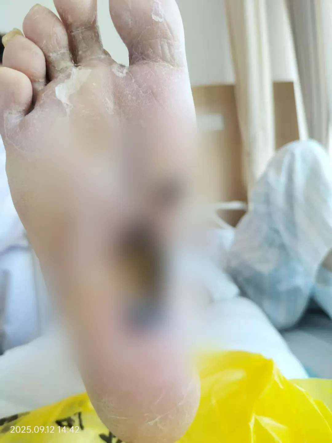

Advantage 1LargerPulse Energy

  • Energy adjustable in five levels, with the highest energy reaching 3.2kV (fixed energy of 3.0kV for similar products).

Gear

Energy

Gear 1

2.8kV

Level 2

2.9kV

3rd Gear

3.0kV

4-speed

3.1kV

5 gears

3.2kV


Advantage 2MoreNumber of electrodes

  • The balloon is equipped with one pair of electrodes every 8mm, and the three lengths of 40/60/80mm have 4/6/8 pairs of electrodes respectively, ensuring uniform energy release. Treatment stitching does not require "Overlapping".

Image

40mm

4 pairs of electrodes

Image

60mm

6 pairs of electrodes

Image

80mm

8 pairs of electrodes


Advantage 3LongerBalloon Length

  • Provides an 80mm length balloon, more suitable for long lesions,Reducing operation time and IVL treatment frequency, cost-effective

Image


Case Brief

Patient Basic Information

Patient:73 years old, female.

Chief Complaint:"Right lower limb claudication for more than 3 years, right foot sole ulcer and gangrene for more than half a month" admitted to the hospital, Day 4.

History of Present Illness:Two years ago, the patient developed intermittent claudication in the right lower limb without obvious cause. Half a month ago, an ulcer appeared on the sole of the right foot with severe pain and poor response to medication. The patient then visited our outpatient department and was admitted to the hospital with a diagnosis of "atherosclerotic occlusion of the lower extremity arteries and diabetic foot." During the course of the disease, the patient had poor appetite and sleep, general mental state, reduced urine output, normal bowel movements, and reported slight weight loss.

Past Medical History:Type 2 Diabetes Mellitus (>10 years, insulin + oral medication), Chronic Renal Insufficiency, Coronary Heart Disease with Chronic Heart Failure, Refractory Hypertension (left renal artery stent half a month ago), Extensive Atherosclerosis.

Physical Examination:Patient is conscious, appears anemic, and in fair spirits, reports poor appetite: Abdomen is soft, with mild tenderness in the right/upper middle abdomen, no rebound tenderness; low skin temperature and pale skin below the right knee, gangrene on the sole of the right foot, obvious tenderness on touch, exudate present but no significant foul odor; lower limb arteries: bilateral femoral arteries palpable, left popliteal artery ++ right -, left posterior tibial artery ++ right -, dorsalis pedis arteries bilaterally -; decreased muscle strength and superficial/deep sensation in the right toes.

ABI:Right 0.46; Left 0.59.

Rutherford Classification:Level 6.

Image
Image

Preoperative

Diagnosis and Surgical Strategy

Preliminary Diagnosis:Diabetic Foot.

Proposed Surgical Strategy:Debulking (Shockwave Balloon) + DCB.

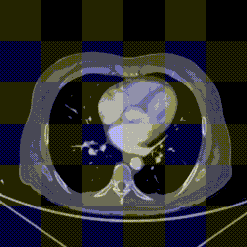
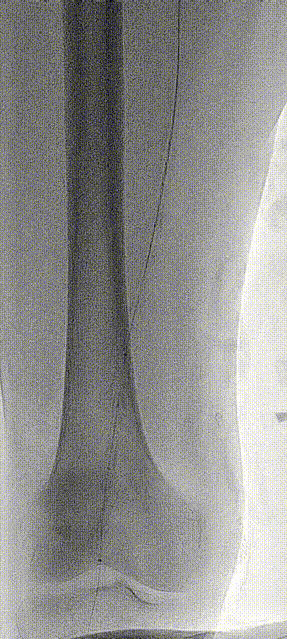
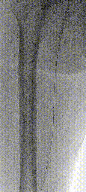
Baseline Angiography

The patient was in the supine position, routinely disinfected and draped. Under ultrasound guidance in the left inguinal region, a retrograde puncture of the left femoral artery was successfully performed, and a 5F vascular sheath was inserted. Under DSA, a guidewire + catheter was advanced to the bifurcation of the abdominal aorta for angiography: multiple calcified plaques were visualized in both common iliac arteries, and blood flow in both iliac arteries was unobstructed.

Image

Lesion Clearance Process

The C2 catheter was advanced across to the right deep femoral artery, and a super-stiff guidewire was left in place. A 7F 55cm long sheath was then positioned in the common femoral artery. Angiography through the sheath revealed extensive calcification throughout the superficial femoral artery, with an approximately 5cm occlusion in the mid F2 segment, and mild-to-severe stenosis in the remaining lumen. Delayed imaging showed collateral circulation from the deep femoral artery to the popliteal artery and infrapopliteal arteries. Severe calcification and stenosis were observed in the P1 segment of the popliteal artery. The rest of the popliteal artery and the tibioperoneal trunk demonstrated patent blood flow. The anterior tibial artery showed good contrast filling, moderate stenosis in part of the posterior tibial artery with slightly sluggish flow, and the peroneal artery filled well with patent vessels below the ankle.

VR catheter + CXI catheter + guidewire successfully passed through the lesion segment in the antegrade direction, establishing an antegrade pathway. The guidewire was exchanged and placed in the proximal segment of the peroneal artery. A 4*120mm 0.14 balloon was used for pre-dilation of the lesion segment. Angiography showed significant improvement in blood flow with elastic recoil in multiple calcified segments.

Image
Image
Image

Shockwave BalloonLesion Preprocessing

InDEEPQUAKE Shock Wave BalloonThe 6*80mm segmental dilation P1 to the opening segment of the superficial femoral artery was performed using level 5 energy, with a total of 17 cycles and 510 shocks, completing the treatment of the entire lesion segment. No contrast agent leakage was observed upon reexamination.

Image
Image
Image
Image

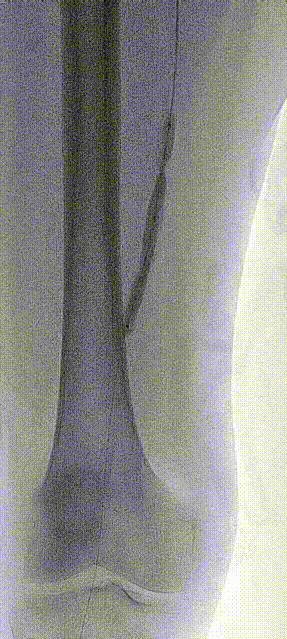
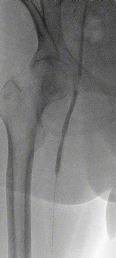
Lesion Treatment Process
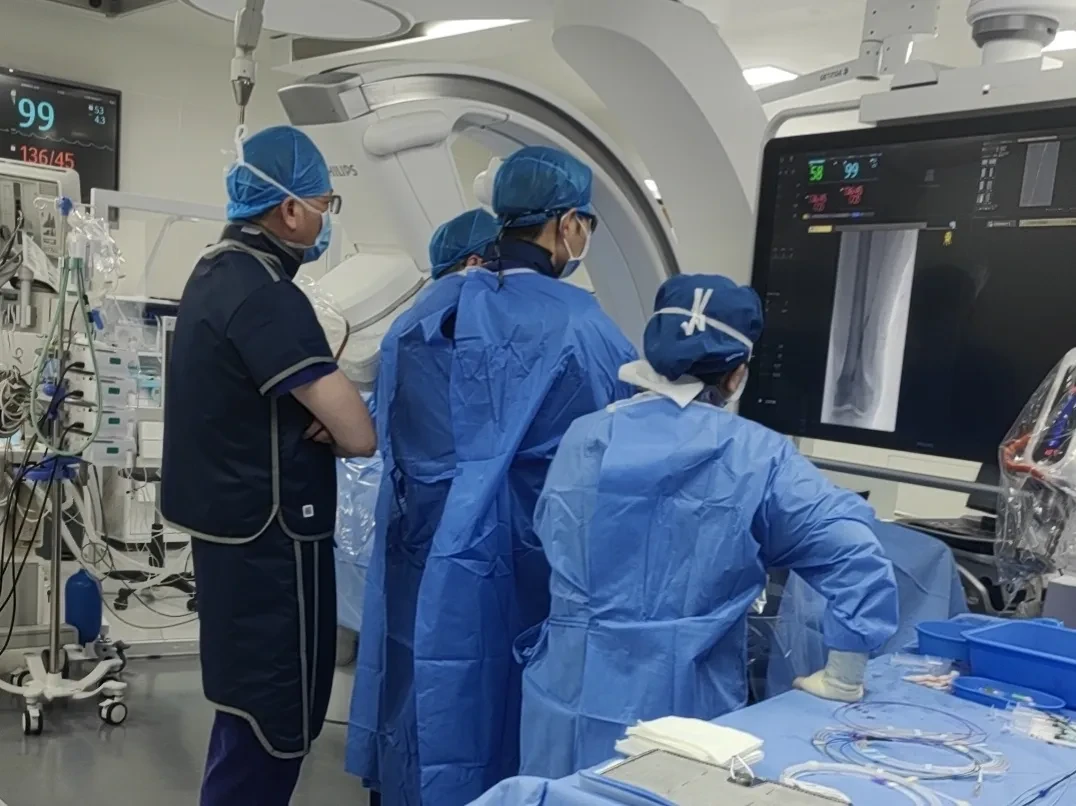
A short segment dissection was formed in the distal segments of P1 and F1, without flow limitation. After exchanging for an ultra-hard guidewire, sequential dilation was performed on the entire segment from P1 to the superficial femoral opening using a 5*220mm drug-coated balloon and a 6*150mm drug-coated balloon, lasting for 3 minutes. Repeated angiography showed significant improvement in blood flow, with partial healing of the P1 segment dissection. Follow-up angiography revealed smooth blood flow throughout the superficial femoral artery, good visibility of the infrapopliteal branches, palpable dorsalis pedis artery pulse, and no change in the iliac artery.

Image
Image

Withdraw the catheter sheath, suture the puncture site with a suture, apply local pressure for several minutes, and then apply pressure dressing.

Image


Operator Evaluation

  1. DEEPQUAKE Shock Wave System offers higher energy levels for efficient resolution of calcified lesions;

  2. More treatment sessions and stable energy output are required to ensure the effectiveness of treating long-segment calcified lesions.

  3. Diabetic patients experience overall reduced arterial compliance, with challenges in achieving patency, low long-term patency rates, and poor peripheral perfusion. The use of shockwave balloons not only decreases the likelihood of rescue stent implantation but also improves the endovascular treatment prognosis for such patients.


Expert Introduction

Image

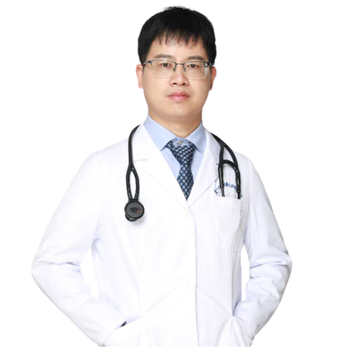
Professor Hui Zhuang

Director of the Vascular Surgery Department, Cardiovascular Hospital Affiliated to Xiamen University.

Specializes in endovascular individualized customized treatment for aortic diseases, specializes in surgical treatment of peripheral arterial occlusive disease and diabetic foot, excels in endovascular treatment of infrapopliteal arteries, and excels in endovascular treatment of pelvic venous insufficiency.

Led multiple provincial-level research projects, completed numerous national-level papers and SCI papers.

Participated in the compilation and translation of several vascular surgery books and contributed to the development of multiple domestic vascular specialty guidelines. Completed numerous invention patents and utility model patents.

Currently:Deputy Chairman of the Youth Committee of the Vascular Surgery Professional Committee of the National Cardiovascular Disease Expert Committee; Member of the Lower Limb Artery Committee of the Vascular Surgery Professional Committee of the National Cardiovascular Disease Expert Committee; Member of the Endovascular Professional Committee of the Chinese Medical Doctor Association;Committee Member of the Visceral Artery Group, Vascular Surgery Physician Branch, Chinese Medical Doctor Association;Member of the National Peripheral Vascular Intervention Quality Control Professional Committee.
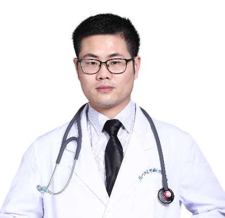

Image

Professor Sun Hu

Leader of the Vascular Surgery Medical Team at the Cardiovascular Hospital Affiliated with Xiamen University, engaged in vascular surgery with extensive clinical experience in various vascular diseases. Specializes in the treatment of varicose veins of the lower extremities, arteriosclerosis obliterans of the lower extremities, diabetic foot, ulcers of the lower extremities, deep vein thrombosis of the lower extremities, pulmonary embolism, and abdominal aortic aneurysms.

Member of the Vascular Surgery Physicians Branch of the Xiamen Medical Association, Member of the Vascular Surgery Branch of the Xiamen Medical Association, Member of the Intervention Branch of the Xiamen Medical Association, Youth Member of the Vascular Surgery Branch of the Fujian Medical Association, Member of the Vascular Surgery Professional Committee of the Fujian Strait Medical and Health Exchange Association, Member of the Vascular Collateral Disease Branch of the Fujian Integrated Traditional Chinese and Western Medicine Association, Member of the Endovascular Science Popularization and Humanities Committee of the Chinese Medical Doctor Association, Member of the Vascular Malformation and Access Professional Committee of the China Health Science and Technology Promotion Association, Member of the Expert Committee on Pressure Group of the Peripheral Vascular Disease Professional Committee of the Chinese Society of Microcirculation, Member of the Wound Treatment Group of the Plastic and Reconstructive Surgery Professional Committee of the Chinese Rehabilitation Medical Association.


Image

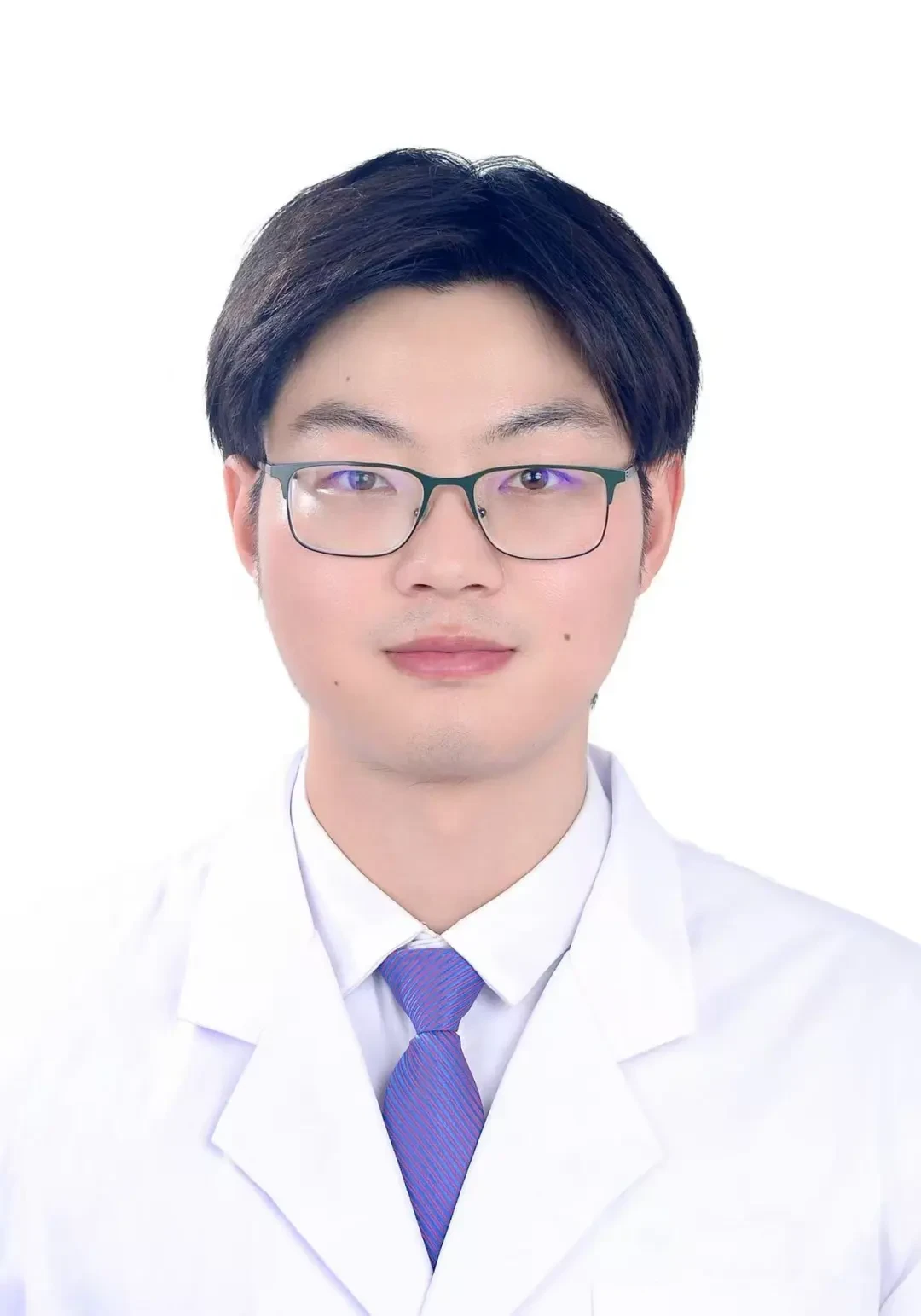
Professor Shen Xuwei

Attending Physician, Department of Vascular Surgery, Cardiovascular Hospital Affiliated to Xiamen University; Master of Medicine.

Professional Direction and Specialties:Proficient in the diagnostic and treatment protocols for common and frequently-occurring diseases in vascular surgery;Diagnosis and Treatment of Lower Extremity Arteriosclerosis and Embolic Diseases, Carotid Artery Diseases;Diagnosis and treatment of lower extremity varicose veins, thrombosis, and chronic venous insufficiency, as well as interventional diagnosis and treatment of various emergency and peripheral vascular diseases.


Image

Professor Li Lin

Resident Physician of Vascular Surgery, Cardiovascular Hospital Affiliated to Xiamen University.

2014 Bachelor/Undergraduate Nanjing Medical University; 2019 Master/Graduate Nanjing Medical University First Affiliated Hospital; 2023 Doctorate/Graduate Heidelberg University, Germany.

2020-2021 BenQ Medical Center, Nanjing Medical University;2021-Affiliated Cardiovascular Hospital of Xiamen University.


Previous Recommendations

【Leave Nothing Behind】Professor Jia Nan's Team: DEEPQUAKE Peripheral Shock Wave System First Application in China — Angioplasty for Dialysis Patients with Lower Limb Arterial Disease

[Breakthrough Limits, Conquer Calcification] Professor Zhang Yan / Professor Guo You Collaboration: DEEPQUAKE Peripheral Shock Wave System First Applications in China — A Case of Long-Segment Severe Calcification Lesion in the Superficial Femoral Artery

@CAVS 2025 | Grand Pharmaceutical Group Limited's Peripheral Shock Wave Academic Symposium Successfully Convened

[More Complete Models, Comprehensive Coverage] Professor Fang Xin's Team: DEEPQUAKE Peripheral Shock Wave System First Application in China -- A Case of Infrapopliteal Artery Calcification

[Five-level Treatment, Safe and Effective] Professor Li Xin's Team: The First Application of DEEPQUAKE Peripheral Shockwave Balloon in China — A Case of Long-segment Calcified Lesions in the External Iliac, Common Femoral, and Superficial Femoral Arteries

【Piercing the Clouds, Splitting the Stones – To the Utmost Perfection】Director Tian Hongyan / Professor Zhang Junbo's Team: First Application of DEEPQUAKE Peripheral Shockwave Balloon in China - A Case of Severe Lower Limb Calcification

【Piercing the Clouds, Splitting the Stones, Achieving Excellence】Professor Guan Sheng's Team: A Powerful Impact to Save Limbs — The Healing Journey of a Severely Calcified Patient

【Piercing the Clouds, Splitting the Stone – Striving for Excellence】Director Chen Zhong / Professor Wang Sheng Team: DEEPQUAKE Peripheral Shockwave Balloon First Applications in China – Long-Segment Calcified Lesions of the Superficial Femoral Artery

【Piercing the Clouds, Splitting the Stone, Achieving Excellence】Director Chen Zhong / Professor Yang Yaoguo Team: DEEPQUAKE Peripheral Shockwave Balloon First Applications in China — Long-Segment Calcified Lesions in Femoropopliteal and Below-the-Knee Arteries

Grand Pharmaceutical Group Limited's DEEPQUAKE Peripheral Shock Wave System Debuts at the 4th ICCD Cardiovascular and Cerebrovascular Device Conference


Copyright Statement: This platform aims to help healthcare professionals better understand the latest developments in relevant disease areas. The information published on this platform does not imply agreement with its descriptions or viewpoints; it is solely for providing more information. If there are any copyright issues, we kindly request the rights holders to contact us, and we will address them as soon as possible. The information is intended for healthcare professionals to use as a reference, and such information cannot replace professional medical guidance in any way, nor should it be regarded as medical advice. If such information is used for purposes other than understanding updates, this platform and its authors assume no related responsibility.Contact Email for Cooperation:vascular@edoctor.work。

Image