Home "Guidewire Fenestration + Cerebral Protection Innovation": Prof. Huang Jinqi's Team Treats Complex Thoracic Aortic Aneurysm with LSA Involvement Using Novel Fenestrated Thoracic Stent Graft and Custom Cerebral Embolic Protection Device

"Guidewire Fenestration + Cerebral Protection Innovation": Prof. Huang Jinqi's Team Treats Complex Thoracic Aortic Aneurysm with LSA Involvement Using Novel Fenestrated Thoracic Stent Graft and Custom Cerebral Embolic Protection Device

Oct 23, 2025 07:30 CST Updated 07:30
Percutek Therapeutics

Developer of Minimally Invasive Cardiovascular Treatment Devices

Today, I will share a case byProfessor Huang Jinqi's Team from the First Hospital of Putian City, Fujian ProvinceA Highly Challenging TEVAR Surgery Case. The patient was diagnosed with a thoracic aortic aneurysm, combined with complex lesions of the left subclavian artery (LSA): poor anatomical conditions of the aortic arch, with the proximal part of the aneurysm involving the origin of the LSA, resulting in insufficient proximal anchoring zone.At the same time, there is aortic atherosclerosis, plaque, mural thrombus, and multiple ulcers in the ascending aorta and aortic arch, with an extremely high risk of cerebral infarction during surgery, making the condition very complex.


Treating this case not only requires effectively isolating the lesions in the aorta and the LSA branch while reconstructing the LSA, but more importantly, it is crucial to focus on cerebral protection to reduce the risk of cerebral infarction caused by emboli detachment during surgery. After thoroughly analyzing the patient's condition, Professor Huang Jinqi’s team developed a meticulous surgical plan: using a 250T guidewire to perform in-situ fenestration on the Percutek Therapeutics thoracic aortic stent graft to achieve rapid LSA reconstruction and lesion isolation. During the operation, the team innovatively applied a self-made simplified and improved cerebral embolic protection device, significantly reducing the possibility of complications such as cerebral embolism in elderly patients.


Thanks to the team's sophisticated techniques and precise operations, the surgery was successfully completed. Postoperative angiography confirmed that the lesion area was completely isolated, with smooth blood flow in the aorta and its arch branches, achieving satisfactory surgical outcomes.


Image


Medical Condition Introduction


Gender:Male

Age:81 years old

Chief Complaint:Cough, expectoration, and hemoptysis for 3 days.

History of Present Illness:Three days before admission, the patient developed cough, expectoration, and hemoptysis without obvious cause. The symptoms occurred in paroxysmal bouts of consecutive coughs, with a small amount of white mucous sputum. The patient visited a local clinic and received medication treatment (details unknown), but the symptoms did not improve significantly. For further diagnosis and treatment, the patient came to our hospital and was admitted to our department with a preliminary diagnosis of "pneumonia."

Admission Diagnosis:Pulmonary infection; Mediastinal mass (to be investigated); Hypertension.

CTA Details:The proximal end of the thoracic aortic aneurysm is closely adjacent to the ostium of the LSA, with a maximum diameter of approximately 49.9mm and a length of about 48mm; the LSA shows atherosclerosis, multiple ulcers, plaques accompanied by localized dissection, with the lesion being approximately 21mm away from the origin of the left vertebral artery; the distance between the LCCA and the LSA is about 12mm, and the aortic diameter at the posterior edge of the LCCA ostium is approximately 33.7mm.


Image

Preoperative Three-dimensional Reconstruction


Image

Preoperative CTA Cross-Section

Image

Condition of the Aortic Arch

Image

LSA Lesion Condition


Image


Treatment Challenges


The proximal end of the thoracic aortic aneurysm lesion is adjacent to the root opening of the LSA, with a significantly insufficient proximal anchoring zone, necessitating the extension of the anchoring zone proximally and the reconstruction of the LSA.

The patient is elderly and in poor physical condition. Atherosclerosis is present at the proximal opening of the LSA, with multiple ulcers and plaques observed, accompanied by localized dissection and mural thrombus formation. During the operation, caution should be exercised to minimize arch manipulation, ensure proper brain protection, and prevent complications such as cerebral embolism.

Large aortic aneurysm requires thoracic aortic stent grafts with excellent sealing, flexibility, and conformability.


Image


Surgical Plan Strategy


01

Endovascular Repair of Thoracic Aortic Stent Graft + Left Subclavian Artery Chimney Technique: The surgical procedure is relatively simple, but there is a higher risk of endoleak and occlusion, with suboptimal mid- to long-term outcomes.

02

Endovascular Repair of Thoracic Aortic Stent Graft with In Vitro Fenestration: Excellent sealing of the lesion, preservation of original hemodynamic characteristics, but complex operation. Preoperative stent modification is required based on measurement results, which is time-consuming; intraoperative precise alignment and ultra-selective window positioning are required, posing higher risks.

03

Endovascular Repair of Thoracic Aortic Stent Graft + In-situ Fenestration: Excellent sealing of the lesion, no need for preoperative stent modification. However, traditional in-situ fenestration of aortic stent grafts requires advanced interventional instruments such as in-situ fenestration needles, lasers, biopsy needles, and other specialized membrane-piercing devices.


The Hua Mai Thoracic Aortic Stent Graft can perform in-situ fenestration using the soft tip of a CTO guidewire. Considering the mid-to-long term treatment outcomes and the simplicity of intraoperative manipulation, after comprehensive evaluation, Professor Huang Jinqi's team chose Hua Mai • Tian Yi.®Endovascular repair of the thoracic aorta with a stent graft and reconstruction of the left subclavian artery using the in-situ fenestration technique via guidewire.


Image


Surgical Procedure


 01. After the patient was fully anesthetized, they were placed in the supine position, and the area was disinfected and draped. Following systemic heparinization, the left brachial artery was punctured, a 5F vascular sheath was inserted, and a pigtail catheter was advanced to the ascending aorta with the guidance of a malleable guidewire for standby use.

Image

Puncture of the Left Brachial Artery

Image

Insertion of a Muddy Guidewire into the Left Brachial Artery

 02.Puncture the right femoral artery, insert a 5F vascular sheath, and after pre-placing 2 sutures, exchange for an 8F vascular sheath.

Image

Puncture of the Right Femoral Artery

 03.Successful puncture of bilateral common carotid arteries using minimally invasive needles under ultrasound guidance, introducing minimally invasive sheaths bilaterally. Under fluoroscopy, one self-made modified cerebral embolic protection device was placed in each corresponding internal carotid artery via the bilateral minimally invasive sheaths.

Image

Homemade Improved Cerebral Embolism Protection Device

Image

Bilateral Internal Carotid Artery Placement of Modified Cerebral Embolic Protection Device

Image

Placement of Modified Cerebral Embolism Protection Device Imaging


 04. A pigtail catheter was introduced through the right femoral artery to the ascending aorta, and exchanged for an ultra-hard guidewire. Angiography showed significant dilation of the thoracic aortic aneurysm adjacent to the ostium of the left subclavian artery (LSA). Additionally, there were complex lesions near the proximal LSA, including atherosclerosis, ulceration, plaque, and localized dissection. This condition poses a risk of mural thrombus or plaque detachment during the placement of the main stent and guidewire/catheter manipulation, significantly increasing the perioperative risk of cerebral infarction.

Image

Implantation of Percutek Therapeutics Thoracic Aortic Stent Graft, Angiographic Positioning

 05.Under the self-made modified cerebral embolization protection device, the Percutek Therapeutics thoracic aortic stent graft PTBS4036180 was implanted, with the proximal end of the graft positioned distal to the LCCA opening. After confirming no displacement in the first three segments during slow release, the remaining part was quickly released, followed by the post-release of the bare stent. The stent was well-expanded.

Image

Slow-Release Stent Front Three Sections

Image

Post-release of bare stent


 06.A support catheter was exchanged and placed at the LSA ostium along the left brachial artery, with its tip fixed on the outer layer of the Percutek Therapeutics thoracic aortic stent graft. The C-arm angle was adjusted bilaterally to determine the fenestration position. A 250T guidewire combined with a 4F VER catheter successfully punctured through the membrane for fenestration.

Image

250T Guidewire Successfully Penetrates Membrane and Opens Window

 07.After inserting a 3×100mm balloon through the brachial artery and dilating to create an opening, the balloon was withdrawn, and a 0.018" CXI was introduced over a 250T guidewire.®The support catheter and 4F VER single-bend catheter successfully entered the thoracic aortic stent graft. Afterward, a 0.035" guidewire was exchanged and advanced into the descending aorta, selecting the delivery system sheath for the thoracic aortic stent graft. During the withdrawal of the delivery system, the guidewire was pulled out from the right femoral artery to create tension. Subsequently, an 8×80mm and a 10×40mm balloon were sequentially introduced via the femoral artery along the tensioned guidewire, and the fenestration was dilated antegradely.

Image

3×100mm Balloon Dilation at the Fenestration

Image

Guidewire Selection for Delivery System Sheath

Image

Guidewire withdrawn through the delivery system

Image

8×80mm Balloon Dilation Fenestration

 08.A 12×60mm self-expanding covered stent was deployed at the LSA via a guide wire from the femoral artery, and then post-dilated with a 10×40mm balloon. The LSA blood flow was smooth without endoleak, thus completing the supra-aortic reconstruction of the LSA.

Image

LSA placement of 12×60mm self-expanding covered stent and release

Image

10×40mm Balloon Post-Dilation LSA Stent

 09.Postoperative angiography showed: the aortic aneurysm, LSA dissection, and ulcer were effectively isolated; the blood supply to the three branches above the aortic arch was good; the stent position was satisfactory. The cerebral protection umbrella was retrieved under fluoroscopy. The surgery proceeded smoothly. All guidewires, catheters, and sheaths were removed, the puncture site was closed, and pressure dressing was applied for complete hemostasis. The incision was closed layer by layer, and the patient was safely transferred to the intensive care unit for further treatment post-surgery.

Image

Angiography after Thoracic Aortic Surgery

Image

Good blood supply in the right cerebral artery

Image

Good blood supply in the left cerebral artery


Image


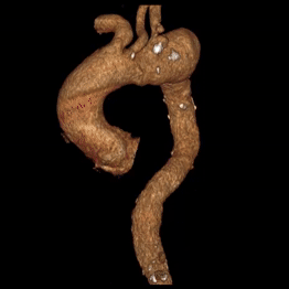
Comparison between 11 days post-operation and pre-operation


Image

Preoperative/Postoperative 3D Reconstruction Comparison

Image

Preoperative/Postoperative CTA Axial Comparison


Postoperative review showed good recovery, with smooth blood flow in all branches, no signs of cerebral infarction or cerebral ischemia, complete isolation of the aortic aneurysm, LSA dissection, and ulcerative lesions, no endoleak, and the stent was in good position and shape.


Image


Summary of Case Experience


This is a case of an elderly patient with a thoracic aortic aneurysm accompanied by a proximal ulcer near the LSA and a localized dissection. The proximal end of the thoracic aortic aneurysm has extended to the root of the LSA, resulting in insufficient length of the proximal anchoring zone. Therefore, it is necessary to perform supra-arch branch reconstruction of the LSA and occlude the diseased area of the LSA.


After thorough and meticulous analysis and careful consideration, Professor Huangjin Qi's team ultimately decided to adoptPercutek Therapeutics New Technology Thoracic Aortic Stent GraftPerform surgical treatment. During the surgical procedure, the team relied on their superb technical skills,Successfully completed the puncture and in-situ fenestration of the thoracic aortic stent graft using only a 250T guidewire.Percutek Therapeutics' new technology thoracic aortic stent graft is easy to penetrate and expand, with simple and safe balloon expansion operations, allowing for efficient arch reconstruction of the LSA. Meanwhile, during balloon expansion and branch stent implantation,Through the traction technique, it is inserted from the femoral artery, reducing the damage to the brachial artery and avoiding the risks caused by vascular injury.. More notably, the teamInnovatively Apply Self-made Improved Cerebral Embolism Protection Device, which played an important role during the operation,Minimizes the risk of severe complications such as cerebral embolism caused by surgical procedures, strongly safeguarding the patient's life.


Postoperative imaging examinations showed significant closure of the lesion, smooth blood flow in each branch above the aortic arch, good stent morphology, no signs of cerebral infarction or cerebral ischemia, and no complications such as endoleaks. This indicates that the surgical plan was successfully implemented.The actual surgical outcome was highly consistent with the preoperative expectations.`, providing valuable clinical experience and reference cases for the treatment of such complex lesions.`


Image


Expert Introduction


Image

Professor Huangjin Qi


Director of the Department of Interventional Vascular Surgery, First Hospital of Putian City, Associate Chief Physician, Associate Professor, Master's Graduate Supervisor.

Currently serves as the Director of the Peripheral Vascular Intervention Training Base at Putian First Hospital, National Health Commission; Director of the Quality Control Center for Comprehensive Interventional Technology in Putian City; Member of the Vascular Surgery Branch of the Fujian Medical Association’s Surgery Division; Standing Committee Member of the Vascular Surgery Branch of the Fujian Strait Medical and Health Exchange Association; Deputy Director of the Vascular Collateral Disease Branch of the Fujian Integrated Traditional Chinese and Western Medicine Association; Member of the Lower Extremity Arterial Disease Committee of the Endovascular Society of the Chinese Medical Doctor Association; Member of the Portal Hypertension Professional Committee of the Chinese Anti-Cancer Association; Member of the Portal Hypertension Professional Committee of the Zhongguancun Technology Innovation Strategy Alliance; Member of the Peripheral Vascular Intervention Committee of the Interventional Radiology Branch of the Chinese Medical Association.


Image

Professor Zheng Jingda


Attending Physician of the Department of Interventional Vascular Surgery, Putian First Hospital.

Member of the Youth Committee of the International Union of Angiology (IUA) China Branch, Standing Committee Member of the Vascular and Endovascular Surgery Specialty Committee of the Fujian Province Grassroots Health Association, Member of the Vascular Surgery Branch of the Fujian Strait Medical and Health Exchange Association, Member of the Cardiovascular Surgery Branch of the Fujian Strait Medical and Health Exchange Association, Member of the Committee of the Putian Comprehensive Intervention Quality Control Center.

Image


Image


Department Introduction

Department of Interventional Vascular Surgery, Putian First Hospital


The Department of Interventional Vascular Surgery at Putian First Hospital is a key clinical specialty in Putian City. It was one of the earlier departments in the province to establish an Interventional Vascular Surgery ward. Since its establishment in March 2012, with the support of hospital leadership and under the leadership of Director Huang Jinqi, the department has developed rapidly. All operations have steadily increased year by year, and it has now become a well-known comprehensive minimally invasive interventional diagnosis and treatment center within the province, integrating medical treatment, teaching, and research.

In 2016, the department became a council unit of the Chinese Venous Interventional Alliance. In 2018, it became a council unit of the Chinese Hemorrhage Center Alliance. In 2019, it became a council unit of the Fujian Province VTE Prevention and Treatment Alliance. In 2022, it was selected as a peripheral vascular interventional training base by the National Health Commission's Capacity Building and Continuing Education Center. In 2023, it was selected as the deputy council unit of the Fujian Innovation Alliance of the National Interventional Medicine Sub-Innovation Alliance (IMIA).

Image


Image

END


Copyright Statement: This platform aims to help healthcare professionals better understand the latest developments in relevant disease areas. The information published on this platform does not imply agreement with its descriptions or viewpoints, but is solely for providing more information. If there are any copyright issues, we kindly request the rights holders to contact us, and we will address them as soon as possible. The information is intended for healthcare professionals to stay informed and should not replace professional medical guidance in any way, nor should it be considered as diagnostic or treatment advice. If such information is used for purposes other than staying informed, this platform and the author shall not bear any related responsibility.Contact email for cooperation:vascular@edoctor.work。

Image