Case Study: Prof. Zhu Jun's Team at Yibin Second People's Hospital Treats Hepatocellular Carcinoma with Vispearl® Radiopaque Drug-Eluting Microspheres

H&H Healthcare
R&D and Producer of Interventional Medical Devices for Heart Disease
Basic Information:Male, 57 years old
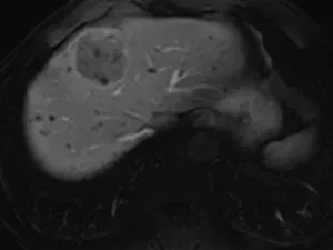
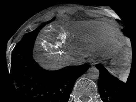
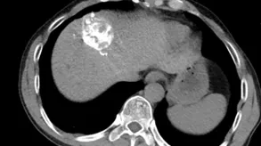
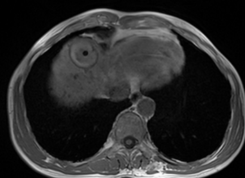
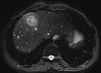
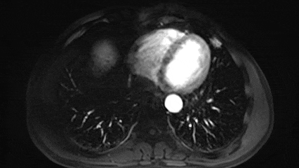
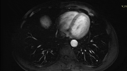
History of Present Illness:The patient visited a local hospital one month ago due to discomfort in the throat. During an abdominal color Doppler ultrasound examination, an intrahepatic space-occupying lesion was discovered, with no accompanying symptoms such as abdominal pain or diarrhea. On August 11, 2025, for further diagnosis, the patient came to our hospital and underwent an enhanced upper abdominal magnetic resonance imaging (MRI) scan, which showed: 1. An abnormal signal mass was found in the left inner lobe (segment IV) of the liver, approximately 5.5cm × 5.2cm × 4.1cm in size. The enhanced scan showed a "fast-in fast-out" enhancement pattern, suggesting a high possibility of hepatocellular carcinoma (HCC); 2. Multiple small round abnormal signal foci were diffusely distributed within the liver, suggesting a high possibility of bile duct hamartoma, with some being cysts. Since the discovery of the intrahepatic space-occupying lesion, the patient has not yet received relevant treatment. For further diagnosis and treatment, the patient visited the hospital today and was admitted under the outpatient department with a diagnosis of "malignant liver tumor." Since becoming ill, the patient's mental state, sleep, and diet have been acceptable, with no significant recent weight changes.
Past Medical History:A history of hepatitis B for more than 10 years, with a self-reported use of interferon treatment (specific regimen and course unknown). No other medical history.
Abdominal Examination: No special positive signs.
Laboratory Tests:Abnormal Prothrombin: 2259.15 mAU/ml; Hepatitis B Small Three Positive
Admission Diagnosis:Liver Cancer
Surgical Strategy:Hepatic V-TACE Procedure
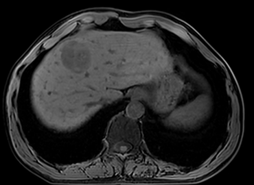
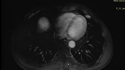
Preoperative T1 Plain Scan
Preoperative T2 Plain Scan
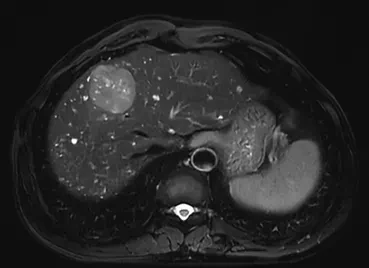
Arterial Phase
Venous Phase
Delay Period
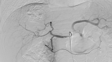
A conventional femoral artery vascular sheath was used to introduce a guidewire for placement of a 5F catheter, which was shaped in the aortic arch. The catheter was positioned in the hepatic artery for DSA examination. Angiography revealed thickening of the common hepatic artery and a mass-like tumor stain in the hepatic region. The left medial hepatic artery was the main blood supplier, with an increase in the number of feeding arteries.Thickened, disordered, and tortuous. A 2.5F microcatheter was placed and super-selectively advanced to the first tumor vessel for angiography.
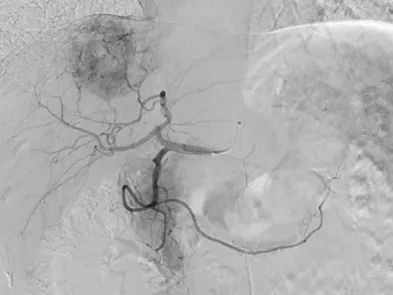
The CBCT arterial enhancement area is within the MRI-indicated tumor enhancement region.However, there is still a defect area, identified as one of the feeding arteries of the tumor.
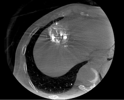
40-90μm Vispearl microspheres (2g) were loaded with pirarubicin (60mg), mixed, and allowed to stand for 10 minutes before the supernatant was removed. Contrast agent (iodixanol) was added to dilute the mixture to 18ml. Under fluoroscopy, 6ml of the drug-loaded microsphere solution was slowly injected through a microcatheter. After embolization of the target vessel reached stasis, a CBCT scan was performed, revealing dense deposition of imaging microspheres in the local tumor area and clear delineation of the tumor capsule.
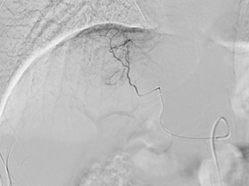
Subsequently, the microcatheter was superselected to the second tumor vessel for angiography. Microcatheter CBCT showed that the enhanced area of the artery was within the tumor enhancement area indicated by MRI, and it completely filled the defect target area, confirming it as another feeding artery of the tumor.
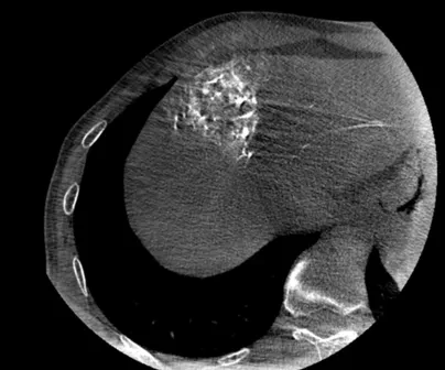
Under fluoroscopy, 9ml of drug-loaded microsphere suspension was slowly injected. CBCT plain scan was performed after the target vessel was embolized to stasis.
5F catheter hepatic artery follow-up angiography showed that tumor staining had almost disappeared. Immediate postoperative CT plain scan revealed a large amount of visualized drug-loaded microspheres deposited in the tumor target area.
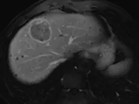
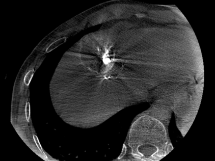
CT plain scan one month after surgery: A large amount of high-density shadow and a small amount of gas accumulation within the mass, scattered spot-like and linear dense shadows around the tumor (mostly vascular shadows). MRI one month after surgery: Abnormal signal mass in the original left medial lobe (segment IV) of the liver, compared with before: complete necrosis of the lesion, slightly smaller than before, no obvious enhancement, a small amount of gas accumulation visible within the tumor, inflammatory edema band visible around the tumor. Abnormal prothrombin decreased to 41.75 mAU/ml.
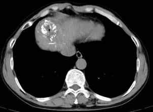
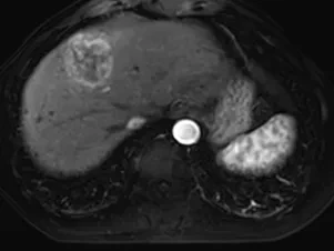
Postoperative T1 Plain Scan
Postoperative T2 Plain Scan
Arterial Phase
Venous Phase
Delay Period
Professor Zhu Jun stated: The use of large-diameter microspheres for embolization in interventional treatment of liver cancer can easily lead to premature occlusion of the blood-supplying arteries, often resulting in insufficient tumor microcirculation and collateral embolization, affecting the coverage of chemotherapeutic drugs, and increasing the recurrence rate after TACE. The diameter of the terminal blood vessels in the liver is generally less than 50µm. Small-diameter microspheres have the advantage of penetrating target tissues at a higher spatial density, thereby achieving greater drug coverage. Previous studies have also shown that compared with 100-300µm microspheres, the use of small-diameter (70-150µm) microspheres in TACE treatment for hepatocellular carcinoma results in a better objective response rate. However, small-diameter microspheres can cause more severe tissue necrosis and increase the risk of ectopic embolization, thus reducing safety compared to large-diameter microspheres. Vispreal®The excellent imaging properties of the spheres can precisely "protect" small-sized microspheres, providing accurate intraoperative feedback and enhancing the safety of small-sized microspheres. Visualizable drug-loaded microspheres with small particle sizes will become a key factor in the development of precise TACE.


Zhu Jun Professor
Yibin Second People's Hospital
Member of the Interventional Physicians Branch of the Chinese Medical Doctor Association, Member of the Tumor Ablation Study Group
Member of the Tumor Minimally Invasive Treatment Committee of the Chinese Anti-Cancer Association
Member of the Chinese CSCO Tumor Ablation Committee
Standing Committee Member of the Hemorrhage Special Committee of the Chinese Research Hospital Association
Vice Chairman of Southwest Comprehensive Intervention Specialty Alliance
候任主任委员 of the Interventional Medicine Special Committee of Sichuan Medical Association
Standing Committee Member of the Interventional Physicians Branch of the Sichuan Province Medical Association
Vice Chairman of the Tumor Intervention Special Committee of the Sichuan Anti-Cancer Association
Vice Chairman of the Interventional Medicine Committee of Sichuan Province International Medical Promotion Association
Areas of Expertise: Comprehensive interventional treatments (vascular and non-vascular) for various solid tumors, interventional portosystemic disconnection and shunt procedures for portal hypertension due to cirrhosis, minimally invasive integrated diagnosis and treatment of pulmonary nodules, etc.