AI in Medical Imaging Thrives on Collaboration, Quality, and High-Dimensional Data: An Interview with Professor Dinggang Shen

United Imaging
High-end Medical Device Developer
As a key application of computer vision, AI-based medical image recognition is continuously evolving toward greater depth and diversification. Traditional 2D imaging is advancing toward 3D volumetric (and even 4D) imaging, with AI playing an increasingly significant role in the image analysis process. AI-assisted image interpretation has become a new norm and is gradually gaining wider adoption.
So, exactly how much can AI enhance medical image analysis? What will be the future development direction of “AI + Medical Imaging”? We may gain insights from Professor Dinggang Shen’s speech.
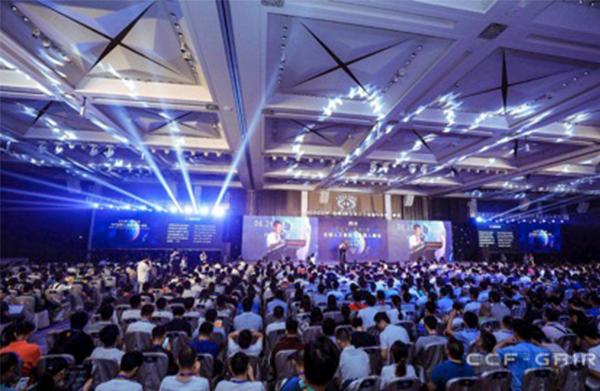
On June 29, the “CCF-GAIR 2018 Global Artificial Intelligence and Robotics Summit,” hosted by the China Computer Federation (CCF) and co-organized by Leifeng.com and The Chinese University of Hong Kong, Shenzhen, was grandly held in Shenzhen. The conference brought together 140 renowned experts from around the world in the field of artificial intelligence to conduct in-depth discussions across 11 AI-related domains.
Guests delivering keynote speeches and presentations at the conference include Turing Award winners, members of the Chinese and U.S. Academies of Sciences, and members of the Chinese and U.S. Academies of Engineering from academia, as well as top professors from prestigious universities such as Carnegie Mellon University (CMU), the Massachusetts Institute of Technology (MIT), and Stanford University; also featured are AI executives from industry leaders including Microsoft, Intel, and Tencent.
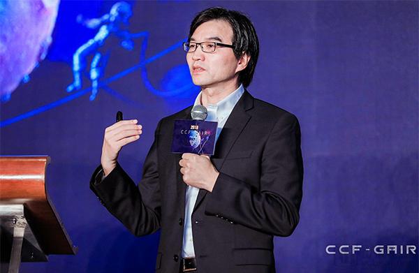
At this summit, Professor Dinggang Shen, a professor at the University of North Carolina, Co-CEO of United Imaging Intelligence, IEEE Fellow, and Chair of MICCAI 2019, delivered a speech titled “Deep Learning in Medical Image Analysis.”
VCBeat attended the presentation and conducted an exclusive interview with Professor Shen Dinggang, compiling the following summary based on his conference talk.
“Having spent nearly 20 years in the United States, I worked alongside physicians in the radiology departments of Johns Hopkins University, the University of Pennsylvania, and the University of North Carolina at Chapel Hill. This experience gave me a thorough understanding of their entire workflow and insight into how to better integrate our artificial intelligence technologies into the appropriate segments of their clinical processes. It is not feasible to apply AI across the entire workflow from start to finish—at least not at present,” stated Professor Dinggang Shen during his presentation.
He explained that AI companies in the field of medical imaging must have a clear sense of their R&D direction when developing products: their role is to assist physicians, not replace them. Developers must also clearly understand the pain points and needs of hospitals, and then implement corresponding technologies to address these issues. Of course, technological implementation is equally important; only with qualified technology can successful collaborations with hospitals be established.
The process by which medical imaging AI companies test their products in hospitals is inherently a collaborative one. However, this collaboration should not be limited to exchanges regarding data and equipment operation. To develop high-quality medical AI products and deliver the best possible experience to physicians, medical imaging companies must first understand what constitutes an optimal user experience. This requires them to be thoroughly familiar with the entire clinical diagnosis and treatment workflow. In other words,Researchers should not limit themselves to the field of medical imaging; instead, they must immerse themselves in clinical settings, closely observing every step and detail of clinicians’ workflows. By gaining a profound understanding of real-world clinical application scenarios, they can identify effective pathways for integrating their work with clinical diagnosis and treatment., only in this way can we determine how to embed AI technology into existing clinical workflows to streamline processes and enhance efficiency; only then can we develop products that are accepted by physicians and remain competitive.
Deep collaboration requires returning to the product itself, namely the performance of AI products. Professor Shen Dinggang believes that in today’s era where AI products are emerging in large numbers, the core competitiveness of a product still lies in its corresponding performance and application scenarios.
At present, many products can achieve an accuracy rate of over 90% or even higher during the testing phase. However, in reality, this may not be the result of a single detection run (or tests conducted on completely independent samples). This is because many algorithms continuously adjust their parameters based on training data during the development process. Consequently, the accuracy rates obtained in this manner (particularly those derived from the development database) are difficult to replicate in practical applications.
Moreover, many companies are keen on showcasing the superiority of AI through human-versus-machine competitions; however, starting from the data preparation stage, it is no longer a fair contest.
“During data preparation, any data with quality issues (such as motion artifacts, poor image quality, or missing imaging modalities) were discarded by the competition organizers. This curated dataset is highly advantageous for AI algorithms.However, in real-world practice, it is not feasible to withhold all diagnostic information from patients due to such issues, nor is it possible to directly obtain pre-screened data. Consequently, results derived from such data hold limited practical significance. Furthermore, this approach can be misleading to both physicians and society to a certain extent.”
Therefore, assessing the accuracy, sensitivity, and specificity of an AI product requires the use of fresh, unprocessed data collected from hospitals, rather than curated datasets used in competitions. Even the most impressive test results are futile if they cannot withstand validation in clinical practice, fail to align with physicians’ workflows, or cannot be deeply integrated into hospital systems. Ultimately, regardless of how compelling a company’s product presentations or how meticulous its marketing efforts may be, the core determinant remains the quality of the product itself.
“What we aim to achieve is a full-chain, full-stack artificial intelligence solution that spans imaging, screening, diagnosis, prognosis, as well as subsequent treatment and follow-up. Throughout this entire process, AI can assist physicians, thereby optimizing the workflow and achieving optimal diagnostic outcomes,” explained Professor Shen Dinggang.
This stems from the inherent advantages of United Imaging Intelligence as a subsidiary of Shanghai United Imaging Healthcare Co., Ltd. (hereinafter referred to as “United Imaging”). “United Imaging is currently the largest manufacturer of high-end medical imaging equipment in China. Its imaging devices require extensive AI technologies, which are applied across pre-imaging, intra-imaging, and post-imaging stages.”
He cited as an example that while high-quality imaging equipment is available in remote primary care hospitals, there is often a shortage of skilled technicians capable of accurately positioning patients for scanning. In such cases, computer vision can assist with precise patient positioning, enable one-click scanning, and select the most appropriate scanning protocol. This approach ensures image quality and the integrity of acquired data at the source.
“Not only that, full-stack imaging AI can deliver ‘forward-looking and backward-integrating’ AI.”After being equipped with an AI system, a scanning device can proactively identify the source of pathology, perform more detailed scans of areas with potential lesions, and generate high-resolution images for physicians’ reference.。
Many companies are currently developing pulmonary nodule detection solutions, but these typically operate on pre-acquired images. Imagine integrating the imaging process with subsequent AI-based diagnosis. This would be akin to having an experienced radiologist present during the scan: as each set of slice images is reconstructed, the “radiologist” would indicate whether any pulmonary nodules are present. If suspicious nodules are detected, the system would reconstruct images with higher density (i.e., thinner slices) in those regions, while normal areas would be reconstructed at standard slice thicknesses. In other words, the imaging equipment would automatically increase reconstruction density in areas with suspicious pulmonary nodules. In this example, combining AI methods with the imaging process has the potential to enhance the accuracy and effectiveness of subsequent pulmonary nodule screening.
Professor Shen Dinggang believes that the integration of artificial intelligence and medical imaging should reach such a level. Furthermore, the targeted diseases should not be limited to just one, two, or even five or six popular areas, but should encompass a wider variety of disease types. Achieving this goal requires not only the efforts of United Imaging Intelligence but also the joint collaboration of many like-minded partners. “It is impossible for United Imaging Intelligence alone to achieve this. Therefore, we aim to build an ecosystem that allows us to work with a broad community of physicians, particularly those willing to learn AI, to scale up these efforts. Meanwhile, we will engage in open collaborations with third-party companies. By sharing United Imaging Intelligence’s AI modules, we seek win-win cooperation to extend AI solutions to rare diseases, thereby creating broader benefits for society and patients.”
Beyond their relentless pursuit of precision, medical imaging companies are also continuously innovating in other areas, with many new models and technologies steadily evolving.
Most domestic AI technologies for medical imaging remain at the 2D level. While they currently meet certain clinical requirements, advancements in technology are enabling 3D and even 4D imaging AI to provide clearer and more intuitive information, accurately reflecting the distribution and evolution of patients’ conditions. This represents a significant breakthrough for physicians from both research and therapeutic perspectives, thereby opening up new possibilities for the development of “AI + medical imaging.”
Meanwhile, this also places new demands on the development of computer hardware. As an application of computer vision, medical image AI research already requires substantial computational power at the current stage. However, to gain insights into subtle changes in patients’ lesions and explore the underlying causes of these changes, it is necessary to employ 4D imaging technology for continuous image acquisition, which in turn necessitates more powerful computer hardware support.
Everything is still evolving; “AI + Medical Imaging” holds significant future potential, yet there is still a long way to go.