Can Pathologists Independently Develop AI Applications for Pathological Diagnosis? See How Chengdu Knowledge Vision Tech Is Doing It

Knowledge Vision
Intelligent Decision-Making Platform Provider
Cancer, the King of All Cancers. In 2018, China ranked first globally in both cancer incidence and mortality rates. As the aging process in China intensifies, cancer incidence continues to rise. Cancer will become a challenge that every family may face in the future. China has entered a stage of nationwide cancer prevention and control.
Clinically, pathological diagnosis is the gold standard for cancer assessment. As the diagnosis and treatment of malignant tumors have advanced to the molecular level, pathological diagnosis can effectively evaluate protein expression and gene amplification in tumor cells using techniques such as immunohistochemistry and fluorescence in situ hybridization. However, the difficulty in achieving precise quantitative pathological evaluation, coupled with a significant shortage of pathologists in China, severely constrains the development of precision medicine in the country.
To address the aforementioned challenges, experts propose that integrating abundant digital pathology image data with emerging computer algorithms, such as AI, and leveraging powerful computational capabilities will lead to the development of numerous computer-aided diagnostic software tools for digital slides. These systems enable computers to automatically detect lesion regions in digital slides and quantitatively assess various parameters, thereby assisting pathologists in making rapid, accurate, and highly reproducible pathological diagnoses.
“Currently, the product development efforts of most AI companies are concentrated in the areas of assisted analysis of cervical cytology and quantitative analysis of a few popular immunohistochemistry markers, with relatively little R&D dedicated to other fields. However, in actual pathological diagnosis and scientific research, there remains a substantial amount of unmet demand for pathological analysis.” Xiang Fei, founder of Chengdu Knowledge Vision Technology Co., Ltd., stated in an interview: “Therefore, we are committed to building a no-code cloud platform for pathological AI application development, addressing challenges such as high technical barriers, significant hardware investment, and elevated costs associated with communication and data annotation. This enables pathologists to conduct pathological AI research tailored to their actual needs without any coding expertise.”
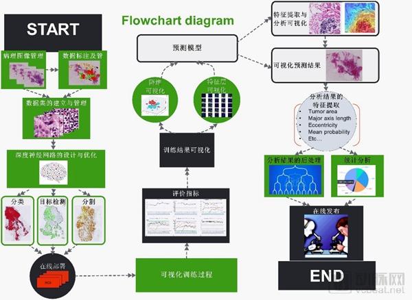
“AI VIEWER utilizes data encryption and authorized access mechanisms to both safeguard user data security and facilitate multi-party collaboration, communication, and data sharing. By identifying common clinical application requirements for AI-assisted pathological analysis, we have standardized and modularized the technologies involved in the development of pathology AI applications. This enables end-to-end management of data, image annotation, algorithm development, and application deployment, as well as qualitative, locational, quantitative, visualized, and digital analysis of histopathology,” said Dr. Wang Yizhe, CTO of Chengdu Knowledge Vision Technology Co., Ltd., in an interview. “Pathologists can simply drag and select the corresponding modules and functions to generate AI applications that assist in pathological diagnosis.”
AI VIEWER
In the field of R&D for pathological AI applications, AI VIEWER boasts comprehensive and unique technological expertise:
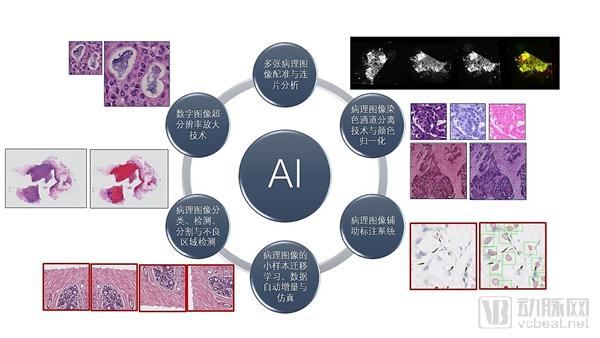
1. Deep Learning-Based Pathological Image Classification, Object Detection, Image Segmentation, and Abnormal Region Detection.
2. Deep learning-based super-resolution magnification technology for digital pathology images enables the practical application of using a 20x objective lens instead of a 40x lens for whole-slide imaging, thereby reducing storage space and transmission time by 75% and significantly enhancing teleconsultation capabilities.
3. Registration and Mosaic Analysis of Multiple Digital Pathology Images Based on Deep Learning, Addressing the Clinical Challenges of Multiplex Staining and Joint Biomarker Analysis
4. Deep learning-based stain normalization and color channel separation techniques eliminate variations in staining intensity of pathological slides caused by differences in reagent batches, staining duration, temperature, and scanners.
5. The deep learning-based AI-assisted pathological image annotation system significantly improves annotation efficiency and reduces time costs.
6. In view of the specific characteristics of small-sample pathological images, transfer learning, automatic incremental learning, and simulations were conducted to ensure the accuracy of results on small-sample datasets.
Technical Accumulation of AI VIEWER
“With the assistance and support of Roche Diagnostics and the Pathology Research Laboratory of West China Hospital, we have completed the development of AI VIEWER 1.0, a no-code cloud platform for pathological AI application R&D,” said Dr. Wang Yizhe, CTO of Chengdu Knowledge Vision Technology Co., Ltd., in an interview. “Currently, AI VIEWER 1.0 has been applied to AI-assisted quantitative evaluation studies for breast cancer HER2 and bladder cancer PD-L1.”
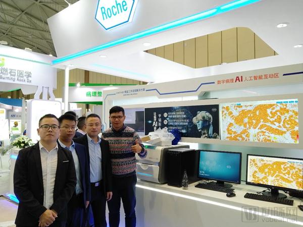
From October 11 to 14, 2018, the 24th Academic Conference of the Pathology Branch of the Chinese Medical Association and the 8th China Pathology Annual Conference were held in Chengdu, Sichuan Province. The Pathology AI Application Development Cloud System Version 1.0, independently developed by Chengdu Knowledge Vision Technology Co., Ltd., made its public debut at the Roche Diagnostics booth during the China Pathology Annual Conference. This launch received unanimous acclaim from the industry and peers.
Knowledge Vision, in Partnership with Roche, Launches Its First Cloud Platform for R&D of Pathology AI Applications
AI VIEWER can be widely applied in the pathological analysis involved in new drug development, dedicated to improving the work efficiency and quality of CROs in drug R&D. VCBeat has learned that currently, China's CROs account for only 2% of the global market, but there is still significant room for growth.
“We hope to leverage Roche Diagnostics’ brand and distribution channels to promote our platform and services globally,” said CEO Xiang Fei. “In China, we have already established business collaborations with Roche Diagnostics, Roche Pharmaceuticals, Guanhe Medicine, the Pathology Research Laboratory of West China Hospital, and the Department of Pathology at Fudan University Shanghai Cancer Center.”
AI VIEWER proves valuable across multiple scenarios: AI-assisted PD-L1 analysis in bladder cancer, quantitative HER2 analysis in breast cancer, automated localization and qualitative analysis of thyroid tissue, detection of intratumoral hemorrhage locations and area calculation in neuroblastoma, histological analysis of bone marrow smears, and assessment of hepatocyte abnormalities within hepatic lobules.
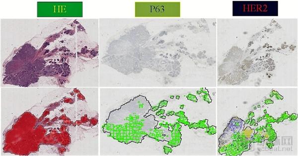
AI VIEWER for HER2 Research in Breast Cancer
During the new drug development process, AI VIEWER enables assisted quantitative analysis of digital pathology images, providing drug researchers with visualized predictive data. This ensures traceability, quality assurance, and quality control in the pathological analysis process, thereby reducing drug development costs and clinical trial risks while enhancing the market competitiveness of pharmaceutical products.
Meanwhile, pathology diagnostic results based on quantitative analysis provide intelligent decision support for new drug researchers by determining the correlation between pathological data features and prognosis, improving the efficiency and pace of quantitative analysis of pathology slides, and ensuring consistency in histopathological analysis. This approach can be used for subject screening and the determination of cutoff values for companion diagnostics.
Building on this foundation, we aim to achieve in-depth mining of patterns and quantitative features in pathological images, thereby fostering the emergence of multi-omics cross-disciplinary research. By integrating pathomics with genomics and radiomics, we will establish a multi-omics interactive diagnostic system that compares and fuses pathological morphological information with genomic molecular data, as well as anatomical and functional imaging information. This approach enables the discovery of details imperceptible to the human eye, along with new features, patterns, and rules that are difficult to summarize through subjective experience, ultimately advancing the development of precision diagnosis.
Looking to the future, Knowledge Vision Technology firmly believes: “With a steadfast focus on innovative R&D and dedicated accumulation of expertise, we will ultimately unleash our potential and emerge as pioneers in the field of AI-assisted pathological diagnosis.”