Seven Groundbreaking Scientific Advances of 2018 That Are Truly Remarkable
Editor’s Note: This article is reprinted from BioExplorer, authored by Chen Moyi. Republished by VCBeat with authorization.
On December 24, The Scientist magazine announced its selection of the top technological breakthroughs of 2018.
“Bipaternal” mice, tumor-destroying nanorobots, “AI-powered” liquid biopsies, and revolutionary technologies disrupting gene expression analysis…
This Year, Scientists Are Still “Awesome”!
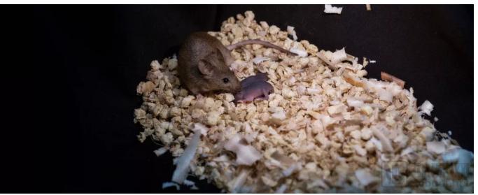
A healthy adult bimaternal mouse has produced its own offspring, whereas bipaternal mice die shortly after birth.
(Image source: LEYUN WANG)
Same-sex reproduction is not uncommon in the animal kingdom. For instance, parthenogenesis—whereby females produce offspring without mating with males—has been observed in reptiles such as lizards, amphibians such as frogs, and various fish species. In contrast, androgenesis, the male counterpart to parthenogenesis, is extremely rare; to date, scientists have documented this phenomenon in only one species of zebrafish.
However, neither parthenogenesis nor androgenesis occurs in higher mammals. Imprinted genes, which are absent in reptiles and amphibians but evolved in mammals, are considered a major factor hindering same-sex reproduction in mammals.

Image source: Cell Stem Cell
In a study published this October in the journal Cell Stem Cell, scientists from the Chinese Academy of Sciences successfully produced offspring from two male mice for the first time, generating androgenetic mice with two paternal genomes. These androgenetic mice appeared normal and were capable of independent respiration, but all died within 48 hours after birth.
As early as 2015, scientists successfully created “two-mother” mice; these parthenogenetic mice appeared to develop normally and were capable of producing offspring.
Researchers believe that this new finding confirms the feasibility of androgenesis in higher mammals; however, the inability of all androgenetic mice to survive to adulthood suggests that androgenesis faces more obstacles than parthenogenesis.
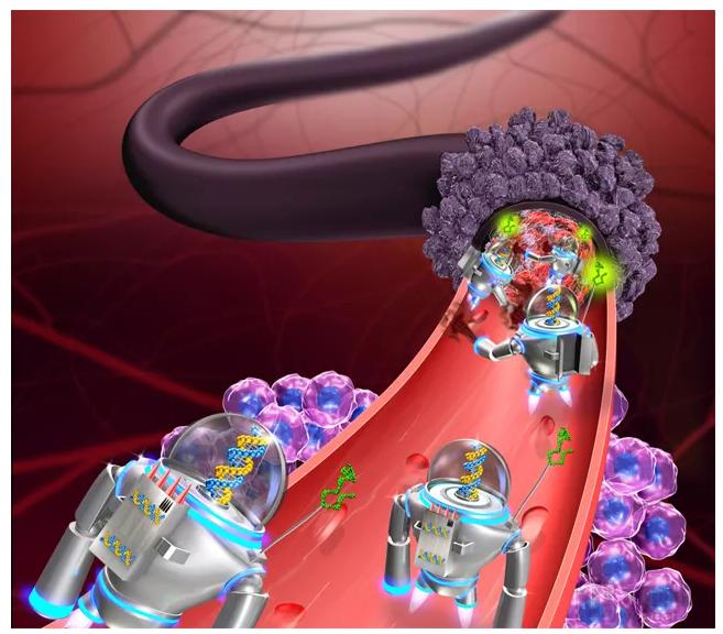
DNA Nanorobots Deliver a Drug That Cuts Off Blood Supply to Tumors in Mice (Image credit: Baoquan Ding and Hao Yan)
Precisely delivering drugs to tumor cells via nanorobots, thereby selectively killing cancer cells without endangering surrounding healthy tissue, is the dream of many scientists.
In February this year, scientists from the National Center for Nanoscience and Technology of China reported a type of DNA-based nanorobot capable of “traveling” through the bloodstream to locate tumors and release thrombin, a protein that induces blood coagulation, thereby triggering cancer cell death in mice.
Researchers chose DNA as the raw material for constructing nanorobots primarily due to its inherent biocompatibility and biodegradability.
Experiments have demonstrated that this novel nanorobot can precisely target and effectively kill tumor cells, achieving favorable outcomes in various mouse tumor models. The related findings were published in the journal Nature Biotechnology.

Image source: Nature Biotechnology

Machine learning algorithms for detecting cancer DNA in blood are poised to pave the way for personalized cancer treatment. (Image credit: ISTOCK, VITANOVSKI)
“Detecting cancer at its earliest stages” and “providing personalized, dynamic treatment for patients” are the two major challenges in modern oncology. To find solutions, some laboratories and biotechnology companies are turning to artificial intelligence (AI). They aim to develop machine learning algorithms capable of deciphering faint signals in the blood. Based on these signals, scientists can identify cancer at an early stage and determine in real time whether patients are responding to their treatment.
To date, machine learning algorithms for detecting trace amounts of tumor DNA in blood samples have performed well in clinical validation studies, but no self-learning algorithm has yet been approved for clinical use. By directly identifying mutations in DNA, RNA, and proteins from blood, these technologies hold the potential to surpass imaging and tissue biopsy techniques in cancer detection and monitoring.

Software-Based Chemical Screening Can Minimize Animal Testing (Image Credit: iStock, Niderlander)
Globally, millions of animals are used for toxicity testing of compounds. Toxicologists have now developed software capable of accurately predicting the outcomes of these tests. The software predicts animal experiment results with an accuracy of 87%, whereas repeat experiments themselves reproduce the original results only 81% of the time. This finding was published in July this year in the journal *Toxicological Sciences*. The authors hope that this software will help reduce the use of laboratory animals.

Drones enable researchers to collect vast amounts of biological data more easily than ever before, at lower cost and with higher resolution.
(Image source: ROHAN CLARKE)
Some researchers aim to minimize the use of laboratory animals, while others seek to gather as much information as possible about wildlife. When used appropriately, drones can serve as valuable “allies.” Some scientists are already leveraging drones for such purposes, including collecting whale snot and assessing the size and health status of entire sea turtle populations.
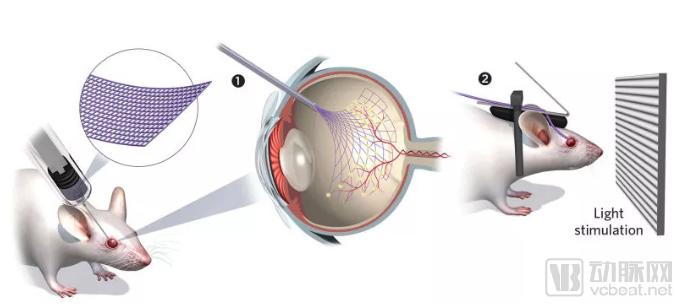
Ultraflexible mesh electrodes monitor intact, fully functional eyes in awake animals. (Image credit: GEORGE RETSECK)
Previously, to record the activity of retinal cells, researchers often had to enucleate the eyes from animals, dissect the retina, and place it flat on an array of microelectrodes. Under such conditions, the retinal cells could respond to light for several hours.
This June, Harvard University nanotechnology expert Charles Lieber and his colleagues developed a novel mesh electrode. This ultra-flexible mesh electrode can remain within the eyes of living animals, recording retinal cell activity for up to several weeks. Notably, the mesh structure has minimal impact on vision and detaches from the retina after a few weeks.
Marla Feller of the University of California, Berkeley, described it as a “remarkable” innovation. The related findings were published in the top-tier journal Science.

Image source: Science
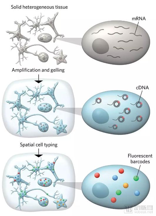
STARmap enables simultaneous analysis of multiple RNAs in intact, volumetric samples.
(Image source: GEORGE RETSECK)
Cells of a specific type or organization may appear similar but behave differently. For instance, in the brain, neurons of the same subtype can play vastly different roles depending on their location and connectivity. In other words, spatial information is absolutely critical when it comes to specific cellular functions. Therefore, researchers are developing tools capable of detecting the expression of multiple genes within tissue sections.
STARmap, developed by scientists at Stanford University, is one such tool. STARmap involves converting tissue samples into hydrogels to better detect “barcodes” that reveal the locations of target RNAs. The research team used this technique to analyze the simultaneous expression of up to 28 genes in 150-micrometer-thick mouse brain tissue sections, as well as more than 1,000 genes in 8-micrometer-thick sections. These analyses revealed differences in the distribution of excitatory and inhibitory neuronal subtypes across the cerebral cortex.
Sten Linnarsson, a molecular systems biologist at the Karolinska Institute in Sweden, believes that STARmap is an important step toward true three-dimensional gene expression analysis. The related findings were also published in Science magazine.

Image source: Science