ECR 2019: The Convergence of Medical Imaging Devices and AI – Innovations from Global Giants and the Strategic Direction for Chinese Enterprises

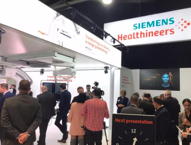
Siemens Healthineers
Integrated Healthcare Service Provider
The 2019 European Congress of Radiology (ECR) drew to a close amidst a vibrant interplay of theoretical discourse and technological innovation. During the event, more than 30,000 representatives from diverse sectors across 137 countries worldwide participated in the conference.
The theme of ECR 2019 was “the bigger picture,” aiming to review and summarize the development path of European radiology while ushering in a new chapter with emerging technologies. In his opening address, Congress President Prof. Lorenzo E. Derchi highlighted discussions sparked by the application of artificial intelligence (AI) in radiological sciences, including AI’s role and impact on radiologists’ daily workflows, its utilization in patient care, and its relationship with the evolving role of radiology researchers.

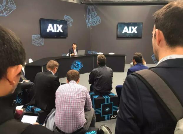
The widespread application of artificial intelligence (AI) technology in radiology has undoubtedly become a major highlight of this exhibition. To underscore its growing importance in the field of radiological sciences, the European Congress of Radiology (ECR) has specially organized a three-day Artificial Intelligence Exhibition (AIX).
So, what flagship capabilities did major medical device manufacturers showcase at this conference? What new advancements have been made in artificial intelligence technology?
As a fundamental modality in medical imaging, the key constraint on the service delivery of MRI, DR, and ultrasound equipment remains efficiency. Although these products have undergone decades of iteration, the exhibits showcased today indicate no sign of slowing progress in their evolution. Improvements in image quality, reductions in scan time, and the expansion of various application values are gradually shifting the focus of traditional devices toward clinical assistance and enhancing patient experience.
Siemens Healthineers showcased its Multix Impact DR system and the Mobilett Elara Max mobile X-ray unit. The Multix Impact DR system features a height-adjustable floating tabletop and an in-room touch-based user interface. Equipped with a 17 x 15-inch digital detector, the tabletop can be installed adjacent to the imaging device, enabling radiologic technologists to remain by the patient’s side during examinations, thereby enhancing the patient’s psychological comfort.
The Mobilett Elara Max mobile X-ray system is specifically designed with an antimicrobial coating to reduce the risk of hospital-acquired infections. This coating covers the entire system, while cables are concealed within the unit’s articulated arm. Siemens has also upgraded the machine’s safety software package and created a “virtual workstation,” enabling patients to conveniently access relevant websites directly at the bedside without compromising IT network security.
In late 2018, Philips launched the 1.5T Ingenia Ambition “helium-free” MRI scanner. During manufacturing, after liquid helium is added to the magnet, it is completely sealed. Even in the event of a quench, the liquid helium is not vented outside the magnet but remains contained within it, allowing the system to continue operating after appropriate procedures. This design eliminates the need for refilling with new liquid helium throughout the magnet’s service life. The product has now seen further advancements: optimized to require no vent pipe, this fully sealed magnet is approximately 900 kilograms lighter than its predecessor, thereby reducing both site-selection challenges and construction costs associated with traditional magnets.
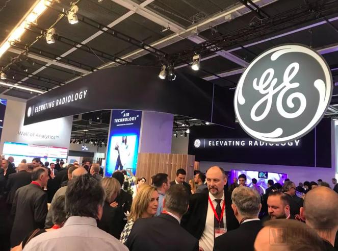
GE continues to refine and develop its Adaptive Imaging Receive (AIR) coil components. According to industry insiders, these flexible, lightweight components can more closely conform to the area being imaged, thereby improving image quality, while imposing a lighter burden on frail patients and neonates.
For MRI systems, magnetic field homogeneity is a critical factor. The term "critical factor" here refers not to the magnetic field homogeneity in the absence of a subject, but rather to the homogeneity maintained after a subject has entered the bore. At this event, Hitachi prominently showcased its Echelon Smart Plus system. To preserve magnetic field homogeneity even after a subject enters the scanner, the system is equipped with the HOSS (High Order Shim System).
In addition to the global shimming system for wide fields of view, Echelon also provides high-precision local shimming for specific regions such as the knee joint and breast, thereby realizing the HOSS (High Order Shimming System) local shimming function, which is expected to be widely adopted. By utilizing HOSS, image quality can be significantly improved for scanning sequences with higher demands on magnetic field homogeneity, such as BASG-EPI. Furthermore, fat suppression effects are markedly enhanced in areas off the magnet center, including the shoulder and knee. This capability will also support advanced functional examinations, such as MR spectroscopy, in the future.
Compared with other companies, Samsung places greater emphasis on the application of artificial intelligence technologies. Showcased at this conference, Bone Suppression is an algorithm that reduces interference from bone signals in chest X-ray examinations, thereby extracting lung tissue that might otherwise be obscured. SimGrid is another technology designed to facilitate the replacement of X-ray grids while reducing scatter artifacts to improve image quality.
Another software, S-Detect for Breast, can evaluate breast lesions on ultrasound images and assist in standardized reporting and classification of suspicious lesions. In the field of digital imaging, Samsung showcased its Auto Lung Nodule Detection application, SimGrid grid replacement technology, bone suppression technology, and an AI-based application for classification and diagnostic notification of intracranial hemorrhage.
Canon has launched the Vantage Orian, a wide-bore 1.5T scanner. This product made its debut at the 2018 ECR congress. For this release, the vendor redesigned the MRI digital architecture by relocating the electronics onto the scanner gantry, enabling each of the system’s 128 channels to utilize an independent analog-to-digital converter.
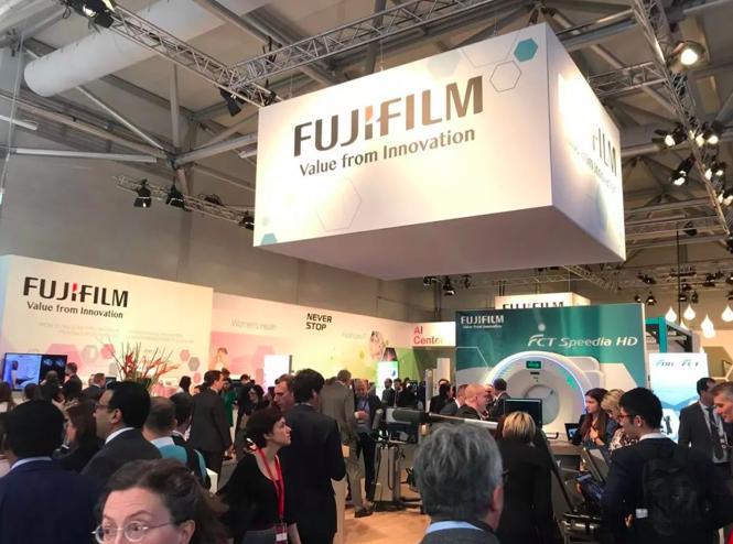
Fujifilm is promoting its FDR Smart X, a DR system available in ceiling-suspended or floor-mounted configurations, compatible with the FDR D-Evo II and FDR es digital detectors.
The highlight of this year’s conference is undoubtedly the Artificial Intelligence Exhibition (AIX). This exhibition hall provides a platform for small and medium-sized life sciences companies to engage in mutual exchange, and attendees can apply for AI-related training offered by the NVIDIA Deep Learning Institute. Meanwhile, traditional medical device giants are continuing to deepen their development of AI-powered products, with these industry leaders showcasing their latest digital offerings in this exhibition hall.
From an overall trend perspective, medical device manufacturers are attempting to accelerate equipment imaging through artificial intelligence technology on one hand, in order to reduce the use of contrast agents and improve imaging speed. On the other hand, they are applying artificial intelligence technology to image analysis, using algorithms to segment images, reducing doctors' workload, and obtaining more useful information.
At the conference, GE showcased software applications and smart devices based on Edison. Edison is General Electric’s artificial intelligence platform that connects existing AI partners and products. This system demonstrates GE’s core strategy to open up its third-party software market, presenting an excellent opportunity for small and medium-sized enterprises in attendance.
At the same time, ultrasound equipment manufacturers have re-emphasized their systems’ versatility and usability, sending a clear message to major ultrasound users in the imaging industry: not only should these modalities be made easier to use, but tools optimized for specific applications must also be developed.
At the conference, GE added its latest product, the Revolution Apex, to its Revolution series of CT scanners. As a next-generation intelligent CT scanner, the Apex utilizes the Edison deep learning image reconstruction algorithm to generate TrueFidelity images, which are characterized by high spatial resolution, natural texture, and low-contrast detectability.
GE has also integrated AI into its ultrasound equipment. The Logiq E10 ultrasound scanner showcased this time employs artificial intelligence technology similar to that used in autonomous vehicles and 3D video games. The scanner’s method of acquiring and reconstructing data is akin to MRI and CT systems, enabling automatic identification of the image focus.
Philips has launched IntelliSpace Discovery 3.0, a software platform designed to facilitate the development and deployment of radiology AI algorithms in clinical research. Recently, this product was implemented at the First Hospital of Jilin University (hereinafter referred to as “Jida Yiyuan”), where it assists radiologists in providing precise diagnosis and treatment through intelligent image post-processing, while also supporting clinical research and translational applications. Furthermore, Philips’ previously released IQon Spectral CT has long been renowned for its robust data acquisition capabilities. Recent studies indicate that software-based technologies can reduce subsequent scanning workload, lower contrast agent usage, and decrease X-ray radiation doses, thereby enhancing hospital operational efficiency.
Siemens has also made extensive and in-depth investments in artificial intelligence technology. How to continuously provide innovative digital healthcare solutions, build a digital healthcare ecosystem, and fulfill its corporate value commitment to digital healthcare has become the giant’s foremost pursuit at present.
At AIX, Siemens stated that it is striving to create an AI-powered digital “twin” model capable of simulating the physiological conditions of individual patient organs to predict changes and treatment outcomes. The company added that these digital models can also help optimize workflows by forecasting the impact of such changes.
Fujifilm showcased its REiLI medical imaging and informatics AI project. The company is currently developing a suite of artificial intelligence technologies, such as region recognition technology for identifying and extracting organ regions. Other applications under development within the REiLI portfolio include computer-aided detection systems designed to support radiologists’ clinical decision-making by reducing image interpretation time. Another new Fujifilm AI application, FCT Pixel Shine, provides image processing for low-dose CT scans.
Fujifilm showcased a 64-slice CT scanner equipped with a console featuring an embedded Synapse 3D reconstruction module. The company also highlighted its FCT Pixel Shine technology, which leverages AI algorithms to achieve efficient image processing at ultra-low radiation doses.
Samsung is showcasing the integration of artificial intelligence technology into its imaging modalities. Samsung’s S-Detect for Breast software evaluates breast lesions on ultrasound images and assists in standardizing reports and classifying suspicious lesions. In digital radiography, Samsung demonstrated its Auto Lung Nodule Detection application, SimGrid grid replacement technology, and bone suppression technology. It also launched an AI-based diagnostic application for the classification and notification of intracranial hemorrhage.
Samsung has launched a software package called SonoSync for ultrasound. The software provides web services that enable real-time ultrasound examinations, allowing clinicians to view images in a timely manner. SonoSync can also connect to smartphones, tablets, laptops, and desktop computers.
Professor Luis Donoso Bach is the Director of the Department of Clinical Diagnostic Imaging at Hospital Clínic de Barcelona and a Professor of Radiology at the University of Barcelona. As a leader in European radiology, his research spans multiple areas of radiological practice, including liver imaging, digital imaging, and IT applications and product development in diagnostic radiology.
Professor Luis Donoso Bach stated that with the introduction of technologies such as CT, MRI, and interventional radiology, the role of radiologists has shifted from the periphery to the center of healthcare services: “Today, no medical institution can function without an imaging department, after all, patients undergo at least one imaging examination before being discharged.”
Radiology has struck a balance between technology and clinical practice, emerging as a robust discipline. To sustain its development, it must engage more deeply in clinical care, moving beyond a purely auxiliary diagnostic role to bridge the gap with patients. Meanwhile, radiologists must transcend the mere detection of lesions and interpretation of images, as machines now perform these tasks more effectively than humans.
“The role of radiologists should be to resolve clinical problems by integrating imaging data with clinical information, tailored to specific contexts. To continue evolving, radiologists must become not just radiologists, but ‘experts in imaging information.’ The development of artificial intelligence has only underscored the necessity of this transition.”
To date, radiologists have only explored “narrow AI,” using simple algorithms to perform straightforward tasks. Machines still require human assistance to learn how to execute these tasks. The next major trend in artificial intelligence is deep learning, a process in which machines can learn autonomously without human intervention.
Meanwhile, standardized big data will hold immense value, and radiologists will continue to play a vital role in this field. “We can help validate AI applications. Since AI penetrates medicine through imaging, we are in an advantageous position. We must embrace technology and assist in the development and implementation of AI tools, as they are currently in the validation phase. Physicians, especially radiologists like us, are indispensable,” said Professor Luis Donoso Bach.
Professor Valentin Sinitsyn is the Head of the Department of Radiology at the Faculty of Medicine, Lomonosov Moscow State University. A pioneer in the field of cardiac imaging in Russia, he has helped drive its development; however, he believes that considerable efforts are still required to increase the utilization of magnetic resonance imaging (MRI) and computed tomography (CT).
He candidly expressed his disappointment at the slow development of cardiac MRI and CT in Russian clinical practice. While the benefits of cardiac imaging are well known, cardiac CT and MR examinations account for only about 1% of cardiac imaging procedures. He believes that the primary reasons for this situation are the insufficient knowledge and motivation among radiologists and cardiologists.
So, in which direction will artificial intelligence develop? We must answer the following questions.
For MRI, the value of artificial intelligence lies in replacing imprecise manual imaging measurements and accelerating the application of quantitative imaging in routine clinical practice. This extends from automated size measurements, such as the long and short axes of tumors, to more advanced quantitative phenotypic characterization of tumors, including structure, organ location, and heterogeneity. To advance beyond manual methods, AI must address the following key areas:
1. Enable more informed decision-making and improve diagnostic accuracy;
2. Ability to compare changes between prior and current imaging studies and assess disease progression;
3. More personalized treatment plans;
4. Ability to predict patient treatment response.
The trend toward quantitative imaging is still in its early stages, and several market barriers need to be addressed: First, there is low awareness of quantitative imaging in the commercial market; second, there is a lack of reproducibility among results generated by quantitative imaging software from different vendors; third, for radiologists to use this technology in their workflow, quantitative imaging results must be displayed during the initial reading rather than processed afterward; and fourth, most PACS systems do not support the search and mining of quantitative imaging data.
Of course, these issues are not insurmountable. As public understanding of artificial intelligence deepens and inter-company exchanges become more robust, these barriers may be gradually overcome in the coming years.
The benefits of using artificial intelligence in hybrid imaging are evident for decision support, prediction, and other applications; however, radiology experts must adopt practical approaches to implement AI in their work, which can only occur when it is integrated into the core workflows of hospitals.
The combination of artificial intelligence (AI) and hybrid imaging (HI) will give rise to a new field of research. “We will benefit greatly from artificial intelligence,” said Professor Thomas Beyer from Vienna, Austria, President of ESHIMT.
Hybrid imaging (HI) combines anatomical images with molecular imaging. PET/CT and PET/MR have already provided readers with more extensive information than CT alone. “For HI, especially PET/MRI, radiologists and nuclear medicine specialists will begin to leverage dynamic imaging modes, which is akin to observing the functional changes of lesions over time,” said Thomas Beyer. However, the more parameters added to tumor observation, the more difficult it becomes for physicians to determine the phenotype of a disease. Therefore, the deep learning capabilities of artificial intelligence and other technologies can serve as a decision-support mechanism.
Furthermore, histopathological imaging (HI) is closely related to pathology, and an increasing number of researchers regard AI as a tool to mediate between medical imaging and histopathology. Current studies have combined digital histopathology with PET/MRI to establish machine learning-based prediction models for differentiating benign from malignant prostate tumors without the need for biopsy.
Antoine Leimgruber, a nuclear medicine physician, radiologist, and physicist from Switzerland, stated that oncology may require the integration of imaging data through radiomics, proteomics, genomics, pathology, and therapeutic and radiation oncology more than any other field to advance toward personalized medicine. Radiology can help radiologists and nuclear medicine physicians obtain the maximum amount of disease-related information.
To date, the majority of data collected using radiomics has focused on lung cancer. Half of the studies have concentrated on characterizing lesions as either malignant or benign. Antoine Leimgruber believes that these are merely preliminary findings. “It is relatively straightforward to leverage existing scientific knowledge and technology to determine whether intervention is required for pulmonary nodules.” Although this remains a complex issue, it represents the first step toward more integrated radiomics or radiomics-plus approaches.
More comprehensive impactomics approaches are emerging, but their implementation in clinical settings remains highly challenging due to the rapid daily influx of data into every hospital; the crux of the issue lies in how healthcare teams can effectively integrate this information.
“A reliable starting point is for artificial intelligence to integrate simple, standardized data extracted from reproducible protocols into hospital workflows in as automated a manner as possible, thereby enabling practical use and access to the data. Otherwise, the old adage ‘garbage in, garbage out’ will also affect radiology, which benefits no one,” concludes Antoine Leimgruber.
In the field of artificial intelligence, Chinese technology has consistently remained in the world’s first tier, with Chinese influence frequently featured at major international conferences.
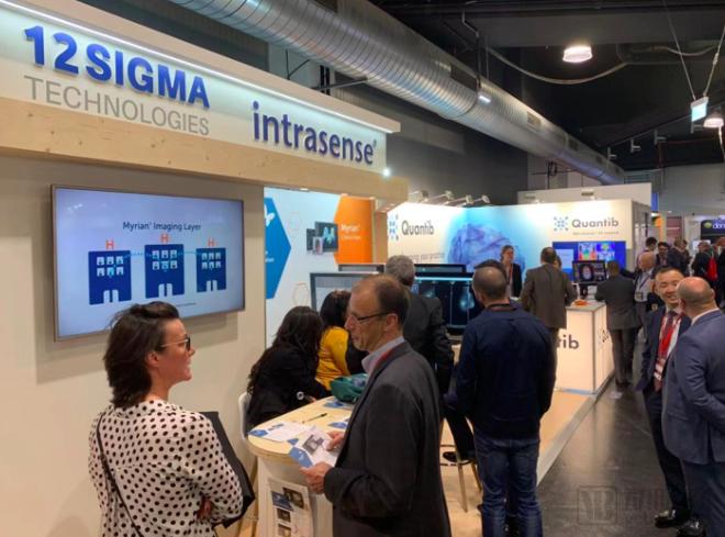
At this conference, we can see the booths of AI companies such as InferVision, 12 Sigma, and Biomind, as well as device manufacturers including Jiangsu Magspin, Prelove Medical, JuSha Display, Shenzhen Beacon, and SIUI, showcasing their outstanding technologies.
As a pioneer in AI for cardiovascular and cerebrovascular diseases in China, Shukun Technology shared its mature experience in applying AI to assist in the diagnosis of such conditions with authoritative professors from the British Society of Radiology and the Chinese Society of Radiology, as well as global equipment manufacturers, pharmaceutical companies, and solution providers at the conference.
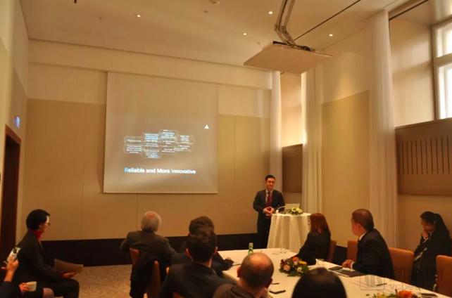
Shukun Technology Shares AI Technologies for Cardiovascular Care at ECR
At ECR 2019, TumorDeep and Intrasense, a long-established French expert in medical imaging software solutions, jointly showcased three new intelligent diagnostic products for medical imaging, including an Intelligent Diagnostic System for Pulmonary Nodules, an Intelligent Diagnostic System for Chest X-rays, and an Intelligent Diagnostic System for Mammography.
After the conference, Tumashenwei deeply felt that imaging-related enterprises and departments have shifted from a wait-and-see stance to a willingness to experiment, making it increasingly easier for AI vendors to find partners for implementation. In light of this trend, artificial intelligence vendors should strive to enhance product value to better meet physicians’ expectations.
In line with the theme of the event, SIUI demonstrated the intelligent operational workflows of its MAI Smart Remote IoT Platform and the IBUS breast ultrasound system, which is dedicated to women’s health, at the ECR exhibition. These demonstrations sparked considerable interest among attendees. Visitors flocked to the SIUI booth to learn about the technical features of new products and applications from sales representatives and personally experienced the convenience brought by SIUI’s smart-era innovations.
As the standard-setter for C-arm systems in China, Perlove Medical has served over 10,000 medical institutions worldwide with its C-arm products. At ECR 2019, Perlove Medical showcased its currently best-selling dynamic flat-panel mini C-arm. Since its market launch in 2017, this flat-panel C-arm has gained market recognition for its clear imaging and convenient clinical operation. The upgraded version presented at the exhibition is more mature and better aligned with clinical needs.
Academically, China has never lagged behind. As a Chinese radiologist attending the European Congress of Radiology (ECR), Dr. Wang Ming from the Department of Radiology at Peking Union Medical College Hospital presented an innovative achievement related to artificial intelligence at the ECR site, titled “Preliminary Application of AI-Based Image Optimization Combined with Iterative Algorithms in ‘Dual-Low’ Aortic CTA.” This presentation showcased the research outcomes of a collaborative project between Peking Union Medical College Hospital and Neusoft Medical Systems, focusing on improving low-dose CT image quality through AI-combined iterative algorithms. It validated the unique technological innovations and patented technologies of China’s high-end CT equipment (NeuViz 128 Jingrui CT), realizing value that “goes beyond imaging.”
Opportunities abound, yet challenges must also be acknowledged. Today, industry giants such as GPS, Canon, and Hitachi are deploying artificial intelligence through ecosystem-based strategies, leveraging their substantial resources to secure leading positions. If domestic manufacturers fail to introduce more robust and market-validated products in a timely manner, they risk falling behind at the starting line of commercialization.
Therefore, the application and exploration of medical AI must continue to accelerate without any letup. We look forward to witnessing more surprising advancements in the integration of AI and radiology at the 2020 ECR, and even more so to seeing a greater Chinese presence on the international stage, showcasing cutting-edge achievements in this academic arena.