China Medical Imaging AI White Paper Officially Released in Beijing
On March 26, 2019, the VB100 New Year Strategy Launch Day, hosted by VCBeat (WeChat official account: vcbeat), was grandly held at LSPACE in the 813 Creative Industrial Park in Beijing. As a key segment of the launch event, the “White Paper on AI in Chinese Medical Imaging,” drafted under the leadership of the China Medical Imaging AI Industry-Academia-Research-Application Innovation Alliance and bringing together imaging experts and research specialists from top-tier (Grade A tertiary) hospitals across China as well as leading AI medical companies, was officially released.
At the event, Liu Shiyuan, Chairman of the China Medical Imaging AI Industry-Academia-Research-Application Innovation Alliance, took the stage to deliver a speech. He stated that the Alliance’s work aims to promote industry development, with the "White Paper on Medical Imaging AI in China" being one of its key initiatives. The White Paper encompasses six major areas, reflecting the collective efforts and expertise of stakeholders across all sectors—industry, academia, research, and application—and represents the most authoritative voice in China’s current AI landscape.

Address by Liu Shiyuan, Chairman of the China Medical Imaging AI Industry-Academia-Research-Application Innovation Alliance
Subsequently, Qian Dahong, Vice Chairman of the China Medical Imaging AI Industry-Academia-Research-Application Innovation Alliance, introduced key highlights from the “White Paper on Medical Imaging AI in China” to the attendees, covering aspects such as application scenarios and policy trends. Finally, members of the White Paper Editorial Committee and representatives from leading artificial intelligence enterprises took the stage to participate in the white paper launch ceremony, jointly witnessing this exciting moment.

Qian Dahong, Vice Chairman of the China Medical Imaging AI Industry-Academia-Research-Application Innovation Alliance, introduced the contents of the white paper to the attendees.

“China Medical Imaging AI White Paper” Officially Released
The “White Paper on AI in Chinese Medical Imaging” is the most authoritative report and serves as a strategic blueprint for the development of enterprises in the medical imaging AI sector. The report provides an in-depth analysis across six key areas: applications of artificial intelligence in healthcare, recent advancements in medical imaging AI algorithms, market demand assessments, current status and future prospects of clinical adoption, policy landscape, and challenges and recommendations.
This report also analyzes the developmental value of AI in medical imaging across 15 major clinical applications, providing stakeholders with a deep understanding of industry development models and opportunities. It introduces over 40 representative AI-in-medical-imaging companies, offering enterprises a comprehensive strategic framework and direction for growth. Furthermore, it provides an in-depth analysis of the challenges and opportunities posed by current policies to AI in medical imaging, putting forward constructive recommendations to help industry participants decipher policy trends and identify appropriate development strategies.
Meanwhile, answers are provided to the following six major questions:
1. Beyond medical imaging, how is artificial intelligence applied across seven major healthcare domains, including vital sign monitoring, genomics and proteomics, clinical data, and medical video?
2. Open AI computing platforms have enabled the cloud-based collaborative sharing of high-quality medical resources. What qualitative leaps have algorithms for medical imaging achieved?
3. What are the issues with structured data in medical imaging AI in terms of academic research, product development, and regulatory registration?
4. Based on over 5,000 survey questionnaires on AI for medical imaging needs collected from 2,135 hospitals across 31 regions in China, what are the specific requirements of physician teams and academic research institutions?
5. What opportunities and challenges does AI in medical imaging face across 15 major clinical applications, including musculoskeletal disorders, cardiovascular diseases, neurological imaging, and ultrasound?
6. How can China's medical AI imaging sector leverage policy to break through bottlenecks within the constraints of entrenched traditional healthcare infrastructure and regulatory frameworks?
1. Image Reconstruction in Imaging Equipment
Image reconstruction via AI algorithms, which generates high-quality images equivalent to those from high-dose CT using low-dose CT and PET data, represents a significant advancement in deep learning applications for image reconstruction. Notably, its speed is markedly superior to that of traditional fully iterative reconstruction methods, demonstrating strong potential for clinical application.
2. Chest X-ray Interpretation
By leveraging AI for preliminary assisted reading and analysis of chest X-rays, the system helps physicians conduct medical imaging screening for various diseases or intelligently prioritizes the image review sequence, thereby enhancing physicians' reading efficiency and diagnostic accuracy.
3. Fundus Examination
By learning from fundus images, AI enables effective diagnosis of serious ophthalmic diseases such as glaucoma, diabetic retinopathy, and age-related macular degeneration, thereby promoting the widespread adoption of fundus disease diagnosis and advancing the treatment of ocular conditions.
4. Brain Region Segmentation
AI-based segmentation of brain region MR images yields more accurate results than previous algorithms. By precisely segmenting over 100 brain structures and analyzing them along a temporal axis, clinicians can clearly observe time-dependent changes in the architecture of gray matter, white matter, and various brain nuclei.
5. Diagnosis of Brain Diseases
Medical imaging is the primary method for diagnosing cerebral hemorrhage. Early detection, diagnosis, and treatment can significantly save patients' lives and improve survival rates.
6. Organ Segmentation/Target Delineation
In a treatment planning system (TPS), the correct localization and accurate delineation of target organs constitute the foundation and one of the key technologies for TPS operation. The accuracy of segmentation directly impacts the precision of subsequent radiotherapy plan design and the efficacy of radiation therapy. Furthermore, organ delineation is a prerequisite and critical step for numerous medical imaging applications, including computer-aided diagnosis, three-dimensional visualization of medical images, image-guided surgery, and virtual endoscopy. In terms of delineation accuracy, the consistency between fully automated intelligent delineation results and expert manual delineation can exceed 97%.
7. Orthopedic Injury Assessment
Visualize bone damage through AI algorithms, intelligently detect signs of various types of fractures, automatically annotate suspected fracture sites, and clearly display fractures from multiple angles and layers, thereby assisting physicians in making rapid and accurate diagnoses while reducing the risk of missed diagnoses.
8. Diagnosis of Breast Diseases
AI technology can accurately segment the breast and dense glandular tissue, precisely quantify breast density, objectively assess breast cancer risk, and accurately detect and localize masses and microcalcifications, thereby improving lesion detection rates.
9. Ultrasound-Assisted Diagnosis
Ultrasound imaging is widely adopted due to its advantages, including non-invasiveness, real-time capability, and safety. By integrating AI technology, ultrasound imaging can facilitate the auxiliary diagnosis of benign and malignant breast lesions and thyroid nodules. Furthermore, the migration of ultrasound devices to cloud computing has enabled unlimited expansion of technical processing resources, effectively enhancing system processing speed, optimizing resource allocation, and achieving interconnectivity across various terminals. Currently, the average diagnostic accuracy among physicians at tertiary hospitals ranges from 60% to 70%, with even lower rates at primary care institutions; in contrast, AI-assisted diagnostic systems now achieve an accuracy rate exceeding 85%.
10. Pathological Slide Analysis
AI-based analysis of pathological slides can reveal details that are difficult for the human eye to detect. By learning cellular-level features from pathological slides, the knowledge systems of pathologists and digital pathology diagnostics can be continuously refined. Furthermore, by integrating immunohistochemistry, molecular testing data, and clinical information, a comprehensive final pathological diagnosis report can be generated, providing patients with prognostic information and precise guidance for pharmacological treatment.
11. Bone Age Analysis
There is a significant shortage of radiologists, particularly pediatric radiologists, leading to heavy individual workloads and a strong desire to be freed from the mechanical and burdensome task of interpreting bone age images. The demand for bone age assessment in children’s hospitals is enormous. If relying solely on physicians, it takes 1 to 2 hours to evaluate a single bone age image; even with partial assistance from computer software, each image still requires 15 to 30 minutes. The introduction of artificial intelligence technology enables all steps of the TW3 method to be completed by machines at second-level speed, automatically identifying epiphyses in X-rays, assigning ratings, and applying formulas to calculate bone age numerically.
1. Intelligent Manufacturing Paves the Way for AI
In May 2015, intelligent manufacturing was mentioned for the first time in “Made in China 2025.” In January 2016, the State Council issued the “13th Five-Year Plan for National Science and Technology Innovation,” listing intelligent manufacturing and robotics as one of the major projects under the “Science and Technology Innovation 2030” initiative.
2. Accelerating “Internet+”
In May 2016, the National Development and Reform Commission, the Ministry of Science and Technology, the Ministry of Industry and Information Technology, and the Office of the Central Cyberspace Affairs Commission jointly issued the “Three-Year Action Plan for ‘Internet Plus’ Artificial Intelligence,” which explicitly set a target to establish a domestic AI market application scale worth hundreds of billions of yuan by 2018. The plan outlined support for AI development across six key areas: funding, system standardization, intellectual property protection, human resource development, international cooperation, and implementation arrangements.
3. AI Integrated into National Strategic Planning
In March 2017, “artificial intelligence” was included in the Government Work Report for the first time during the Fifth Session of the 12th National People’s Congress. In the report, Premier Li Keqiang stated that China would fully implement the development plan for strategic emerging industries, accelerate the research, development, and commercialization of technologies such as AI, and build larger and stronger industrial clusters.
1. Chest AI for Pulmonary Nodules, etc.
AI products are fundamentally developed by constructing diverse neural network models to perform image classification, segmentation, and detection, thereby achieving desired functionalities based on the prioritization of clinical needs for pulmonary nodule and fracture assessment, as well as existing technical capabilities. Furthermore, extensive exploration has revealed that ensuring product robustness, usability, and safety is essential for the successful transition of AI products from the laboratory stage to clinical implementation.
The widespread adoption of early screening and treatment for lung cancer has significantly increased the workload on radiology departments, driving strong demand for AI-based solutions for pulmonary nodules. Currently, most commercially available AI products for pulmonary nodules primarily offer detection capabilities, providing quantitative data to support clinical differential diagnosis, such as nodule size, volume, and location. Some advanced AI systems can even precisely localize nodules to specific pulmonary segments, while a few also provide assessments of benign versus malignant likelihood along with integrated text-and-image reports.
2. AI for Intelligent Reporting of DR Images
Direct Digital Radiography (DR) systems offer advantages such as rapid imaging, low radiation dose, high spatial resolution, and low noise. Coupled with relatively low equipment costs, they are widely used in routine health examinations, preliminary disease assessment, and admission physicals. In comprehensive tertiary hospitals, the high volume of outpatient visits and health checks means that drafting DR reports for normal cases consumes a significant portion of radiologists’ energy and time. There is an urgent need for intelligent diagnosis combined with structured preliminary diagnostic reports to enhance diagnostic efficiency. In primary healthcare institutions, despite increased investment in infrastructure and the widespread availability of DR equipment in township health centers, there is a shortage of radiologists qualified to make diagnoses. This has led to a prominent issue where images are acquired but no reports are generated. Some regions have attempted to address this by establishing telemedicine platforms, enabling remote diagnoses by county-level or higher-tier hospitals within medical consortia. However, this approach increases the workload of upper-tier hospitals and raises the risk of misdiagnosis and missed diagnoses.
At this stage, DR intelligent reporting products primarily target the acquisition and diagnostic workflows of digital radiography (DR), offering functionalities such as image preprocessing, image quality grading, preliminary classification of DR disease risk levels, lesion detection, identification of abnormal findings, and automated report generation.
3. AI for Osteoarticular Diseases
DR is a routine examination for orthopedic diseases, offering convenience, practicality, and cost-effectiveness. Achieving early detection of osteoarticular disorders through DR to enable timely intervention represents a significant clinical priority for physicians.
By leveraging deep learning technology, more precise detection and localization of feature points in DR images can be achieved, enabling measurement and visualization of the hip joint. Osteoarthritis can be classified based on segmentation and measurement results, yielding predictions with superior accuracy and severity assessment. A single imaging diagnosis can provide more comprehensive medical information; for instance, convolutional neural networks and software tools can be employed for lesion detection, severity grading, angular and linear measurements, and the application of flexible adjustment tools. This approach further addresses key pain points in clinical practice, such as the high volume of DR diagnoses for physicians, the time-consuming and labor-intensive nature of clinical measurements, and the difficulty in supporting clinical decision-making.
4. AI for Cardiovascular Diseases
With the continuous advancement of clinical science and technology, CT and MRI techniques have demonstrated increasingly significant value in the diagnosis of cardiovascular diseases. The diagnostic utility of imaging technologies for coronary heart disease continues to improve in assessing myocardial perfusion, cardiac function, coronary artery plaque characteristics, coronary artery stenosis, and myocardial motion.
AI technology enables automated interpretation and image reconstruction of cardiovascular medical images, providing radiology and clinical departments with abundant and effective auxiliary diagnostic and therapeutic information. This enhances the work efficiency of healthcare professionals and improves the diagnostic and treatment capabilities of frontline general practitioners. Leveraging high-quality, large-scale training data and high-performance computing environments, AI algorithms employ machine learning methods to classify imaging features, achieve self-optimization of algorithmic parameters, and continuously improve recognition accuracy as data volume increases. Currently, AI can intelligently identify cardiac structures in images, perform automatic vessel segmentation, and generate three-dimensional image reconstructions, thereby aiding in disease diagnosis and treatment. Over the past two years, international efforts have increasingly adopted multicenter study data for machine learning, using AI to augment prognostic information within traditional risk stratification, comprehensively assess preoperative and postoperative patient risks, enhance the predictive capability for cardiovascular event risks, and increase the application value of AI in health economics.
5. AI for Neurological Imaging
With the continuous updating and iteration of algorithms and the growing clinical demands, some products related to the central nervous system have emerged, which can provide more accurate and effective information for radiology departments and clinical departments.
Currently, AI can rapidly segment central nervous system images and simultaneously analyze regions of interest. By refining segmentation results to assess whole-brain function, and integrating clinical symptoms, signs, and laboratory findings, a comprehensive evaluation of diseases can be achieved. This has gradually become an indispensable tool in physicians’ daily practice, while AI-derived analytical results continue to uncover new data value and research directions.
6. Ultrasound AI
The integration of medical imaging with AI has become a focal point of industry development, and the application of AI in ultrasound is receiving significant attention. With breakthroughs in AI technology, the research, development, and application of ultrasound products will enhance the accuracy of imaging diagnosis, conserve medical resources and reduce societal costs, alleviate issues such as the uneven quality of current ultrasound diagnostic techniques and the shortage of skilled physicians at primary care levels, and support the national strategy for tiered diagnosis and treatment.
Assisting primary care physicians with real-time diagnosis through computer-aided diagnostic systems may help alleviate the shortage of medical specialists at the grassroots level. In recent years, heightened public health awareness has driven rapid growth in the health checkup industry. Compared with radiological examinations such as CT and MRI, healthy or subhealthy individuals prefer noninvasive, radiation-free ultrasound as their initial screening modality. This trend will inevitably spur robust growth in the ultrasound equipment market and a continuous increase in the number of ultrasound examinations, thereby intensifying physicians’ workload. The emergence of computer-aided diagnostic systems is poised to mitigate this pressure.
7. Breast Imaging AI
Digital mammography offers excellent contrast and resolution, enabling the differentiation of subtle variations in tissue density. It is simple to perform, relatively affordable, well-accepted, and demonstrates high diagnostic accuracy. It is internationally recognized as an effective measure for opportunistic screening and early detection of breast cancer.
Among the auxiliary tools for mammography interpretation, computer-aided detection (CAD) software emerged earliest and has been adopted by many hospitals in China. However, traditional CAD systems suffer from limited functionality and inadequate performance, characterized by excessively high false-positive rates in lesion detection, which quickly led to a performance bottleneck. In contrast, AI-assisted diagnostic systems offer more robust functionality, stable performance, and the capacity for continuous iterative improvement.
8. Intervention
In the application of radiotherapy treatment planning software, the United States boasts mature products and well-established physician collaboration mechanisms. In China, foreign software currently dominates the market; however, due to vendor concentration and high product prices, widespread adoption remains challenging. Furthermore, in clinical practice, the dose calculation accuracy of Treatment Planning Systems (TPS) is influenced by physicists’ and physicians’ understanding and judgment of regional dosimetry, making it susceptible to multiple sources of discrepancy, including systematic errors, setup errors, and human errors. This lack of guaranteed precision often compromises actual therapeutic efficacy.
By leveraging the feature extraction capabilities of innovative convolutional neural networks, combined with data-driven training, advanced algorithms, and traditional medical logic algorithms, adaptive radiotherapy systems have been developed. These systems enhance the capture of diverse organ features, enabling more precise and personalized radiotherapy planning.
9. AI for Bone Age Assessment
Several companies have entered the AI-based bone age assessment sector and launched related products; however, few have achieved a high level of product maturity, successful clinical implementation, and rapid iterative capabilities. The primary challenges in AI-driven bone age assessment lie in the selection of bone age interpretation standards and training datasets, as well as the comprehensive integration of AI technologies, including computer vision, natural language processing (NLP), and deep learning algorithms. Clinicians not only seek to leverage AI products for rapid and precise bone age interpretation but also expect these tools to facilitate growth and development assessments based on bone age. Such capabilities would more accurately reflect individual growth patterns, assist in disease diagnosis, and evaluate treatment efficacy, thereby providing richer clinical decision support. Ultimately, these advancements aim to address practical issues in clinical practice, such as imprecise bone age interpretation, time-consuming processes, and the cumbersome preparation of growth and development assessment reports.
10. AI for Pediatric Diseases
Although automated EEG recognition technology has led to the development of software for the automatic detection and alerting of spikes and sharp waves based on time-domain analysis, traditional electronic signal processing techniques fail to fully address the identification and removal of artifacts. This results in a high false-positive rate. Furthermore, certain specific abnormal electrical activities lack typical discharge characteristics, making them difficult for conventional software to recognize due to their subtle nature, which often leads to false-negative results and hinders genuine clinical application. By integrating deep learning techniques with signal processing methods to extract features from EEG data, it is possible to localize periods of abnormal waves with second-level precision. This approach enhances the capture of abnormal discharges, enables precise localization of discharge sources, accurately calculates the duration of abnormal discharges, and derives corresponding discharge indices. It thereby overcomes the limitations of traditional software, such as poor recognition performance and difficulty in localization. AI can serve as a powerful tool to improve epilepsy detection rates; for instance, in February 2018, the FDA approved Embrace, a wrist-worn device that predicts seizures based on electrodermal activity.
11. Brain Imaging AI
The application of AI in hemorrhagic stroke enables the immediate detection of lesions, significantly reducing image interpretation time. The diagnosis of acute ischemic cerebrovascular disease using non-contrast CT has long been a major challenge for both radiologists and clinicians. Furthermore, lesion localization and the prediction of penumbra volume remain significant clinical difficulties. Cerebrovascular detection based on head CTA is gradually becoming a key direction for AI development in neuroimaging. AI holds promise not only for advancing the diagnosis and treatment of cerebrovascular diseases but also for fostering research and product development in conditions such as intracranial tumors, thereby providing valuable support to departments of radiology and clinical neurology.
12. Pelvic Imaging AI
Current AI products for colorectal cancer primarily leverage artificial intelligence technologies such as image recognition and deep learning, integrated with digestive endoscopy, to assist clinicians in the real-time detection of colorectal polyps and the immediate characterization of their nature. Operating at a speed of 10 images per second, these systems provide real-time alerts regarding conditions such as non-adenomatous polyps, adenomatous polyps, and adenocarcinoma, thereby enabling clinicians to diagnose colorectal tumors more accurately and efficiently. In prostate applications, AI can automatically segment the prostate from imaging data, detect lesion locations, and simultaneously analyze regions of interest; commercial products are largely focused on the diagnosis of prostate pathology images, with most achieving high overall diagnostic accuracy. AI products for cervical cancer are also predominantly centered on the analysis of pathological slides. Intelligent analysis systems for cervical smears can diagnose cervical cancer and precancerous lesions, as well as assess and predict the degree of malignancy and disease progression trends.
13. Fundus Image AI
Given the severe shortage of ophthalmologists, the lengthy training cycle required, and the continuous improvement in national economic development and living standards, new demands have been placed on the overall supply of healthcare services across society. Healthcare services are facing new opportunities and challenges, creating an urgent need for AI to inject new momentum into the screening and prevention of national eye health issues and fundus complications associated with chronic diseases.
Across various departments, an open attitude has been adopted toward AI applications, with ophthalmology taking the lead. In recent years, research findings in ophthalmology have been consistently published in top-tier academic journals. The field was the first to secure FDA clearance, introducing AI products capable of single-disease recognition, multi-disease recognition, and full-fundus coverage. These innovations have gained widespread recognition among ophthalmologists and practitioners and are now being implemented on a large scale.
14. Pathology AI
In recent years, with the increasing maturity and widespread adoption of digital pathology technologies and products, along with significant improvements in AI computing power and algorithms, pathological AI has acquired sufficient conditions for development in terms of software, hardware, and data. Consequently, pathology has become one of the key R&D directions in medical imaging AI. Current research and development efforts are primarily focused on cervical cytology screening, histopathological assisted diagnosis, quantitative immunohistochemical analysis, and blood smear analysis.
15. AI for Large Vessel Diseases
In the field of large vessel diseases, AI can currently assist physicians in accurately and rapidly identifying and segmenting lesion areas, thereby reducing the workload of radiologists and lowering the probability of missed or misdiagnoses. Furthermore, by automatically segmenting the aorta and its branches to confirm the location of intimal tears, AI systems can provide clinicians with reference-value surgical plans based on expert systems, while also enabling postoperative risk prediction analysis and disease course management.
16. Dermatology AI
AI technologies have become increasingly mature in visual feature extraction, in-depth mining and analysis of the correlation between symptoms and etiologies, and inference consistency. In 2017, Stanford University published an AI model for dermoscopic images in Nature, whose diagnostic concordance and accuracy rivaled those of senior dermatologists. Based on the categories of dermatological images and their application scenarios, current research and development efforts are primarily focused on skin cancer diagnosis using standardized images, diagnosis of common and frequently occurring diseases using clinical digital images, and self-screening for a wide range of conditions using patient-captured mobile images.
17. Analysis of AI R&D Pipeline and Strategy
From the perspective of AI application practices, there are two strategic approaches. The first approach focuses on specific diseases; for instance, developing a comprehensive AI solution for breast cancer diagnosis by integrating mammography, ultrasound, and MRI. The second approach centers on diagnostic modalities; for example, leveraging CT scans to diagnose various conditions affecting the lungs, liver, brain, and other organs. Product lines following this latter strategy typically involve expanding across anatomical sites or extending capabilities to similar disease categories.Another R&D direction involves expanding the boundaries of AI applications by transcending traditional clinical mindsets. By applying commercial thinking to healthcare, AI can be deployed in non-clinical areas such as marketing, patient recruitment, and patient management.
1. Small Data
Deep learning is a technique that heavily relies on data, requiring a large number of labeled samples to function effectively. In the real world, many problems lack such extensive labeled datasets, and the cost of acquiring labeled data is prohibitively high. These issues are collectively referred to as few-shot problems. The primary challenge lies in identifying previously unseen novel classes during training using only a few labeled samples per class, without modifying the already trained model. In recent years, several methods have emerged to address few-shot problems, collectively known as Few-shot Learning (or One-shot Learning if only one labeled sample is available). Wang et al. [31] constructed numerous model libraries using source data, enabling target data to directly regress onto these libraries, with the aim of mapping the weights of one classifier to another.
2. Distributed
Medical AI diagnostic models require a sufficient number of multi-center samples for training. However, healthcare institutions often store patient data separately and do not support data sharing. In response to this challenge, an effective solution is distributed data training. Currently, there are three categories of methods for distributed data training.
(1) From an optimization perspective, optimized gradients can be contributed during multi-center training. Dean et al. [37] proposed the Downpour SGD method (Figure 1).
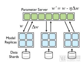
Figure 1
(2) From a model-level perspective, the goal of model sharing is achieved through model ensembling after the completion of model training. Dluhoš et al. [39] performed weighted averaging on the trained weights to further improve accuracy.
(3) Taking into account both the model and optimization levels, Chang et al. [40] integrated these two aspects while considering the correlation between model optimization and performance across different centers. They explored two distinct strategies: one involves training the model at a single center and then transferring it to the next center after convergence, as shown in Figure 2.
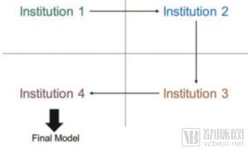
Figure 2
3. Multimodality
Imaging examinations encompass multiple modalities, such as CT, MRI, and DR. In addition to these, there are non-image data sources, including clinical information and laboratory reports. Rational utilization of multimodal data can significantly enhance system performance. In the field of medical image processing, multimodal data primarily improves outcomes through information fusion, which includes two approaches: Early Fusion and Late Fusion.
4. Adjuvant Therapy
Adjuvant therapy generally includes radiotherapy, chemotherapy, hormone therapy, targeted therapy, or biological therapy. From the perspective of standard clinical workflows, AI enhances the quality and effectiveness of clinical treatment in the following areas: (1) AI in patient condition assessment and treatment planning: By integrating medical imaging data with clinical, pathological, and genetic data, AI helps determine the most appropriate treatment regimen. (2) AI in pre-treatment planning: During the treatment process, images used for surgical planning are first preprocessed as needed, primarily involving conversion between different imaging modalities (using U-Net, VGG), denoising (using CNNs), and registration. (3) AI in treatment management: Deep learning can be used to monitor organ motion caused by respiration during surgery, improving prediction accuracy and significantly reducing computation time compared to traditional methods. (4) AI in post-treatment follow-up: After adjuvant therapy, imaging features and responses to tumor markers gradually change over the course of treatment; combining this information with clinical characteristics allows for the evaluation of therapeutic efficacy.
5. Universality (across different devices)
The universality (or generalization ability) of machine learning algorithms refers to the algorithm's adaptability to novel samples outside the training set. The acquisition process of novel samples is influenced by factors such as different imaging angles, imaging noise, and reconstruction algorithms, resulting in new samples having different characteristics from those in previous training datasets.
6. Open AI Computing Platform
With the implementation and widespread adoption of AI technology, the AI industry has transitioned from traditional “small-scale” algorithmic software to “large-scale” application platforms, fostering tighter integration between AI algorithms and cloud computing platforms. Numerous internet companies now offer AI cloud computing platforms and related services. Building on this foundation, medical AI platforms designed for hospitals primarily focus on two directions: imaging AI platforms for processing medical imaging data, and data AI platforms leveraging natural language processing (NLP). By integrating cutting-edge technologies such as the internet, cloud computing, AI, and big data analytics, these platforms facilitate cloud-based collaborative sharing of high-quality medical resources and enable in-depth mining and application of massive volumes of clinical-grade big data. They provide tailored cloud-based intelligent solutions for governments, hospitals, research institutions, and individuals. Furthermore, these platforms offer foundational services—including medical AI model building, training, and open applications—to researchers and developers, thereby driving the robust growth of the medical AI industry.
(The above is an excerpt from the White Paper on AI in Chinese Medical Imaging. For details, please refer to the White Paper on AI in Chinese Medical Imaging.)

Scan the QR code to view the full report.
“White Paper on AI in Chinese Medical Imaging” Features a Robust Editorial Board:
Editor-in-Chief:Liu Shiyuan
Associate Editor:Qian Dahong, Shen Dinggang, Zhang Huimao
Editor-in-Chief:Gao Hong, Xiao Yi
AI Application Module Author:
Author of the AI Application Review: United Imaging Intelligence
Ultrasound AI: Deshang Yunxing Medical Technology
AI Quality Control for Imaging: UESTC Jinpan
Pathology AI: Hengdao Pathology
Pelvic Imaging AI: Huiyi Huiying
AI for Large Vessel Diseases: Huiyi Huiying
AI for Intelligent DR Imaging Reports: iFlytek
Neuroimaging AI: Deepwise Medical
Brain Imaging AI: Deepwise Medical
Cardiovascular AI: Shukun Technology
Fundus Image AI: Tisu Technology
Skin Disease AI: Tisu Technology
Chest AI such as Pulmonary Nodules: Infervision
AI for Osteoarticular Diseases: Xingmai Technology
Intervention: Xingmai Technology
Pediatric Disease AI: Xingmai Technology
Breast Imaging AI: Yitu Healthcare
Bone Age Assessment AI: Yitu Healthcare