Can the Billion-Dollar Digital Pathology Market Become the New Frontier for AI Startups?

Landing Med
Developer of Cervical Cancer Diagnostic Robots

Thorough Images
AI Pathology Image Diagnosis Service Provider

Paige.AI
AI Cancer Diagnosis Technology R&D Provider

Lunit
Artificial Intelligence Software Developer

Knowledge Vision
Intelligent Decision-Making Platform Provider

Dipath
AI-Assisted Diagnostic Tool Developer

iDeepWise
Brain AI Medical Technology R&D Developer
Recently, Paige.AI, a U.S.-based digital pathology startup, received the FDA’s “Breakthrough Device” designation for its AI-powered cancer diagnostics. The company, which is less than two years old, has gained access to over four million archives containing pathological information and digital pathology slides, exclusively licensed from Memorial Sloan Kettering Cancer Center (MSKCC). These data provide it with the potential to change the world. Clearly, it has seized this opportunity.
In China, medical imaging, as a sub-application of computer vision, has been widely adopted in the field of radiology. AI-driven medical imaging companies targeting radiology, such as Yitu Healthcare, Infervision, Deepwise Medical, and VoxelCloud, have expanded their operations overseas.
As a cornerstone of precision medicine, the AI pathology market holds immense potential, with its scale reaching tens of billions of RMB. However, startups dedicated to this field are few and far between; the few existing companies have secured funding only up to Series A, standing in stark contrast to the robust development of medical imaging in radiology.
Why Does This Seemingly Sweet Fruit Remain Unpicked? Let Us Analyze from Technical and Industrial Perspectives to See What Kind of Terrain “AI + Pathology” Truly Is.
Pathology was described by William Osler, the “Father of Modern Medicine,” as the foundation of medicine. Indeed, the accuracy of pathological diagnosis directly impacts patients’ health and fate.
The advent of digital technology has enabled pathologists to capture, stitch, compress, and store pathological images using digital tools, thereby preserving high-quality image information. By integrating database technologies, these processes form a digital pathology slide system. This approach overcomes the limitations of traditional pathology in terms of storage, fidelity, and retrieval, facilitating tasks such as pathological analysis, disease diagnosis, remote transmission, and pathological education through image browsing and analysis.
Artificial intelligence represents an upgrade based on digital technologies. Its applications in pathology include digital image-based initial cytological screening, quantitative morphological analysis, histopathological diagnosis, and prognostic assessment. The inherent value is self-evident; the market for pathological diagnosis alone is vast. Taking gastric cancer as an example, over 20 million patient visits annually require multiple gastroscopic biopsies at pathology departments. A rough estimate places the market size at tens of billions of RMB.
Beyond this, pathological slides harbor deeper layers of information yet to be explored. New drug development, genomics, and even emerging third-party service models are all transforming existing pathology departments.
However, the growth pace of these pathological AI enterprises cannot compare with that of imaging AI companies. Although hospitals generate a vast amount of pathological data annually, the quality of this data is uneven, with significant variations in structure and dimensions. To leverage such data for training algorithms, it must undergo processes such as de-identification and cleaning, the challenges of which are self-evident.
Simply put, the reason no company has been able to replicate Paige.AI’s success is that no domestic enterprise currently possesses pathology data on a scale and with the standardization comparable to that of MSKCC.
This issue is gradually being resolved. The research needs of third-party medical testing centers and hospitals are driving data to flow into artificial intelligence in a rational format, and practitioners at all levels are placing increasing emphasis on the structuring of medical data. Meanwhile, advancements in more specialized fields are increasingly reliant on the processing of pathological information. Consequently, the demand for artificial intelligence in related scientific research is rising sharply, with a gradual transition toward product commercialization.
In scientific research projects, the morphology of tumor cells, animal samples, and human samples subjected to intervention undergoes corresponding changes, which need to be visualized and quantified through specialized methods.
Previous studies on morphological observation have primarily relied on gross examination and microscopy, with immunohistochemistry or immunofluorescence assays employed as adjuncts for diagnostic clarification when necessary, followed by manual counting or software-assisted quantification of captured images. These methods are highly subjective, prone to false-positive results, and exhibit poor reproducibility, thereby underscoring an urgent need for novel approaches to evaluate morphological changes.
The researcher challenge hosted by ISBI evaluated the potential of deep learning algorithms to detect metastases in pathological slides of lymph node metastases from breast cancer patients. The results showed that the area under the curve (AUC) for diagnosis by deep learning algorithms ranged from 0.556 to 0.994, while the AUC for pathologists was 0.724. Notably, the best-performing deep learning algorithm outperformed pathologists in diagnostic simulations.
The applications of deep learning extend beyond this. In their co-authored work, *Application of Artificial Intelligence Technology in the Assessment of Tissue and Cellular Morphology*, Wang Fei, Wei Peilian, Pan Jun, Wu Qing, and Yu Guanzhen provide a detailed overview of application scenarios based on current research findings. Within the industry, many enterprises have leveraged these research insights to pioneer advancements in genomics and drug development.
Tumor-Stroma Ratio (TSR) refers to the ratio of tumor cells to stromal components within tumor tissue, primarily assessed through postoperative pathological sections.
In solid tumors such as colorectal cancer, non-small cell lung cancer, breast cancer, esophageal squamous cell carcinoma, nasopharyngeal carcinoma, cervical cancer, and hepatocellular carcinoma, the tumor-stroma ratio (TSR) is an independent risk factor affecting patient prognosis. Historically, TSR has been primarily assessed by physicians through microscopic visual inspection, with a threshold of 50% commonly used to define stroma-rich versus stroma-poor tumors.
This assessment criterion has several limitations. First, the accuracy of the Tumor-Stroma Ratio (TSR) depends on physician experience; second, the 50% cutoff value is not necessarily accurate. The application of artificial intelligence technology can accurately quantify TSR, and if tumor cell identification is precise, TSR can be determined with single-digit precision.
The research team led by Wang Fei et al. utilized artificial intelligence technology to interpret the tumor-stroma ratio (TSR) in a histopathological slide of tumor tissue. The TSR determined by visual inspection was 30%–50%, whereas the TSR assessed using AI technology was 27.3%, indicating that AI technology has significant advantages in identifying intrinsic features within tumor samples.
Tumor-Infiltrating Lymphocytes (TILs) refer to lymphocytes isolated from tumor tissue that are rich in tumor-specific cytotoxic T lymphocytes and natural killer cells. The identification and evaluation of intratumoral TILs hold significant value for prognostic assessment and therapeutic guidance.AI technology can deliver significant value in this domain, and iDeepWise, a Chinese enterprise, has leveraged this as a breakthrough to design its artificial intelligence products.
Traditional methods for quantifying tumor-infiltrating lymphocytes (TILs) and analyzing their spatial distribution, which rely on hematoxylin and eosin (H&E) staining or immunohistochemical staining, are highly subjective, time-consuming, labor-intensive, and lack accuracy. In contrast, artificial intelligence (AI) can efficiently and accurately employ convolutional neural networks to calculate lymphocyte counts and spatial distribution. Saltz et al., leveraging The Cancer Genome Atlas (TCGA) database, proposed TIL mapping based on H&E images across 13 TCGA cancer types. These TIL maps were generated through computational analysis using trained convolutional neural networks to classify images, thereby revealing the local spatial architecture of TIL patterns and correlating them with overall survival.
The third qualitative analysis application is using AI to identify lymph node metastasis with perineural invasionCurrently, the evaluation of perineural invasion still relies on microscopic visual inspection, which is prone to missed diagnoses and fails to reflect the status of perineural invasion across the entire slide. The research team led by Wang Fei et al. employed deep learning techniques to separately learn and identify tumor cells and neural tissues in hilar cholangiocarcinoma. Their study revealed the entire process of tumor cell invasion into neural tissue, including the initial aggregation of tumor cells around neural tissue, followed by invasion of the perineurium, subsequent erosion of nerve fibers, and ultimately metastasis along the nerves.
Today, South Korean companies have leveraged this technology to develop AI products targeting breast cancer.
Basic research and clinical efficacy evaluation utilize cell and animal models, wherein the therapeutic effects and adverse reactions of drug or genetic interventions on the body and tumors must be demonstrated and evaluated through morphological methods. Traditional microscopic visual observation and interpretation based on hematoxylin-eosin (H-E) staining or special staining techniques have limitations.
By leveraging deep learning techniques to analyze the morphological features of cellular and animal pathological samples—such as necrosis, hemorrhage, lymphocytic response, fibrous proliferation, tumor formation and count, and angiogenesis—we can capitalize on their highly characteristic and regular patterns. This makes the application of artificial intelligence (AI) for evaluating drug efficacy highly feasible. Our research group previously established an animal model of cholangiocarcinoma and administered various pharmacological interventions. By employing AI technology to learn the disease-specific features, we demonstrated that AI can clearly delineate the disease progression and clinical therapeutic effects.
Cytological experiments serve as the cornerstone of basic and clinical translational research, yet few studies have focused on morphological changes in cells. Professor Chris Bakal and Dr. Julia Sero from The Institute of Cancer Research, London, utilized PerkinElmer’s Opera® high-content imaging and analysis system to acquire images. They analyzed the morphological and physiological characteristics of thousands of individual breast cancer cells under various treatment conditions, employing methods akin to those used in neural network research, and detected changes and trends within mitochondrial populations. This study is poised to play a significant role in phenotypic screening and the investigation of unknown mechanisms of drug action.
A Novel Cell Identification and Sorting System: The Ghost Cytometer Combines Advanced Imaging Technology with Artificial Intelligence for Cell Identification and Sorting. The Ghost Cytometer identifies cells at a rate of over 10,000 cells per second and sorts them at a rate of several thousand cells per second.
Furthermore, the combination of temporal waveforms and random pattern intensity distributions enables the reconstruction of cellular morphology on computers. This allows machine learning algorithms to be applied directly to compressed waveforms without image reconstruction, thereby achieving efficient image-based, morphology-free cell detection. This approach will be used to identify and sort circulating tumor cells in patient blood, accelerating drug discovery and improving the efficacy of cell-based therapies.
When evaluating the efficacy of drug or genetic interventions, in addition to H&E staining, special staining techniques can be employed as auxiliary diagnostic tools, including immunofluorescence and immunohistochemistry. Among these, immunohistochemistry is widely used due to its cost-effectiveness, convenience, rapidity, and high-throughput capabilities; however, its results exhibit poor standardization owing to variations in technical proficiency and limitations in evaluation systems.
Since immunohistochemical staining images are also two-dimensional and exhibit distinct features, artificial intelligence technology is highly suitable for interpreting the results, evaluating consistency, and automatically scoring the staining patterns.
Diagnosis is an intuitive application of artificial intelligence in the field of pathology. In the common stereotype, a physician’s role is to provide diagnostic recommendations to patients, while artificial intelligence is intended to replace physicians.
This impression clearly presents logical issues; however, as a data processing approach, properly trained AI can indeed comprehensively examine pathological data to assist physicians in making clinical judgments.
In fact, AI companies in China specializing in medical image analysis account for half of the “AI + pathology” market. VCBeat (WeChat ID: vcbeat) interviewed several domestic and international enterprises engaged in pathological image diagnosis and summarized their characteristics as follows.

Thorough Images, an AI company specializing in pathology that was founded in 2017, focuses on six pathological scenarios: lung, stomach, intestine, lymph node, prostate, and breast.
Wang Shuhao, CTO of Thorough Images, stated, “The selection of this clinical scenario is driven by market considerations. It remains a blue ocean market, and we aim to benefit more patients from the outset—gastric cancer being a prime example.”
Approximately 20 million patients in China require biopsies annually, with the majority undergoing two or more procedures. The domestic pathology resources are ill-equipped to handle such a massive screening volume. Furthermore, in gastroenterology, physicians face a high rate of repetitive work. Often, patients may merely suffer from enteritis yet still receive cancer-targeted treatments for colorectal cancer, leading to many unnecessary biopsies. Artificial intelligence technology can rapidly identify and address this issue.
Based on the 2017 gastric pathology slide test report from the Chinese PLA General Hospital, Thorough Images AI has achieved a sensitivity of 100% and a specificity of 90% in identifying gastric malignancies. Under current conditions, the screening accuracy has reached a considerably high standard.
Subsequently, the company will proceed to accurately classify the screened cancers and confirm each subtype of gastric cancer to provide more precise diagnostic recommendations.

iDeepWise (iDeepwise.ai) has provided cervical cancer screening services to more than 30 renowned Grade A tertiary hospitals and clinical laboratories across China since its inception. To date, iDeepWise has completed retrospective analyses of nearly 100,000 cervical cytology slides.
During the research process, the sensitivity of its TCT-assisted screening product for detecting precancerous lesions increased from 65% with manual slide review to nearly 100%, while the negative predictive value rose to approximately 80%, effectively reducing the slide review workload of pathologists by 80%.
Yang Zhiming, CEO of the company, stated: “Using the MS-CNN deep learning algorithm for cell classification on the Helerv public cervical cytology dataset, our model achieved the world’s best performance on this dataset under identical evaluation conditions, with all metrics surpassing those of the U.S. National Institutes of Health (NIH) classification results (sensitivity exceeded the NIH results by 1–1.5%).”
Regarding future commercialization, iDeepWise can charge fees based on the existing fee schedule for TCT-assisted screening. According to the latest national regulations on medical service pricing, the price for computer-aided diagnosis of cervical cytology ranges from RMB 100 to RMB 160 per test. Based on the current annual volume of approximately 110 million cervical cancer screenings among women in China, it is estimated that this will generate an annual economic benefit of RMB 10 billion to RMB 20 billion in the future.

Compared with other pathology companies, Dipath’s “AI + Pathology” products are comprehensive, aiming to cover as many cancer patients as possible.
Yang Lin, founder of Dipath, told VCBeat: “China sees nearly 5 million new cancer cases annually, while the volume of cell screenings conducted each year approaches 100 million—a remarkably large figure. Our product portfolio covers all major categories used by pathology departments and addresses over 50% of various lesion types across all classifications, boasting the broadest coverage in the world.”
In terms of product design, Dipath centers on its D-Path AI artificial intelligence pathology-assisted diagnostic system and has developed more than 20 intelligent analysis modules in the field of cytopathology. These modules can assist in the subtyping of cancers such as gastric cancer, lung cancer, bladder cancer, breast cancer, renal cancer, and prostate cancer. In the area of molecular pathology, Dipath utilizes artificial intelligence to interpret probe liquid samples, blood cells, and cervical smears.
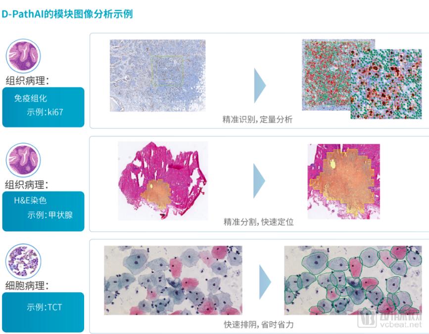
To date, Dipath has employed AI to analyze nearly one million cervical histology slides, with rapid growth also seen in other pathology categories. Recently, at the Zhuoyi AI Challenge, Dipath secured first place in three technical categories: cytopathology (cervical smears), histopathology (thyroid frozen sections), and quantitative immunohistochemistry analysis.
Dipath’s products are derived from over 100 SCI papers published in the field of AI-digital pathology by Dipath’s founders and the Dipath Research Institute, including “Pathologist-level Interpretable Whole-slide Cancer Diagnosis with Deep Learning,” which was published in Nature Medicine (Impact Factor: 30).
In terms of commercialization, Dipath will adopt a modular sales approach, allowing hospital pathology departments to purchase the modules best suited to their needs and expand with additional modules in the future.

Lunit, an AI company from South Korea, has developed a comprehensive suite of AI products for breast cancer. Its chest X-ray and mammography solutions are used for initial disease detection and screening, while its breast tissue pathology slide grading serves as a critical step in the final medical diagnosis.
Although pathological grading plays a crucial role in the diagnostic process, the field still lacks quantifiable objective standards and detailed interpretation procedures. The emergence of digital pathology offers hope for addressing this issue.
Lunit has invested substantial financial and human resources in digital pathology research to objectively interpret diverse morphological features in tissue samples, driving innovations that enhance the accuracy, efficiency, and consistency of histopathological diagnosis.
In 2017, Lunit introduced an artificial intelligence algorithm capable of automated detection and staging assessment of breast cancer metastases in lymph nodes, marking the first attempt to fully automate a specific pathology task from end to end.
The pathological diagnosis of regional lymph nodes (pN-stage: i.e., determining whether breast cancer has metastasized to the lymph nodes) involves an extremely large volume of image data, with resolutions reaching up to 200,000 × 100,000 pixels. This necessitates that pathologists devote substantial time to meticulously reviewing multiple images to accurately determine the pN-stage.
Lunit has leveraged its deep learning technology to develop a highly accurate pN-stage prediction algorithm. This algorithm integrates the detection and classification of tumor metastases across multiple lymph node tissue sections into a single clinical outcome. By utilizing lymph node histology images from the Camelyon17 dataset, Lunit established an algorithm for predicting pN-stage that outperforms most current state-of-the-art technologies worldwide, with the potential to significantly enhance pathologists' efficiency and diagnostic accuracy.
Since we can design software for in-depth analysis of pathological images, why not optimize the images directly during acquisition?
Nowadays, some traditional medical device companies are also attempting to transform their previously rigid instruments into smart devices, leveraging artificial intelligence to enable more precise imaging and faster analytical efficiency.

Fuyi Shares is a medical device company with 15 years of deep expertise in pathology. Its product portfolio covers services including pathological image acquisition, pathological data analysis, and remote pathological diagnosis, providing pathology departments with comprehensive intelligent diagnostic solutions.
The digital pathology intelligent diagnostic system developed by the company enables high-definition digitization of images, with a maximum throughput of 400 slides, achieving 24-hour unattended automatic scanning. It features high-speed scanning and seamless stitching of pathological sections, transforming traditional workflows by digitizing, visualizing, and enabling the storage of pathological specimens, thereby laying a solid foundation for digitalization and informatization.。
Fuyi Co., Ltd.’s digital pathology remote diagnosis system platform, assisted by AI technology, has assembled nearly 2,000 practicing pathologists from public hospitals across China. Diagnoses for the “Remote Pathology Standard Laboratory” are conducted by top-tier pathology experts in each province, with 5–10 associate senior-level or higher experts selected per province. This ensures that diagnostic results carry authoritative weight within their respective regions, eliminates missed diagnoses, and guarantees the authenticity and reliability of the diagnostic outcomes.

Recently, an AI microscope developed by Zhiying Medical—an automated diagnostic system for pulmonary tuberculosis based on microscopic imaging of sputum bacteria—is set to be officially launched for commercial use. The AI microscope utilizes artificial intelligence deep learning algorithms to rapidly scan entire slides and perform mycobacterial counting within three minutes, thereby diagnosing pulmonary tuberculosis.
Traditional rule-based algorithms for medical image processing in sputum smear testing lack adaptability to individual variations, resulting in suboptimal diagnostic accuracy. In contrast, AI-driven image processing, leveraging big data training and deep learning techniques, can significantly enhance detection accuracy.
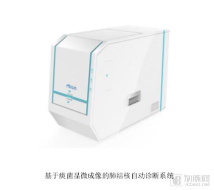
The AI microscope developed by Zhiying Medical integrates artificial intelligence-based image and visual processing technologies to automatically scan sputum-stained smears and perform intelligent detection and analysis. With simple commands from physicians, the AI system automatically identifies and detects targets in sputum-stained smears, quantitatively calculates results, and generates reports. Detection results are displayed in real time on the client interface, providing timely alerts without disrupting clinical workflows, thereby enhancing diagnostic efficiency and accuracy for physicians.
Once basic pathological data are acquired, can we extract deeper insights beyond diagnosis? Forward-thinking companies are attempting to do just that.
Before the advent of artificial intelligence, quantitative analysis of pathological slides was a nearly impossible task, relying solely on physicians’ subjective assessments to roughly estimate the extent of lesions. However, the emergence of artificial intelligence has made precise counting of tissue cells possible.
In this scenario, researchers can rapidly and accurately obtain data on the number, severity, and temporal changes of pathological cells in tissue sections over a given period, thereby enabling easy observation of the impact of new drugs on lesions in clinical trials.
Following this logic, we could leverage AI to monitor histological and cellular changes in animals after drug administration, thereby providing more precise guidance for drug development.

Chengdu Knowledge Vision is engaged in such endeavors, providing quantitative digital pathology image visualization and quantitative analysis for CROs to support clinical drug research. In its collaboration with Roche Diagnostics, the achievements of Knowledge Vision have been recognized by Roche.
However, this concept did not originally target AI-driven drug discovery. In an interview, its founder, Xiang Fei, stated, “We are committed to building a no-code cloud platform for the development of pathology AI applications, addressing challenges such as high technical barriers, substantial hardware investments, and elevated costs associated with communication and data annotation. This enables pathologists to conduct research on pathology AI applications tailored to their actual needs without any coding expertise.”
Foreign companies such as Reveal Biosciences and PathAI are also engaged in similar endeavors. Recently, Reveal Biosciences secured Series A financing led by Intel. Dr. Claire Weston, Founder and CEO of Reveal Biosciences, stated, “To date, Reveal Biosciences’ ImageDx technology has provided information services to more than 300 medical institutions. Our specialized approach to data integration enables the large-scale and rapid generation of pathology AI algorithms. With this funding, we are pleased to expand our ecosystem of researchers, pathologists, and technologists, working together to pioneer new paradigms in AI-driven healthcare.”
In addition to analyzing pathological information itself, studies could correlate relevant data with genetic databases and simultaneously monitor phenotypic and genotypic data of subjects during experiments.
With the advancement of immunotherapy, the spatial localization and quantification of novel tumor immune markers in PD-1/PD-L1 and CAR-T therapies have imposed higher demands on pathological diagnosis, necessitating technological innovation.
Based on traditional information platforms in hospital pathology departments, it is difficult for physicians to conduct large-scale pathological diagnoses. The primary reason lies in the fact that case information, such as text reports and digital images, cannot be manually subjected to big data-driven retrospective research searches, analysis, and management. This naturally hinders the standardization of pathological diagnosis and the evolution of diagnostic standards.
Against this backdrop, Genome Wisdom Inc. has constructed a pathology knowledge graph and developed multiple core technologies, including natural language processing for Chinese pathology reports, bioinformatics analysis of tumor genomics, and artificial intelligence for pathological image analysis.This will provide pathological text, image, and genomic data analysis capabilities for the research plan, while also offering other project participants standardized tools for analyzing their own data.
By integrating pathological staining images, such as H&E and IHC, with genomic and transcriptomic data from genomics, Genome Wisdom Inc. can mine biomarkers with greater precision and efficiency, thereby facilitating drug development.

Third-party medical diagnostic institutions are a key means for the state to implement tiered diagnosis and treatment and promote the reform of public hospitals, as well as an important vehicle for social capital to enter the healthcare industry. The integration of artificial intelligence technology is expected to enhance the medical capabilities of third-party medical testing centers. An increasing number of enterprises are entering this field.
The emergence of cloud pathology has accelerated the development of third-party imaging centers. This model has facilitated remote pathological diagnosis, enabling digital pathology to transition from a theoretical concept of telemedicine to practical application, thereby establishing cloud pathology platforms.
Currently, multiple large enterprises in China are leveraging cloud platforms to deliver AI-based pathological diagnosis technologies to medical testing centers both domestically and internationally.

Traditional cytological screening for cervical cancer relies on medical technicians performing diagnoses under a microscope based on their experience. According to international standards, the daily slide review volume for each technician should be limited to fewer than 100 cases. As humans are not machines, diagnostic errors caused by fatigue or variations in experience are unavoidable. However, the AI-powered cervical cancer diagnostic robot “Landing,” developed by Landing Medical, assists physicians in accurately detecting early precancerous lesions of the cervix, making cervical cancer highly likely to become the first malignant tumor eradicated through preventive screening.
Leveraging this AI technology, Landing Medical has established over 400 “Landing Standard Cytology Laboratories” across China, with coverage extending to tertiary hospitals in provincial capitals, secondary hospitals in small and medium-sized cities, and even grassroots family planning stations in rural areas.
This novel screening model marks the end of an era in which cervical cancer screening relied on expert experience for diagnosis. By leveraging big data to enhance diagnostic quality and modern technology to reduce costs, it improves the efficiency of large-scale cervical cancer screening as well as the detection rates of precancerous lesions and positive cases, playing a significant role in reducing the incidence and mortality of cervical cancer. Furthermore, it provides a practical solution to the common challenge faced by developing countries worldwide: the shortage of specialized personnel for tumor screening at the primary healthcare level. To date, Wuhan Landing Medical High-Tech Co., Ltd. has completed over 2 million cervical cancer screenings.
Landing Medical is steadily expanding its global presence. On April 1, the Landing AI Cloud Diagnostic Platform for Cervical Cancer Screening was officially launched worldwide, enabling women across the globe, particularly in countries along the Belt and Road Initiative, to benefit from high-quality, cost-effective cervical cancer screening services provided by China’s AI cloud diagnostic platform.
Hengdao Pathology adopts a multi-tiered model comprising a “full-time medical and technical team + frontline consultation experts + co-built collaborative platforms.” Leveraging a “digital remote consultation network + physical centers & logistics support,” it provides pathology consultation and diagnostic support to primary care hospitals, focusing on intraoperative frozen sections, complex case consultations, various specialized needle biopsies, and rapid diagnosis of small specimens. Its whole-genome microarray platform offers 20 AI-powered molecular pathology reporting software solutions for multiple cancer types, bringing molecular pathology to the grassroots level.
Leveraging its specialized AI team and technical reserves, Hengdao Pathology actively engages in scientific research collaborations with various renowned Grade A tertiary hospitals to promote the steady development of Pathology Artificial Intelligence (Path AI). By harnessing the advantages of Hengdao Pathology’s big data platform (PathHub™), it aims to build the infrastructure for the entire pathology industry in the AI era, provide intelligent auxiliary tools for pathologists to enhance diagnostic efficiency, facilitate the sharing of pathological big data and technological achievements, and drive industry upgrading.
In October 2018, Ping An Leasing made a significant move, enabling the gradual emergence of 1,000 third-party testing centers with the support of RMB 30 billion in funding. The advantage of Ping An Health (Testing) Centers lies in Ping An’s overall corporate structure. These third-party testing centers not only benefit from patient referrals by Ping An Good Doctor, but also leverage Ping An’s traditional insurance business to provide patients with a wide range of optional medical insurance and commercial insurance services.
With policies now in place, Ping An Testing’s entry has injected vitality into the entire sector. Once the Ping An ecosystem is fully established, China’s third-party medical testing industry may well enter a brand-new phase.
Most of the companies listed above specialize in “AI + pathology.” In reality, many artificial intelligence enterprises focused on medical imaging have already expanded into the pathology sector or established their own medical laboratories. Judging by current trends, the integration of precision medicine and AI within pathology has become an irreversible trend.
This holds true for diagnostics, and even more so for AI-driven drug discovery. It is entirely possible that new drug development will surpass imaging-based products to become the first scenario in healthcare where artificial intelligence achieves commercialization. We hope to see more companies like Genome Wisdom Inc. and Knowledge Vision look beyond the surface of pathological data to integrate genomic data, offering innovative solutions to conquer stubborn diseases such as cancer.
Scientific research is also advancing continuously; deep learning enables researchers to quantify cellular parameters with greater precision and observe changes in tumor cells more intuitively.
Therefore, stakeholders with high expectations for artificial intelligence might consider shifting some of their attention to pathology, where they may uncover unexpected potential.