Scientists Uncover Link Between Membrane-less Organelles and Neurodegenerative Diseases: Exploring Biomolecular Condensates (Part I)
Most biology textbooks state that membrane structures are the most important organizational form within cells. Phospholipid bilayers envelop various organelles, including mitochondria, the endoplasmic reticulum, and lysosomes, thereby separating the distinct proteins found inside and outside these organelles. The remaining cellular components are suspended in the cytosol. Proteins within the cytosol occasionally encounter other bindable molecules, such as substrates and small-molecule drugs.
However, this situation is gradually changing. As scientific research continues to deepen, biomolecular condensates—transient fluid droplets composed of proteins and RNA—have been discovered. These biomolecular condensates are referred to as membraneless organelles. Research into the structural and biophysical properties of these membraneless organelles has advanced rapidly over the past decade. Scientists and drug developers are increasingly focusing on this unique biological structure.
We have compiled and organized a recent article published in Nature Reviews Drug Discovery on membraneless organelles into two parts. This is Part I, which primarily introduces the history of research on membraneless organelles and their relationship with diseases. The forthcoming Part II will analyze the impact of membraneless organelle research on the development of the pharmaceutical industry, as well as the strategic layouts of major pharmaceutical companies and startups in this field.
Preliminary evidence suggests that these, through what is known as liquid- Membraneless organelles formed through liquid-liquid phase separation are closely associated with health and disease. In certain contexts, they appear to function as crucibles that accelerate reactions among their components while preventing these components from interacting with molecules outside their structural boundaries. Genetic mutations affecting the assembly and disassembly of membraneless organelles have also been strongly implicated in neurodegenerative diseases, cancer, and other disorders.
More than a century has passed since membraneless organelles were first reported. As early asIn 1899, Edmund Beecher Wilson, a pioneer in cell biology, published in *Science*Magazinein a review articleThe widespread presence of membraneless organelles has been described, and these structures have been depicted in cellular maps for decades. However, due to the limited understanding of their roles within cells and the biophysical properties of their components, few researchers have focused on this field.
About a decade ago, this situation began to change.
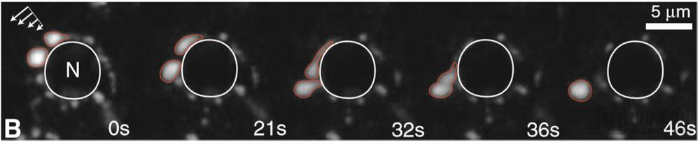
Figure 1: P plasmids extruding from the nucleus (N) (highlighted by red circles)
Image source: Clifford P. Brangwynne, Christian R. Eckmann, et al. Germline P Granules Are Liquid Droplets That Localize by Controlled Dissolution/Condensation.Science.324,1729(2009).
In 2009, Cliff Brangwynne, then a postdoctoral researcher at the Max Planck Institute, and his supervisor Tony Hyman were observing *Caenorhabditis elegans* embryos under a microscope. Their initial aim was to understand the origin of P granules (the germ plasm of nematodes, composed of RNA and RNA-binding proteins). However, during their observations, they discovered that P granules behaved like oil droplets in a vinaigrette: they dripped from the nucleus, fused with one another, and rapidly underwent condensation and dissolution within the cytoplasmic matrix (Figure 1). They reported these findings in the journal *Science* in 2009. This paper became a landmark study as it was the first to apply the concept of “phase separation” to describe specific membraneless organelles.
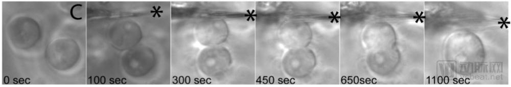
Figure 2: Nucleolar fusion observed under the propulsion of a microinjection needle
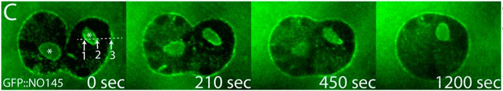
Figure 3: Nucleolar fusion observed under green fluorescent protein labeling
Image source: Clifford P. Brangwynne, Timothy J. Mitchison, et al. Active liquid-like behavior of nucleoli determines their size and shape in Xenopus laevis oocytes.Proc.Natl.Acad.Sci.108,4334(2011)
Two years later,Brangwynne and colleagues further reported in the Proceedings of the National Academy of Sciences, the nucleolus (a structure formed within the nucleus that plays a crucial role in ribosome assembly) exhibits similar fluid-like properties and relies on phase separation. In Xenopus laevis oocytes, they used micropipettes to push nucleoli together. After a brief delay, the two nucleoli began to fuse slowly, ultimately forming a larger sphere (Figure2). Subsequently, they introduced GFP (green fluorescent protein)-labeled NO145, a key component of the perinucleolar filamentous network, into blastocyst cells. Under GFP labeling, nucleolar fusion occurring under physiological conditions was observed via distinct green fluorescent signals at the periphery of the nucleoli (Figure 3).
Soon thereafter, researchers observed similar phenomena in various membraneless organelles, including Cajal bodies, nuclear speckles, stress granules, and RNA transport granules.
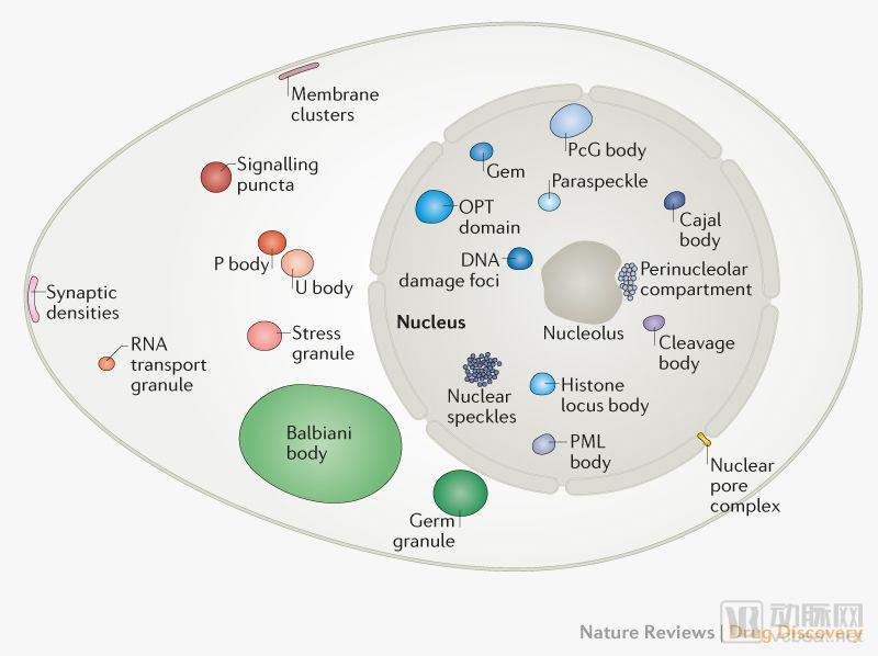
Figure 4: Membraneless Organelles in Eukaryotic Cells
Image source: Salman F. Banani, Hyun O. Lee, et al. Biomolecular condensates: organizers of cellular biochemistry.Nat Rev Mol Cell Biol.18,285(2017)
Meanwhile, researchers are also attempting to decipher the biophysical basis underlying the rapid formation and disassembly of these structures, and have made partial progress. For example, in 2012, Michael Rosen, a biophysicist at UT Southwestern Medical Center, and his colleagues described in Nature how multivalent macromolecules can undergo rapid liquid–liquid phase separation and form micron-scale fluid droplets. Subsequent work has shown that the multivalent macromolecules involved in similar processes are primarily proteins containing intrinsically disordered regions (IDRs—protein domains that cannot fold into stable three-dimensional structures) and RNA molecules.
The link between this phenomenon and the disease was subsequently uncovered step by step. Paul Taylor, a neurologist at St. Jude Children's Research Hospital who has long been dedicated to studying neurodegenerative diseasesatIn 2013, *Nature* reported that mutations occurring in the conserved regions of the intrinsically disordered regions (IDRs) of hRNPA2B1 and hnRNPA1 were associated with amyotrophic lateral sclerosis (ALS). By 2015, research on IDRs had rapidly gained momentum. That year, five independent studies demonstrated that IDRs are critical for the phase transitions of biomolecular condensates.
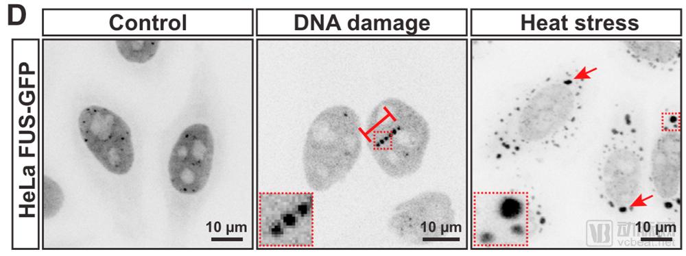
Figure 5: FUS protein forms biomolecular condensates in the cytoplasm under DNA damage and stress conditions
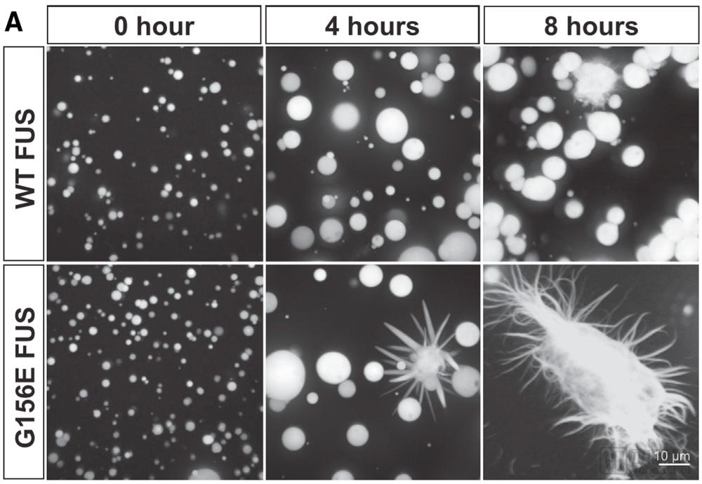
Figure 6: Abnormal aggregation of mutant FUS protein
Image source: Avinash Patel, Hyun O. Lee, et al. A Liquid-to-Solid Phase Transition of the ALS Protein FUS Accelerated by Disease Mutation.Cell.162,1066-1077(2015).
InResearch on membraneless organelles in ALS is rapidly advancing. Hyman, co-founder of Dewpoint Therapeutics—a biotech startup dedicated to the study of membraneless organelles—and his colleaguesIn "published in Cell,The FUS protein forms membraneless organelles at sites of DNA damage and in the cytoplasm under stress conditions (Figure 5), and ALS-associated FUS mutations lead to aberrant phase transitions (Figure 6). The FUS protein plays a crucial role in RNA transcription, splicing, and microRNA processing. Numerous previous studies have confirmed a clear association between the FUS protein and various neurodegenerative diseases. This finding proposes a potential mechanism underlying FUS protein pathogenicity.

Figure 7: HnRNPA1 wild-type and D262V mutant exhibit distinct phase transition behaviors
Image source: Amandine Molliex, Jamshid Temirov, et al. Phase Separation by Low Complexity Domains Promotes Stress Granule Assembly and Drives Pathological Fibrillization.Cell.163,123-133(2015).
In another article published in Cell, Taylor and colleagues stated that hnRNPA1 also undergoes liquid-liquid phase separation, and ALS-associated mutations in this protein affect the phase separation process. When researchers mixed wild-type hnRNPA1 with the D262V mutant form, they observed that the mutant hnRNPA1 exhibited a higher degree of condensation, showing significant differences from the phase transition behavior of the wild-type protein (Figure 7). The results suggest that ALS-related mutations impact the dynamic processes underlying membraneless organelle formation and appear to render certain structures more viscous than under normal conditions. This condition seems to further trigger the fibrillization of inclusions, a hallmark of the disease. Taylor estimates that aberrant phase transition processes account for more than 90% of ALS cases.
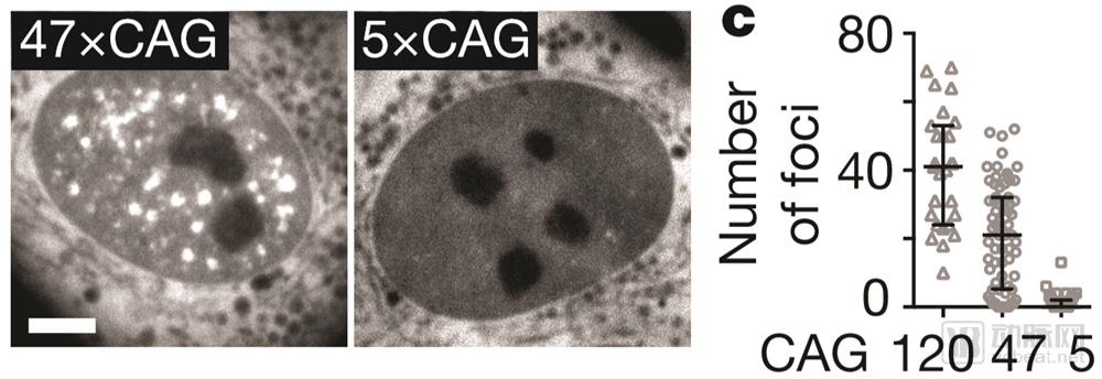
Figure 8: RNA with multiple CAG repeats aggregates into membraneless organelles in the nucleus
Image source:Ankur Jain,Ronald D.Vale.RNA phase transitions in repeat expansion disorders.Nature.546,243(2017).
Other neurodegenerative diseases may also be associated with liquid-related to liquid-liquid phase separation. In 2017, Ankur Jain and Ron Vale published in Naturean article mentions a series of diseases caused by an increase in repetitive sequences, including Huntington's disease, muscular dystrophy, andConditions such as ALS may involve aberrant RNA droplet formation. In this study, researchers found that RNAs containing multiple CAG triplet repeats (the primary pathogenic cause of Huntington’s disease) aggregate post-transcriptionally to form membrane-less organelles, with the extent of aggregation positively correlated with the number of CAG repeats. A similar phenomenon occurs in RNAs with multiple CCCCGG repeats (a major pathogenic cause of ALS).

Figure 9: Spontaneous aggregation of phosphorylated tau441 protein to form membraneless organelles
Image source:Susanne Wegmann,Bahareh Eftekharzadeh,et al.Tau protein liquid–liquid phase separation can initiate tau aggregation.The EMBO Journal(2018).
InIn 2018, Taylor and colleagues reported that soluble tau protein, one of the main culprits in Alzheimer’s disease, also forms condensates.. Whether it is exogenously expressedTau protein, whether recombinant or isolated from patient specimens, undergoes liquid-liquid phase separation. These tau proteins form gel-like condensates within tens of minutes and continue to aggregate and grow larger over the subsequent days.
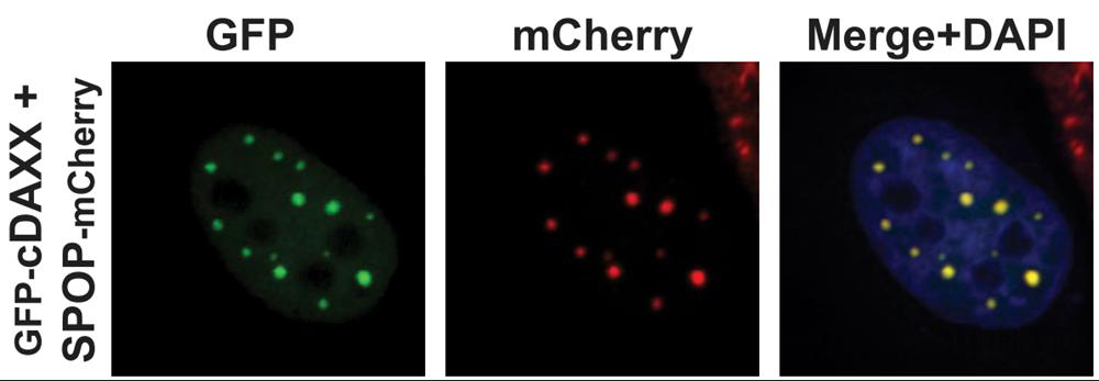
Figure 10: Wild-type SPOP exhibits significant co-localization with cDAXX and condenses into membraneless organelles.
Image source:Jill J. Bouchard, Joel H. Otero,et al.Cancer Mutations of the Tumor Suppressor SPOP Disrupt the Formation of Active,Phase-Separated Compartments.Molecular Cell.72,19-36(2018).
Membraneless organelles also appear to be associated with cancer.In 2016, Massachusetts General Hospital pathologist Miguel Rivera and his colleagues published a paper stating, inIn Ewing sarcoma, phase separation mechanisms may initiate and sustain oncogenic regulatory programs. In 2018, Tanja Mittag, a structural biologist at St. Jude Children’s Research Hospital, discovered that the tumor suppressor protein SPOP is highly active within membraneless organelles. More importantly,Cancer-associated SPOP mutations canInterference with substrate co-localization of the protein affects its phase separation capacity and tumor-suppressive ability (Figure 10).
Taylor added that, given the widespread presence of intrinsically disordered regions (IDRs) across the proteome, these findings have captured the attention of cell biologists worldwide. “Many scientists have suddenly realized that the proteins they have been studying undergo biologically relevant phase transitions, something they had never previously recognized,” he said.
In light of the unique properties of membraneless organelles, pharmaceutical start-ups have begun to establish a presence in this field, while multinational pharmaceutical companies are closely monitoring its next developments. How exactly will membraneless organelles empower the healthcare sector? What challenges are hindering the growth of this nascent field? In our next article, we will continue to analyze the current landscape and future prospects of this domain. Stay tuned.
Original Article Link:https://www.nature.com/articles/d41573-019-00069-w
Cover source:Susanne Wegmann,Bahareh Eftekharzadeh,et al.Tau protein liquid–liquid phase separation can initiate tau aggregation.The EMBO Journal.(2018).