3D Printing and Artificial Intelligence Unlock the 'Black Box' of Precision Oncology
Currently, as artificial intelligence and the healthcare industry accelerate their integrated development, medical AI has become a crucial tool for liberating clinical productivity. It not only frees physicians from mechanical and repetitive tasks but also enables patients worldwide to benefit from standardized medical care characterized by seamless human-AI collaboration.
Particularly in the field of oncology, which emphasizes evidence-based medicine, artificial intelligence is enabling more accurate diagnoses, more standardized treatments, safer minimally invasive surgeries, and shorter operative times, thereby instilling greater confidence in physicians when confronting the medical challenge of cancer. As Alan Perlis, the “father” of the ALGOL language and computer science, once remarked, “A year spent in artificial intelligence is enough to make one believe in God.”
In the field of oncology, sufficient evidence has always been required to support surgical intervention as the appropriate course of action. However, in clinical practice, physicians often lack the opportunity to make direct comparisons across similar cases. When they judge, based on experience, that a lesion is resectable, they proceed with surgery.
During this process, because it is impossible to precisely locate all blood vessels near the lesioned tissues or to finely distinguish the boundaries between cancerous and normal areas, doctors can only determine the success or failure of the surgery by observing the patient's postoperative course.
Dr. Wu Wentao, formerly a neurosurgeon at West China Hospital, reflected deeply on his surgical experiences: “In the past, when orthopedic surgeons performed operations for bone tumors, they had to rely solely on tactile feedback during surgery to determine resection margins. This was because the distinct boundaries visible on preoperative imaging were enhanced by contrast agents, whereas such margins could not be discerned with the naked eye during actual procedures. Visually, obviously protruding areas were certainly tumor tissue; however, much of the surrounding bone that appeared healthy on the surface had already been infiltrated by the tumor internally. No differences could be detected superficially.”
“The original orthopedic surgeon could only attempt to resect a segment and then send the margin tissue for intraoperative frozen section pathology. This process takes half an hour, and after waiting for half an hour, it is found that there are still residual tumor cells at the margin. The orthopedic surgeon then cuts a little further outward—step by step, continuing this process.”
“You can imagine that, first, each small section resected must be sent for intraoperative frozen section analysis, which requires a 30-minute wait; this time consumption is substantial. Second, the bone wound surface exhibits significant bleeding that is difficult to control. Prolonged operative time leads to extensive oozing and fluid evaporation, resulting in massive blood loss and hemoconcentration, and may even cause complications such as pulmonary embolism. Therefore, traditional orthopedic oncology surgeries typically last around ten hours, with blood loss often exceeding 10,000 milliliters. Such procedures can only be performed at major medical centers like West China Hospital; ordinary hospitals simply do not dare to undertake them.”
What if we could precisely differentiate and delineate bone tissue? This is where cutting-edge technology comes into play.
Artificial intelligence has long been extensively applied in medicine. We have observed AI technologies targeting various cancers improving early detection rates from the grassroots level, achieving significant results. However, in China, where early cancer screening remains insufficient, a large number of patients still require surgical intervention for treatment.
Therefore, Dr. Wu Wentao chose to join Baiyang Intelligent Technology as Chief Medical Officer, aiming to leverage technologies such as artificial intelligence, 3D printing, and VR to assist in cancer treatment.
“Cancer treatment is a protracted struggle. Preoperative planning, intraoperative procedures, and postoperative rehabilitation are all critical to cancer care. Baiyang leverages Watson for Oncology, integrated with tens of millions of medical data points, to provide evidence-based solutions and source references for personalized cancer treatment plans. Its newly developed Baiyang Smart Imaging Solution (BïSO) assists physicians in preoperative planning—including surgical planning for neurosurgery and orthopedics—provides intraoperative localization and navigation, and facilitates postoperative efficacy assessment. These technologies have the potential to revolutionize the quality of surgical care.”
“Taking osteoma resection surgery as an example, using BïSO’s CT-MR multimodal image fusion preoperative assistance system, we can analyze imaging data before surgery to understand the extent of the tumor, determine the boundary between tumor tissue and normal tissue, and precisely define the surgical resection range. The gray and purple areas in the images represent the surgical guides. After BïSO scans the patient’s bone, corresponding surgical guides can be rapidly generated through 3D printing technology. With current technology, this personalized and precise design of the guide, tailored to fit the specific bone anatomy, significantly reduces the difficulty for surgeons during the operation.”
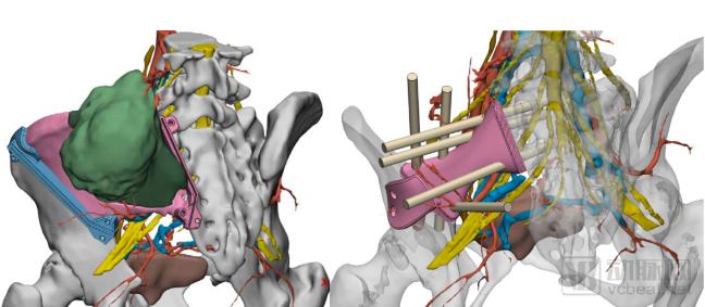
According to practical operational statistics, the use of BïSO’s intraoperative localization and navigation technology can reduce osteoma surgery time to 2–3 hours, with a significant reduction in blood loss from over 10,000 mL to 2,000–3,000 mL. In such cases, patients do not require postoperative admission to the ICU, thereby reducing associated medical costs.
The beneficiaries are not limited to physicians and hospitals. If physicians can present a patient’s tumor status through precise preoperative imaging, they can more vividly describe the procedural details and expected outcomes of each surgical option. This approach enhances transparency throughout the surgical process, turning shared decision-making between physicians and patients from an aspiration into reality.
More importantly, the empowerment of medical resources is key. Complex tumor surgeries that were previously only performed at West China Hospital can now be conducted in county-level hospitals through the BïSO system, allowing more ordinary patients to regain their lives.
Studies have shown that standardized treatment can effectively extend the 5-year survival rate of cancer patients. Today, cancer treatment has entered an era of "chronic disease management." However, the standardization of cancer treatment varies across different regions, with a significant contributing factor being the unstructured nature of imaging reports.
To address this issue, the BïSO system integrates an imaging structured reporting system based on radiology language and a knowledge graph framework, focusing on resolving the problem of non-standardized and inconsistent radiology reports.
To facilitate physician recognition and medical student learning, structured imaging reports often adopt a format combining text and images. When drafting reports, physicians need only annotate areas of concern, allowing the corresponding quantitative measurement results to be directly imported into the report.
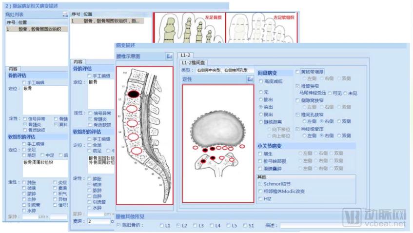
Furthermore, this system is built upon an imaging diagnostic knowledge graph and the International Radiology Lexicon (RadLex), incorporating the radiological diagnostic reasoning logic prevalent in current medical practice to automatically correlate imaging data. For instance, when a physician selects the “lymph node metastasis” option, the system automatically visualizes the metastatic findings on the images and integrates them into the report. In the case of gastric cancer, the system automatically calculates the stage and subtype, populating the report with quantified values compliant with international standards, thereby facilitating accurate tracking of treatment efficacy by physicians.
As of April, more than six months after the launch of the BïSO system by Baiyang Intelligent Technology, the system has assisted physicians in completing over 1,000 neurosurgical procedures and 220 orthopedic surgeries. Related products for applications involving the liver, pancreas, and biliary tract have also reached maturity and are being rapidly advanced.
“This is merely the beginning. In Dr. Wu Wentao’s view, this product suite still lacks sufficient automation and requires occasional physician calibration. In his eyes, a mature product should achieve full automation and intelligence, which will be the focus of his in-depth research.”
Moreover, the greater value of the BïSO system lies in enhancing the surgical capabilities of surgeons in primary and secondary hospitals, while providing more vivid instructional imaging through its visualized reports and procedural recordings, thereby advancing both equipment and human resources in tandem.
Looking ahead, Wu Wentao believes that surgical navigation and surgical robots will be the major direction for the development of medical technology. More intelligent surgical robots and mixed reality-based intraoperative positioning and navigation systems will hold significant market potential.
As part of Baiyang’s BSmartD intelligent physician cloud platform, BiSO is poised to forge deeper connections with Watson for Oncology and genomic solutions in the future. It is conceivable that AI-driven analysis of genomic data will enable more detailed surgical planning down the line. After all, in the age of artificial intelligence, what is truly impossible? Let us give Baiyang some time.