First Imaging Showcases Intraoperative CT and Robotic Imaging Platform at CMEF, Leveraging Cone-Beam CT and 3D Reconstruction Algorithms for Advanced 3D Visualization

First Imaging
Medical 3D Imaging Equipment R&D Service Provider
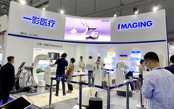
First Imaging's CMEF Booth
May 14–17, VCBeat (WeChat Official Account: vcbeat)InThe 81st CMEF China International Medical Equipment (Spring) FairIt was learned at the exhibition that Jiangsu First-Imaging, a medical imaging company specializing in 3D imaging technology, unveiled two innovative products based on cone-beam CT (CBCT) imaging technology for the first time: a C-arm CT (intraoperative CT) system that provides intraoperative 3D CT images, and Robot Imaging (an imaging robot platform).
In medical CT imaging, cone-beam CT (CBCT) offers significant advantages in terms of radiation dose and spatial resolution for high-contrast structures. Coupled with its flexible and compact design, CBCT is widely applied across various specialized fields, including intraoperative imaging, orthopedic imaging, and dental imaging. The downward diffusion of technology and the deep exploration of niche applications to support precision medicine will emerge as new market growth drivers in the dedicated CT sector.
First Imaging, established in 2015, boasts an R&D team encompassing talent across the entire CT imaging industry chain. Furthermore, its founder, Dr. Xi Yan, possesses extensive experience in innovative CT equipment algorithm research and complete system development.
It is reported that Dr. Xi Yan completed his postdoctoral research at Rensselaer Polytechnic Institute in the United States, and earned both his bachelor’s and doctoral degrees from Shanghai Jiao Tong University. He has long been engaged in the research and product development of X-ray-related equipment. Leveraging the foundational theoretical framework of AI-based 3D imaging and focusing on cone-beam CT (CBCT) technology to drive innovation in 3D imaging products, First Imaging aims to become an industry leader in the field of specialized medical CT. The company’s products provide customers with a comprehensive suite of intelligent digital solutions.
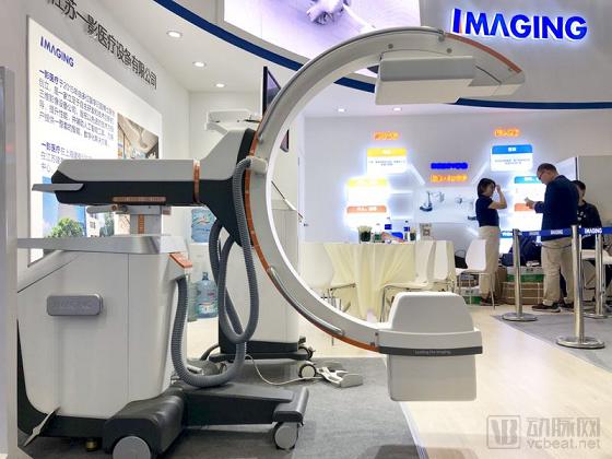
First Imaging Intraoperative CT
Advances in medical technology are driving precision medicine to become the next generation of diagnostic and therapeutic techniques. In the operating room, the integration of surgical robots with three-dimensional (3D) CT imaging has become a prerequisite for performing precise surgeries. Meanwhile, due to the high cost of 3D imaging equipment on the market, most hospitals in China still rely on two-dimensional (2D) X-ray devices. 2D images suffer from single-angle projection and overlap, making them prone to interference. To obtain anteroposterior and lateral views, patients are required to assume multiple positions, which is time-consuming. For instance, in spinal pedicle screw placement surgery, determining the relationship between the screws and the spine often requires appropriate fluoroscopic angles combined with the surgeon’s experience.
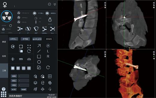
First Imaging Intraoperative CT 3D Spinal Imaging
At this exhibition, First Imaging launched an intraoperative CT system whose performance perfectly meets the 3D imaging requirements of surgical robot-assisted procedures. By acquiring continuous X-ray images through uniform rotation around the patient on a C-arm track, and leveraging powerful software algorithms to reconstruct 3D tomographic images, the system enables direct assessment of the spatial relationship between metallic screws and the spine during spinal surgeries. When integrated with surgical navigation systems, surgical robots, and other equipment, this product provides precise intraoperative 3D CT imaging for a wide range of procedures in orthopedics, interventional therapy, gynecology, and other specialties.
In terms of product performance, the high-power pulsed X-ray source of First Imaging’s intraoperative CT scanner ensures high intensity and strong penetration capability, enabling high-quality image acquisition even for obese patients. This allows both patients and physicians to undergo treatment and operate with lower radiation doses. The system incorporates a suspended three-axis integrated manual-electric C-arm gantry, allowing flexible and rapid positioning of the C-ring to the target location. It is equipped with two types of flat-panel detectors: a high-end CMOS model and a standard a-Si model. Additionally, it features an ultra-large 27-inch imported professional medical-grade monitor (with dual-display functionality on a single screen, equivalent to two 19-inch monitors). With high resolution, high signal-to-noise ratio, and fast imaging speed, the system comprehensively ensures high-quality images throughout the entire process from acquisition to display.
With performance comparable to similar 3D products from top international brands and pricing aligned with domestic 2D products, First Imaging’s intraoperative CT scanner is poised to achieve import substitution in this field, truly facilitating precise surgical treatments in operating rooms across China.
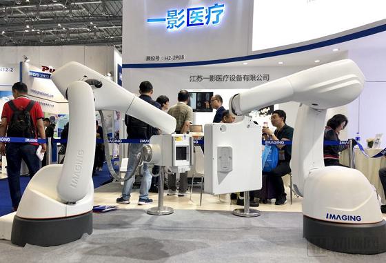
First Imaging Medical Imaging Robot Platform
Another exhibited product, an imaging robot platform, breaks through the limitations of traditional imaging equipment regarding patient positioning and anatomical regions. Its system design emphasizes multifunctionality and scalability by integrating Digital Subtraction Angiography (DSA) capabilities. In addition to conventional DSA functions such as vascular subtraction and 3D imaging, it also offers standing-position 3D imaging. With high resolution and flexible operation, this product provides weight-bearing 3D full-length images for orthopedic diagnosis and treatment, as well as angiography of the gastrointestinal tract, urinary tract, and various blood vessels in any position and at any anatomical site.
“The rich diagnostic information provided by standing 3D full-length imaging will help usher in a new chapter in our understanding of diseases.” Dr. Xi, CEO of First Imaging, stated: “In addition to helping physicians improve their diagnostic and treatment capabilities and reducing radiation doses to enhance patient well-being, the vast imaging data generated by the imaging robot platform will also open up expansive new frontiers for clinical research.”
In orthopedic diagnosis and treatment, imaging robot platforms fill the gap in standing three-dimensional full-length imaging. Image quality is no longer a bottleneck for digital orthopedic technologies, and this approach is highly likely to represent the future of orthopedic imaging. This imaging modality will provide new insights into the etiology and classification of various diseases, and offer more precise evaluations of therapeutic efficacy from a novel perspective. Complex conditions that are difficult to detect with conventional imaging—such as subtle fractures often missed on two-dimensional X-rays and degenerative spinal changes potentially overlooked in supine-position imaging—can potentially be diagnosed using imaging robot platforms.
It is reported that First Imaging’s intraoperative CT scanner is about to obtain the NMPA medical device registration certificate and will enter its first hospital this week to commence clinical applications. Dr. Xi also revealed that the robotic imaging platform will be deployed in hospitals this year for research collaborations, with test reports expected to be obtained this year and the registration certificate anticipated next year. In the future, building on the foundational theories of 3D imaging algorithms, First Imaging will simultaneously develop multiple product lines with core competitiveness for use in both medical and industrial inspection equipment.