RSNA Launches First-Ever AI Course for Radiologists Featuring Andrew Ng as Instructor
Subtle Medical
Medical Imaging Software Developer

Nuance
Healthcare AI Solutions and Services Provider

Infervision
Artificial Intelligence Product Developer
Future physicians will not only need to interpret medical images and make diagnoses, but will also likely need to learn how to collaborate more effectively with artificial intelligence, leveraging technology to further enhance their clinical expertise.
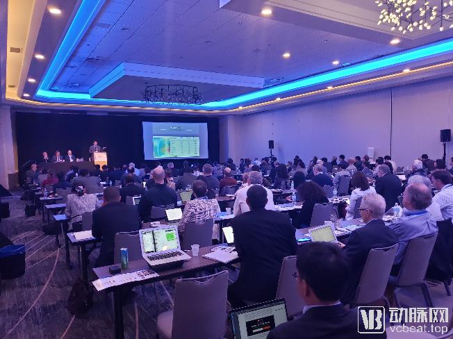
This trend has garnered global attention. From May 31 to June 1, the Radiological Society of North America (RSNA) held its inaugural “Spotlight Course on AI: Radiology in the Age of AI” for radiology professionals. Through this two-day course, the RSNA aimed to introduce the technological origins of the close integration between radiology and artificial intelligence (AI), current applications, and how to interpret academic advancements in AI-based medical imaging, thereby helping physicians adapt to a new era characterized by close collaboration with emerging technologies.
This session of the “AI Lecture Series” comprises several segments, including “A Brief Introduction to AI Technologies in Medical Imaging,” “Exploring Its Impact on Better Safeguarding Human Health,” and “How to Integrate AI Systems into Your Medical Practice.” Each segment features discussions or presentations by leading experts from the AI industry.
This is precisely where the trend lies: the vast amount of data and technological demands faced by the healthcare sector have made it the first to be significantly impacted by large-scale AI technologies, a trend that has also given rise to many of the fastest-adopted applications.
Medical imaging modalities such as CT, MRI, and PET serve as critical resources for clinical diagnosis, while AI’s robust data processing capabilities empower physicians to analyze these images with greater ease and proficiency.
At the conference, Andrew Ng, a world-renowned AI expert and professor at Stanford University, presented the development of AI and deep learning algorithms, as well as recent advances in AI-based medical imaging technology. His laboratory, in collaboration with Stanford Hospital, has completed projects such as ChestXnet and Xray4all, which leverage deep learning to interpret medical images. These deep learning technologies can differentiate eleven distinct pathological findings in chest X-rays, detect abnormalities in knee MRI scans, and identify pathological indicators suggestive of aneurysms in head CT scans, among other applications.
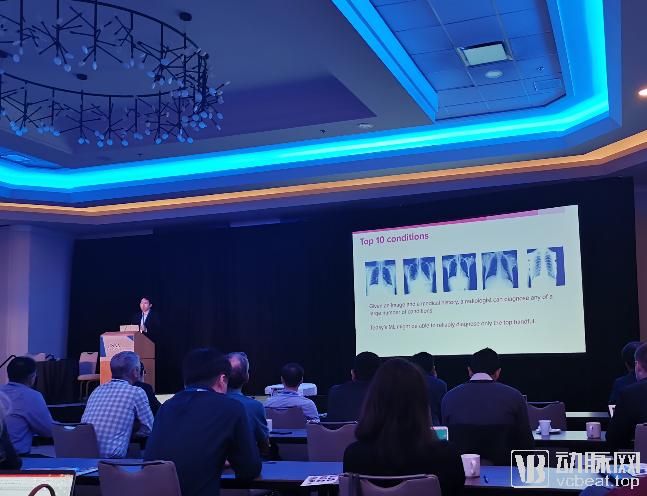
Andrew Ng on the Application of AI in Medical Imaging
“Deep learning can already perform basic tasks that humans complete in a second, but AI still has a long way to go before it can fully replace doctors in making diagnoses, with many breakthroughs yet to be achieved,” said Andrew Ng.
Professor Curtis Langlotz, Vice Chair of the Department of Radiology at Stanford University School of Medicine and one of the organizers of this course, is less pessimistic about the potential crisis of AI replacing physicians. He believes, “Radiologists need to continuously adapt and acquire the most advanced AI knowledge and skills. However, AI is merely another valuable new technology and development in clinical medicine, following other innovations such as CT, MRI, and ultrasound. Clinicians should integrate AI into their clinical practice.”
“Tasks such as measuring lesion size and tracking changes in lesion location and size across different disease stages are often tedious and monotonous, areas where AI outperforms humans. Therefore, from a certain perspective, AI can enhance the work of clinicians; with AI assistance, clinicians can focus on tasks that are cognitively more engaging and challenging.”
In the face of AI’s continuous transformation of the healthcare landscape, how can physicians—who are on the front lines of patient care and provide daily medical services—adapt to this new era?
First, physicians need to gain a deeper understanding of new technologies and learn how to apply them in areas such as clinical diagnosis, surgical prognosis, and early screening. During the course, several researchers in medical imaging AI shared their latest findings in these fields.
“AI will not replace doctors, but doctors who use AI will replace those who do not.” Professor Curtis Langlotz once again cited this golden maxim of the AI era while discussing the clinical application of AI in healthcare.
Andrew Ng also stated, “In the tech world, our work undergoes significant changes every five years. Today, technology is further accelerating the pace of change across all industries. Many tasks performed by radiologists will be automated, but as long as physicians are willing to reflect on the true value of their work, broaden their horizons, and focus on more high-value activities, they have nothing to worry about.”
Second, new technologies themselves are further enhancing physicians’ professional competence.
Dr. Hugh Harvey, a radiology expert at Kheiron Medical in the UK, pointed out that radiologists need to gain a deeper understanding of data science techniques. Radiologists should acquire knowledge in foundational data science and machine learning, particularly in data curation. He noted that while AI technologies such as deep learning demand large volumes of data, discussions often emphasize quantity while overlooking quality. Data extracted directly from clinical systems are far from being suitable for genuine clinical AI research and applications.
General data processing requires at least four layers of operations.
The first layer consists of data directly extracted from clinical systems (PACS, electronic medical record systems). Such data often contain sensitive information, are large in volume but low in quality, and are not suitable for direct use in research.
The second tier consists of data that has undergone ethics committee review and had patient-sensitive information removed. Physicians and researchers can access this data under restricted conditions, but such data generally lacks structure and cannot be directly used for research purposes.
The third layer involves further structured cleaning of these data and visual inspection to ensure issues such as image data quality are addressed.
The fourth layer involves matching these data with corresponding clinical information and labeling the data through manual or automated methods to facilitate AI research and analysis. At this level, researchers must also verify whether the data possess sufficient statistical value and whether there are established standards for labeling. For instance, disease diagnosis should be based on a comparison of imaging interpretations by multiple physicians and confirmed by subsequent disease progression and follow-up results.
For physicians, embracing technology with an open mindset and acquiring proficiency in emerging technologies through courses, activities, and project-based exchanges is likely to yield twice the results with half the effort in future medical services.
Professor Greg Zaharchuk, a neuroradiologist at Stanford University and Director of the Advanced Neurofunctional Imaging Laboratory, who attended the conference, affirmed the importance of such courses. He believes that researchers need to effectively explain AI theories, applications, developments, and limitations to clinicians.
On the other hand, he also emphasized that there is still a significant gap between clinical AI research and the deployment of genuine clinical AI products. Ensuring algorithmic robustness across diverse patient cases, imaging equipment, and scanning parameters remains a pressing challenge that needs to be addressed incrementally during development.
Radiologists face greater opportunities and challenges in the AI era, while for the broader public, technology brings enhanced safeguards and higher standards of medical care.
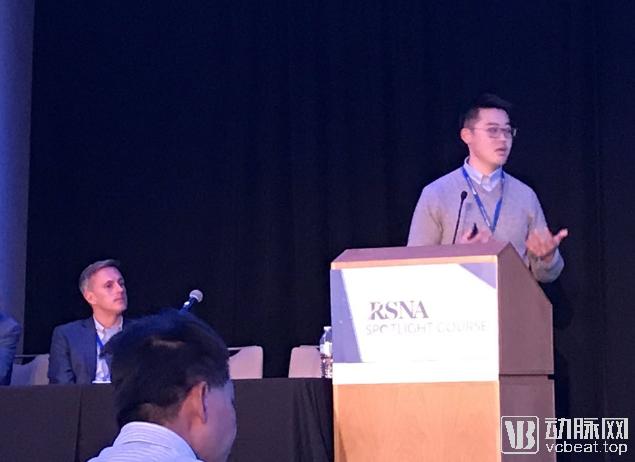
At the event, Pranav Rajpurkar, a Ph.D. student from Andrew Ng’s lab, gave a live demonstration of the Xray4All platform: users upload captured X-ray images, and after approximately one second of transmission, they receive results online. If abnormalities are detected in the image, the platform highlights the affected areas.
“This technology’s application scenarios are particularly well-suited to addressing the shortage of clinicians in developing countries and global health settings,” summarized Pranav.
Arterys, another U.S. AI imaging company that has raised over $45 million, plans to leverage real-world data to support medical decision-making for patients worldwide, automate routine clinical tasks, further promote equity and democratization in healthcare, and provide predictive analytics.
Arterys’ current AI products are all processed based on cloud computing, a model that is far faster, more secure, and more reliable than calculations performed on hospitals’ internal computing systems.
As one of the countries with the highest proportion of healthcare spending in total government expenditure, the United States has consistently been at the forefront of AI technology adoption. Meanwhile, China, a populous nation facing tight average medical resources, also has substantial demand for AI in healthcare.
At this session, Infervision from China, Nuance from the United States, and Subtle Medical, which promotes its solutions through Sino-U.S. collaboration, were invited to deliver presentations. In the closing segment of the conference, these three companies discussed the final critical steps in the industrialization and clinical deployment of AI systems under the theme “Implementing AI: the last mile.”
Infervision has processed millions of medical records in China and is conducting trials at multiple hospitals and imaging centers in the United States. Nuance holds a significant market share in the U.S. for speech recognition and image annotation tools used in clinical imaging, and is also promoting its “Nuance AI Marketplace” for medical imaging AI applications.
Subtle Medical is the only one among the three companies with an AI product approved by the FDA for commercialization. Dr. Enhao Gong, CEO of Subtle Medical, introduced how to clinically deploy its FDA-approved SubtlePET product and conduct clinical trials for products under application, such as SubtleMR.
Subtle Medical’s SubtlePET AI product is the first approved medical imaging enhancement application and the first approved AI application in nuclear medicine. Its value lies in leveraging AI to achieve approximately fourfold acceleration in image acquisition, providing solutions for reducing radiation exposure and contrast agent dosage. This means patients will benefit from more convenient, higher-quality, safer, and smarter clinical imaging examinations.
In the United States, AI must clear stringent hurdles to enter hospitals: it must be deeply integrated with hospital information systems; its efficacy must be validated by clinicians; and a clear return on investment must be demonstrated for hospitals purchasing the AI system.
“During the preparation for deployment, we need to engage in multi-faceted communication with clinicians, heads of information systems, and hospital management and operations teams. Taking Subtle Medical as an example, the company’s clinical and sales leaders collaborate with hospitals to conduct rapid and effective real-world data testing. This allows hospitals to perform clinical tests using their own data in real time, while minimizing any disruption to existing hospital workflows. Through practical testing and demonstrable acceleration of imaging examinations, hospitals can objectively recognize the new clinical and economic value that AI brings, thereby facilitating the progression to procurement and deployment,” Gong Enhao, CEO of Subtle Medical, told reporters.
Jeff Sorenson, CEO of TeraRecon, a medical imaging post-processing company, and also CEO of Envoy, a medical imaging AI platform, and Professor Eliot Siegal, a renowned radiologist and advocate for imaging AI, discussed how to optimize the workflow and deployment process of imaging AI through mutual interviews.
“Rigorous clinical validation of AI algorithms is a critical step in the widespread adoption of medical AI, and we are continuously progressing toward this goal,” emphasized Professor Eliot Siegal.
Although medical imaging is already one of the most suitable and rapidly deployable domains within the AI field, we still face numerous challenges.
First, AI technologies represented by deep learning remain a “black box.” This means that while such technologies can achieve high accuracy in medical imaging detection, AI still struggles to comprehend the true relationships within data and how to classify it.
“At Stanford, we hope to create better attention maps for medical image perception to avoid the black-box effect,” said Dr. Saafwan Halabi, a professor at Stanford University School of Medicine.“Recent studies and reports have discussed how data-driven adversarial attack algorithms can cause AI systems for road sign recognition to malfunction. In the realm of medical AI, ensuring that AI is not misled is a critical component, yet research in this area remains clearly insufficient.”
Dr. Matthew Lungren, Project Lead for Artificial Intelligence in Medical Imaging (AIMI) at Stanford University and co-director of the undergraduate program, discussed “Bias and Implications for Medical Imaging AI.” In actual clinical practice, AI systems are highly susceptible to introducing data bias; for instance, classifiers designed for medical image recognition may inadvertently rely on extraneous markers within the images rather than the pathological lesions themselves.
Current tools do not adequately address bias in data and algorithms. AI deployed in clinical practice must enable users to assess the credibility of its outputs. Incorporating human-computer interaction into system design, along with confidence analysis provided by AI algorithms, can significantly help mitigate potential bias.
Professor Jayashree Kalpathy, co-director of the Machine Learning Laboratory at Massachusetts General Hospital, aims to develop a more robust model and facilitate multi-hospital collaborative projects through transfer learning and federated learning. This approach enables the sharing of trained deep learning AI models without exchanging sensitive data, thereby promoting in-depth cooperation among hospitals.
Overall, artificial intelligence still has many imperfections that need to be overcome, but in the future, AI will undoubtedly become an important pillar of healthy living. Of course, this requires joint efforts from practitioners across the industry to build a more efficient and rational healthcare system.