AI-Driven Transformation of Radiology Workflow: Nanjing Gulou Hospital and Infervision Publish Breakthrough IILS System in The Lancet's EBioMedicine

Infervision
Artificial Intelligence Product Developer
VCBeat (WeChat ID: vcbeat) has learned that a major scientific achievement by the team led by Director Zhang Bing of the Department of Medical Imaging at Nanjing Drum Tower Hospital, in collaboration with the research team at Infervision, has been published in the high-impact academic journal EBioMedicine (a subsidiary journal of The Lancet, IF=6.2). The study proposes an Intelligent Imaging Layout System (IILS) based on artificial intelligence and deep learning for intelligent layout and structured reporting. Leveraging AI technology, the IILS simplifies and optimizes the entire clinical imaging workflow, significantly improving the efficiency and accuracy of radiology departments, and presents pioneering results in the translational application of clinical innovations to the industry.
AI has made substantial progress in the medical field, but most medical AI research focuses on assisted disease diagnosis. However, high-quality standardized images are the foundation of artificial intelligence development, and AI applications can take over tedious, repetitive, low-value administrative tasks.
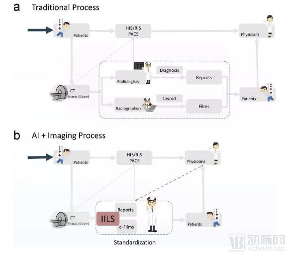
Director Zhang Bing’s team at Nanjing Drum Tower Hospital has joined forces with Infervision, leveraging its powerful AI technology and clinical research capabilities to adopt a more macroscopic perspective,From the perspective of the entire clinical imaging workflow, AI deep learning technology was employed to optimize and resolve issues in the three key stages—image acquisition, image presentation, and disease diagnosis—and this innovative translational achievement has been promoted and applied in clinical practice.。
The publication of this achievement in a *Lancet* subsidiary journal further attests to the high quality of the research and its feasibility for clinical application.
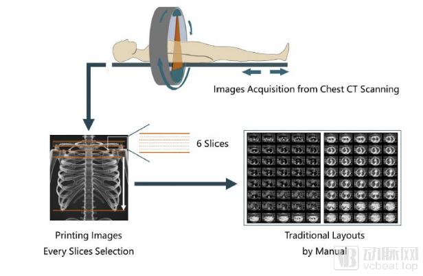
The article highlights a long-standing issue in the image layout phase of clinical imaging workflows: thin-slice chest CT scans (0.625 mm–2 mm) typically comprise more than 250 images, but due to limited space on film, only intermittent printing can be used during film layout and printing, with generally only 40 images retained per sheet.
Therefore,In fact, film loses approximately 82% of image information compared to digital images.Meanwhile, issues such as missed diagnoses, the absence of key slice images, and the lack of standardized imaging reports continue to pose reliability challenges for traditional clinical decision support systems.
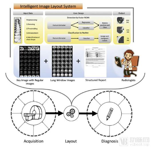
To address this issue, researchers developed and designed an Intelligent Layout and Structured Reporting System (IILS) based on artificial intelligence technology.The machine learning methods employed in this study accurately detected pulmonary nodules (AUC as high as 90.6%) and, following rigorous statistical validation, demonstrated superior performance over six radiologists in both the number of nodules detected and the determination of benign versus malignant status.
By integrating AI with adaptive layout tools, researchers have further developed a precise, effective, and reliable chest CT layout system, suitable for fully automatic or semi-automatic typesetting of medical imaging images.It enables fully automated layout, centralized display, and report generation. The introduction of this system integrates the workflow of image presentation, prevents loss of image information from film, and significantly improves work efficiency.
This research achievement has significantly improved the work efficiency of radiology departments, optimized workflows, enhanced image display quality, and reduced medical costs. It further facilitates the collaborative establishment of a high-quality, standardized diagnosis and treatment system among radiologists, clinicians, and patients, playing a crucial role in optimizing clinical imaging workflows.
The healthcare industry demands a high threshold of expertise and long-term accumulation of experience. How to closely integrate artificial intelligence technology with practical clinical research needs is a challenge faced by every medical AI company. For any medical AI company, robust clinical research capabilities, deep clinical research collaborations with numerous top-tier hospitals, and the production of high-quality research outcomes constitute the “gold standard” for measuring its value.
Infervision boasts a “dual-institute” system for clinical research, comprising its Global Clinical Research Institute (iCR) led by industry luminaries such as Dr. Shen Yun, Dean of iCR, alongside a team of distinguished scientists; this is further empowered by InferScholar® Center, an industry-leading AI-integrated omics research platform. Together, these three pillars constitute Infervision’s top-tier clinical research infrastructure within the industry.
This groundbreaking research highlights the forward-thinking vision, scientific research capabilities, and execution prowess of the team led by Director Zhang Bing at Nanjing Drum Tower Hospital, in collaboration with Infervision. Through the joint efforts of Infervision and its partner hospitals, the application scenarios and value of artificial intelligence technology in the medical field are continuously surpassing expectations.