Kepu Imaging's First Batch of Precision 32 Spectral CT Systems Marks the Dawn of the Domestic Spectral CT Era
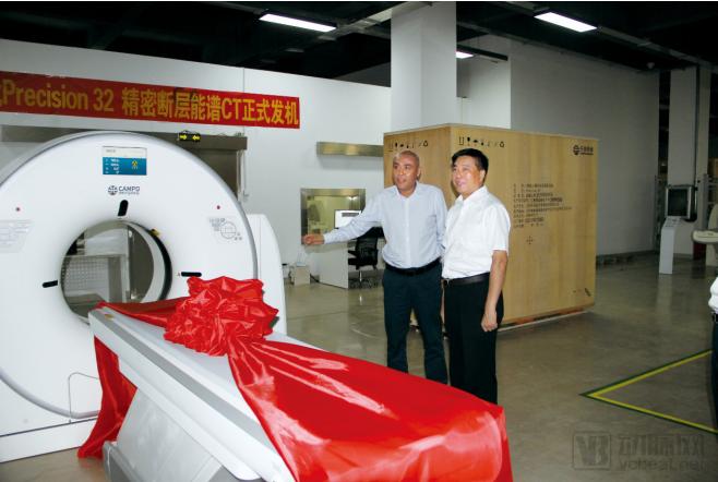
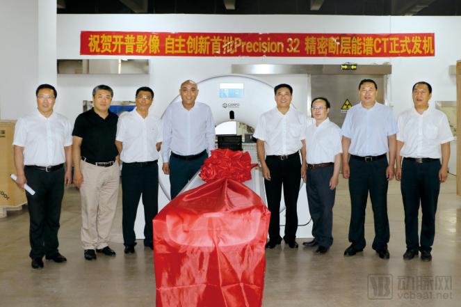
On July 23, Kaipu Imaging, an innovator in China’s high-end medical equipment sector, announced the official delivery of its first batch of Precision 32 Spectral CT scanners, independently developed through proprietary innovation. Tian Shuhuai, Mayor of Benxi City, along with Vice Mayors Wu Shimin, Liu Xudong, Gao Wei, and other officials, visited the site to inspect the facilities and expressed high expectations for Kaipu Imaging’s future development.
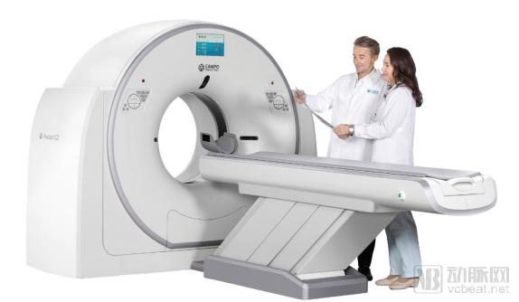
It is reported that Kaipu Imaging’s Precision 32 Precision Tomographic Spectral CT has recently obtained registration certification from the National Medical Products Administration (NMPA), as well as a Medical Device Production License and EU CE certification. This product achieves, for the first time, a high-level integration of the patented P-Axial precision tomography technology with 32-slice spectral imaging technology, and has been widely recognized by customers since its launch. The first batch of Kaipu Imaging Precision 32 CT units will be delivered to multiple user hospitals in Northeast, Central, East, Southwest, and South China. Its extensive future clinical application will comprehensively lead Chinese-made 32-slice CT systems into the spectral imaging era.
The Precision 32 Spectral CT with Precision Tomography is independently developed by Kaipu Imaging, boasting multiple independent intellectual property rights and patents. As Kaipu Imaging’s flagship CT product, the Precision 32 CT features the patented P-Axial precision tomography technology. This innovative scanning method enhances longitudinal sampling for patients and, combined with advanced reconstruction algorithms, achieves ultra-high-resolution longitudinal image reconstruction through both data acquisition and image processing, significantly improving the detection rate of subtle lesions. The globally patented P-Axial precision tomography technology enables the Precision 32 CT to produce ultra-fine images with a resolution of just 0.275 mm, offering distinct advantages in applications such as inner ear imaging and pulmonary nodule detection, thereby comprehensively supporting precise diagnosis. For instance, in inner ear imaging, it not only clearly visualizes structures such as the ossicles and cochlea but also distinctly delineates the spatial relationships among various inner ear structures.
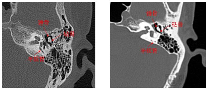
Precision 32-CT 0.275 mm High-Resolution Inner Ear Images Conventional CT 0.5 mm Thin-Slice Inner Ear Images
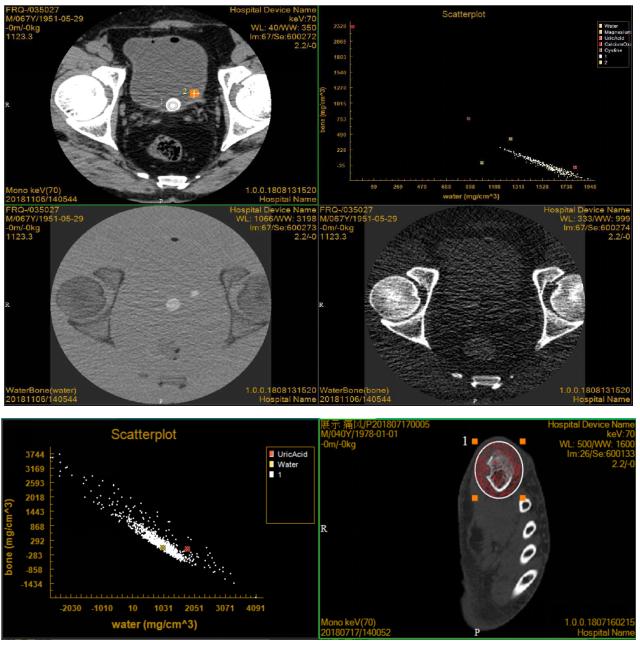
Spectral CT imaging is an emerging technology that originated in the 1970s but failed to achieve widespread application due to hardware and software limitations. The advent of spectral CT imaging, centered on dual-kV technology in the 21st century, has created new possibilities for its clinical application and research. Spectral CT leverages the differential attenuation of materials at varying X-ray energy levels to provide more imaging information than conventional CT. Compared with conventional single-parameter CT images, spectral CT offers a novel imaging paradigm featuring multi-parameter capabilities and quantitative analysis.
Precision 32: For the first time, spectral imaging technology—previously available only on high-end CT systems—is extensively applied to a 32-slice CT scanner. This enables qualitative diagnosis and quantitative analysis of lesions and tissues that are difficult to characterize with conventional CT, thereby enhancing diagnostic accuracy and safety and elevating the success rate of CT imaging diagnosis to an entirely new level.
Precision 32 High-Definition Spectral CT introduces low-dose technologies such as Ultra-Low Energy CT Scanning, P-Dose, and P-IR to minimize patient radiation exposure. It provides dedicated pediatric scanning protocols that combine 60kV ultra-low energy CT scanning with precise dose modulation via P-Dose, along with parameters optimized for appropriate dose levels. Specifically designed for pediatric care, these features can reduce radiation doses in children’s CT scans by 70%. P-Dose intelligently adjusts the scanning dose for different body parts based on their density and morphology. Meanwhile, the advanced iterative reconstruction algorithm, P-IR, ensures high-quality images are maintained across all low-dose scanning modes.
Kaipu Imaging operates two major business segments: the research, development, and manufacturing of medical imaging equipment, and third-party medical imaging diagnostic centers. Committed to building a new ecosystem for medical imaging that comprehensively covers products, services, and platforms—ranging from high-end medical imaging equipment to precise diagnostic services and intelligent imaging cloud solutions—the company serves public welfare through innovative healthcare industry models. In the field of R&D and manufacturing of high-end medical equipment, its core R&D team, led by Chief Scientist Dr. Zou Yu, comprises hundreds of professionals stationed at two major R&D centers in Benxi, Liaoning Province (known as “China’s Pharmaceutical Capital”), and Chicago, USA. Leveraging robust independent innovation capabilities, the team is engaged in the R&D and production of high-end medical devices such as CT scanners.
Sun Zhaochang, Chairman of Kaipu Imaging, stated: “Stay true to our original mission and work diligently.” We officially launched our first independently developed CT scanner at a remarkable pace within the industry, and achieved the initial shipment of units within less than a month after regulatory approval, earning widespread recognition from customers. We are deeply grateful for the opportunities and challenges presented by this era. Under the national macro-strategic plan of “Healthy China,” Kaipu Imaging will join hands with other outstanding domestic medical device enterprises to continuously catch up with and surpass advanced international technologies, striving to promote the broader and deeper application and popularization of high-end technologies, ultimately benefiting the public.