AI-Powered Respiratory Imaging: Groundbreaking Clinical Trial Results from The First Affiliated Hospital of Guangzhou Medical University
Faster speeds, higher precision, empowering primary care, and facilitating the professional growth of young physicians... How much have you heard about the promising future of imaging AI at various medical conferences? Beyond high-quality R&D datasets and pilot hospitals, how effective is AI in real-world applications? To what extent can it truly assist radiologists, particularly junior doctors?
To date, the vast majority of publicly available AI-related findings shared at industry conferences and published in academic journals have been limited to small-scale technical feasibility experiments based on specific algorithms, with performance validation in large-scale, real-world clinical settings being exceedingly rare. It is precisely for this reason that the experimental results presented in this article are particularly valuable.
This study was comprehensively designed and supervised by Professor Zeng Qingsi, Director of the Department of Radiology at the First Affiliated Hospital of Guangzhou Medical University. It was jointly conducted by Dr. Deng Yu and Dr. Liu Yanwen in collaboration with Yitu Healthcare, a leading domestic medical AI unicorn company. Using the care.ai® Intelligent 4D Chest CT Imaging System as the platform and chest CT images from real-world clinical settings as samples, the study investigated the efficacy of AI in authentic clinical work environments, with a particular focus on the supportive role of AI systems for junior physicians in interpreting chest CT scans.

Director Zeng Qingsi, Department of Radiology, The First Affiliated Hospital of Guangzhou Medical University
As the National Clinical Research Center for Respiratory Diseases and the State Key Laboratory of Respiratory Disease, the Department of Respiratory Medicine at the First Affiliated Hospital of Guangzhou Medical University has ranked first in its specialty for nine consecutive years in the Fudan University’s "Best Hospitals in China" ranking. The department performs an average of 400 CT scans per day, with chest CTs accounting for more than two-thirds of the total volume. Each physician reviews up to 30,000 chest CT images daily. Since August 2018, the Department of Radiology at the First Affiliated Hospital of Guangzhou Medical University has introduced an AI-assisted imaging diagnostic system, which has now been integrated into clinical workflows and is changing radiologists’ image-reading habits, particularly in the interpretation of large volumes of lung screening CTs.
The results of this study indicate that the AI system’s ability to detect nodules across different lung lobes and varying densities is comparable to that of senior physicians, while significantly enhancing the detection capabilities of junior physicians. This improvement is particularly notable for ground-glass nodules and small nodules under 4 mm. In terms of detection time, the AI system’s image interpretation efficiency is more than ten times that of the manual group. The “AI + physician” model can also reduce image interpretation time by nearly 30% among junior physicians. However, the AI system is not a panacea; radiologists’ assistance remains essential for differentiating pulmonary nodules from pleural thickening or endobronchial mucus plugs, as well as for assessing nodule density. Furthermore, the detection performance of the AI system is directly affected by image quality.

Dr. Deng Yu, Department of Radiology, The First Affiliated Hospital of Guangzhou Medical University
“Maximizing the detection of early-stage lung cancer, correctly measuring pulmonary nodules, accurately evaluating their morphology, and selecting optimal management strategies have always been core issues in nodule assessment. There is a strong clinical demand to use AI systems to overcome the limitations of visual assessment, perform quantitative analysis of lesions, and reduce the rate of defective reports. This was precisely the original intention behind our trial.” Discussing the significance of the study, Dr. Deng Yu stated, “In this trial, the AI system demonstrated robust detection performance, efficiency, and reliability in real-world clinical settings. Its auxiliary role for junior physicians was particularly notable. These findings hold significant importance for exploring the clinical application methods, scope, and evaluation criteria of AI systems.”
Introduction to the AI System
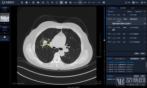
The care.ai® Chest CT Intelligent 4D Imaging System represents a significant breakthrough in the field of AI-driven healthcare. While most AI medical products on the market are limited to detecting pulmonary nodules, this system goes further by providing comprehensive quantitative intelligent analysis of lesions, differentiating between benign and malignant cases, automatically comparing historical images, and generating structured reports, thereby offering full support for clinical practice.
Test Methods
Thin-section chest CT scans (1-mm slice thickness) were randomly selected from individuals who had previously undergone health check-ups. A total of 218 CT datasets were included as study subjects according to the inclusion and exclusion criteria. Cases with severe respiratory artifacts on CT images, incomplete image data, or diffuse lung diseases that could interfere with interpretation were excluded. The gold standard for diagnosis was established through a consensus review by two senior radiologists, whose combined assessments reflected clinical reading practices as closely as possible.
Observer
(1) Senior Physician Group: one senior physician with more than 10 years of experience in thoracic imaging diagnosis;
(2) Junior Physician Group: one junior physician with more than 5 years of experience in thoracic imaging diagnosis;
(3) AI System: Two versions (high-sensitivity version, low-sensitivity version—also known as the “dual-high” version with high sensitivity and high specificity) of the care.ai® Chest CT Intelligent 4D Imaging System.
Test Results
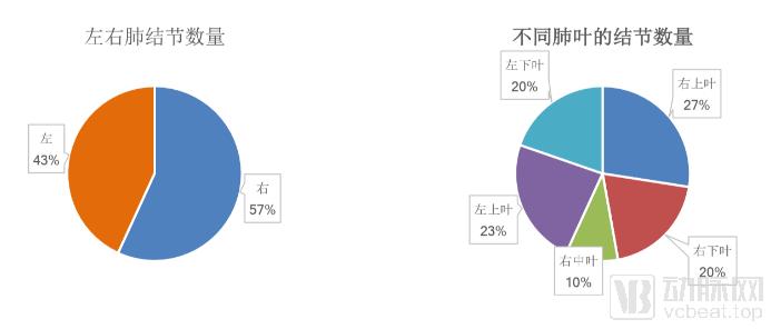
A total of 218 CT scans were included in this study. According to the gold standard, 176 scans were positive and 42 were negative. A total of 619 nodules were detected, with an average of 3.5 nodules per positive scan.
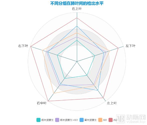
1. The AI system (red line) demonstrated detection performance comparable to that of senior physicians across different lung lobes and significantly improved the detection capabilities of junior physicians.
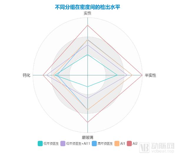
2. The AI system (red line) demonstrated detection capabilities for nodules of varying densities comparable to those of senior physicians, while the “AI + physician” reading model significantly improved the detection performance of junior physicians.
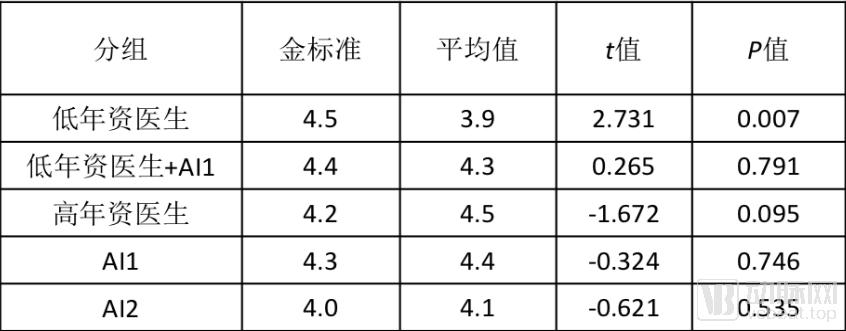
3. The difference between the AI system’s (red curve) assessment of nodule size and the gold standard was not statistically significant, indicating that it can improve the accuracy of nodule size interpretation by junior physicians to a certain extent.
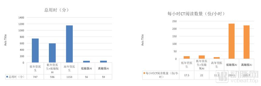
4. AI holds an overwhelming advantage in image interpretation speed. The figure below compares the number of images interpreted by different groups within one hour; the AI system’s efficiency is approximately ten times that of the manual-only group. Notably, adopting the “AI + physician” interpretation model increased the efficiency of junior physicians by 30%.
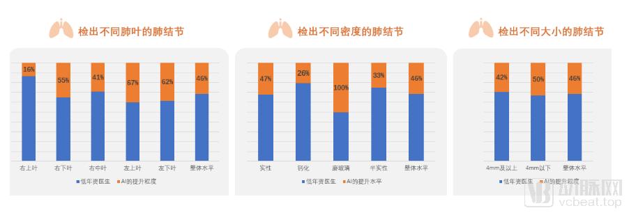
5. Further analysis reveals that the AI system provides comprehensive enhancement of nodule detection capabilities for junior physicians, significantly improving detection across different lung lobes and varying nodule densities. The most notable improvement was observed in the detection of ground-glass nodules, which increased by 100%, while the detection rate for small nodules under 4 mm improved by 50%.
Summary
1. The AI system’s nodule detection performance across all lung lobes comprehensively surpasses that of junior physicians, with detection capabilities for nodules of varying densities comparable to those of senior physicians; the AI system’s “miss-prevention” function is highly robust;
2. The AI system has an overwhelming advantage in the time required for reading lung nodules, taking only 1/10 of the time compared to pure manual reading, while the "AI + doctor" reading mode can improve the efficiency of junior doctors by 30%.
3. The “AI + Physician” image interpretation model provides comprehensive improvement in nodule detection capabilities for junior physicians; under this workflow, the nodule detection sensitivity of senior physicians can be increased to over 90%;
4. AI is not omnipotent; it still requires the assistance of radiologists in differentiating pulmonary nodules from pleural thickening or endobronchial mucus plugs, as well as in assessing nodule density. Furthermore, its detection capability is influenced by image quality.
It is foreseeable that the “physician + AI” model for interpreting chest CT images will become the mainstream approach in radiology. This model will deliver substantial value in large-scale early cancer screening programs, which require the interpretation of massive volumes of imaging studies. Furthermore, AI systems will serve as powerful aids for the professional development of junior physicians, becoming essential tools for radiologists, particularly early-career practitioners.