AI-Powered Lung Cancer Screening: 22 Suspected Cases Identified Among 1,277 Volunteers in YITU Medical's 'AI Anti-Cancer Map' Initiative

YITU
Provider of Full-Stack Intelligent Healthcare Product Solutions
Large-scale early cancer screening initiatives not only facilitate the monitoring of key epidemiological indicators, enable high-value clinical research, and support the formulation of regional public health policies and drug development; they also directly improve the health status and life expectancy of the regional population, reduce life-years lost and mortality due to malignant tumors, save substantial treatment costs, and build an impregnable “Great Wall of Health” for residents.
However, China has long faced a severe shortage of medical resources, particularly radiologists. Healthcare institutions are already stretched to their limits in meeting the daily demand for outpatient imaging diagnostics. How can they cope with the demand for early cancer screening that often involves thousands, or even tens of thousands, of individuals? How can large-scale early cancer screening initiatives be conducted in an economical, efficient, and precise manner?
On this world-class challenge, a joint initiative by the Second Affiliated Hospital of Guangzhou Medical University and YITU Medical is poised to provide an answer.
In November 2018, YITU Medical announced the launch of the “AI Cancer Prevention Map” project, planning to collaborate with hundreds of medical institutions over the next five years to enhance their capabilities in early cancer screening through AI applications and to promote the widespread implementation of AI-based early cancer screening solutions across China.
In the same year, a low-dose spiral CT lung cancer screening program based on AI image interpretation technology was conducted at the Second Affiliated Hospital of Guangzhou Medical University. Among 1,323 high-risk individuals (aged 50–91 years, with a median age of 61.48 years; 606 males and 717 females), 10 cases of lung cancer were detected, 9 of which were early-stage cases, marking an initial success for AI-based early screening.
In 2019, the initiative continued, with more research findings emerging.
Professor Zhang Zhenfeng, Deputy Director of the Department of Radiology at the Second Affiliated Hospital of Guangzhou Medical University and leader of the 2019 lung cancer early screening project, revealed that this year’s lung cancer early screening campaign ran from April to June, enrolling a total of 1,277 volunteers with a mean age of 62.65 years (age range: 50–92 years). Among them, there were 755 females (59.1%) and 522 males (40.9%); the mean age was 62.0 years for females and 63.58 years for males.
Through the "physician + AI" image interpretation model, 22 cases of suspected malignancy were identified, among which 6 patients showed an increase in the solid component of the lesions, indicating a high probability of malignancy.
Notably, supported by AI systems, this large-scale early lung cancer screening initiative did not significantly increase the image interpretation workload for radiologists. With only one primary interpreting radiologist, aided by 1–2 flexible technical staff, the screening task was completed efficiently and accurately.
The screening utilized the care.ai® Chest CT Intelligent 4D Imaging System, independently developed by YITU Medical. This system not only enables efficient detection of nodules but also automatically performs nodule size measurement, benign-malignant differentiation, and automated comparison with historical images, while generating structured reports. These capabilities significantly enhance the work efficiency of radiologists and save time.
When discussing the experience and insights gained from using AI systems to assist in large-scale early lung cancer screening, Professor Zhang Zhenfeng stated that AI has become a valuable assistant and “second brain” for clinicians. Especially in large-scale early lung cancer screening, the new model of “physician + AI” image interpretation has become standard practice. It not only saves physicians nearly 30% of their image-reading time, but its automatic historical image comparison function also greatly reduces manual workload, enabling doctors to easily and clearly review historical image comparisons and doubling-time metrics. This provides strong clinical evidence support for patient follow-up, making diagnoses more reliable and reassuring.
To achieve early screening for lung cancer, physicians need to examine tiny pulmonary lesions through thin-slice image interpretation, which leads to a sharp increase in their workload. Generally, it takes a radiologist at least 3–5 minutes to perform 1 mm thin-slice interpretation for each patient.
Taking this screening program as an example, a total of 1,277 volunteers were enrolled. Without the assistance of AI, the estimated total time required by physicians would exceed 6,400 minutes. This translates to at least 1.2–1.5 hours of overtime per workday over the course of more than three months. If manual comparison with historical imaging is also taken into account, the time commitment could double, reaching 2–3 hours per day.
With the support of the “physician + AI” image interpretation model and leveraging the care.ai® Chest CT Intelligent 4D Imaging System, radiologists participating in this lung cancer early screening initiative not only performed exceptionally well in image interpretation but also did not experience a significant increase in work pressure.
Professor Zhang Zhenfeng revealed: “The ‘Physician + AI’ image interpretation model assists clinicians not only by improving the efficiency of lesion detection but also through its profound understanding of clinical medicine. Features such as simple and efficient automated comparison with historical images, clear differentiation between benign and malignant lesions, and structured report generation that significantly reduces physicians’ documentation workload are deeply aligned with the needs of clinical practitioners. These capabilities enhance both the efficiency and accuracy of image interpretation while boosting radiologists’ confidence, alleviating the psychological pressure associated with high-volume reading tasks. This makes large-scale early cancer screening initiatives less daunting and enables their nationwide implementation across China.”
Furthermore, clinical trial results have empirically validated the efficiency and reliability of the “physician + AI” image interpretation model.
A clinical study demonstrated that the care.ai® Chest CT Intelligent 4D Imaging AI System exhibits nodule detection capabilities comparable to those of senior radiologists across different lung lobes and varying nodule densities. Furthermore, it significantly enhances the detection performance of junior radiologists, with particularly notable improvements in identifying ground-glass nodules and small nodules under 4 mm, while reducing image interpretation time by nearly 30%. In terms of detection efficiency, the “physician + AI” workflow achieves an interpretation speed more than 10 times faster than that of the manual-only group.
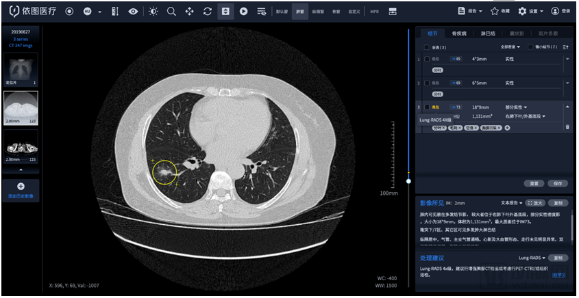
Patient Information: Male, 73 years old, 3 positive nodules (nodule diameter ≥3mm). The AI system classified the findings as Lung-RADS Category 4B based on the Lung-RADS criteria. System benign/malignant risk stratification result: High risk.
Diagnostic Opinion:
1. Right lung ground-glass nodule, increased in size compared to previous imaging; PET/CT or biopsy is recommended to rule out malignancy.
2. Emphysema and pulmonary bullae in both lungs
3. Fibrotic foci in the posterior basal segment of the left lower lobe
4. Aortic sclerosis, coronary artery calcification
5. Pancreatic Calcifications
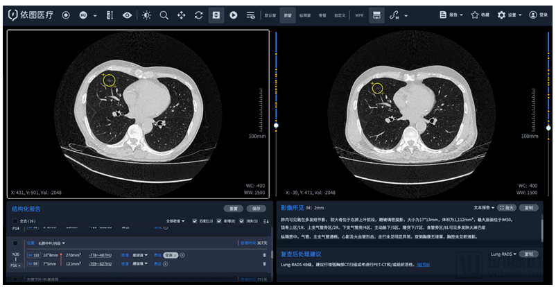
Commentary: The historical image comparison function of the care.ai® Chest CT Intelligent 4D Imaging System leverages YITU’s proprietary YIF technology to automatically register lesions in prior images, thereby assessing temporal changes. This includes automatically calculating doubling time and providing comparative analyses of size, volume, density, and imaging features before and after, even serving as a basis for evaluating treatment response in solid tumors according to RECIST criteria.
Patient Information: Female, 62 years old, with two positive nodules (nodule diameter ≥3mm). The AI system classified them as Lung-RADS Category 4X based on the "Lung-RADS" criteria. The system's benign/malignant grading result: High Risk.
Diagnostic Opinion:
Two nodules in the dorsal segment of the right lower lobe show no significant change compared to previous imaging; PET/CT or needle biopsy is recommended for definitive diagnosis.
Review: The care.ai® Chest CT Intelligent 4D Imaging System automatically extracts the size, volume, and density of pulmonary nodules and can automatically analyze six morphological features, including lobulation, spiculation, pleural indentation, cavitation, vacuole sign, and calcification. By opening the AI “black box,” it provides clinicians with evidence-based support for decision-making, serving as a unique productivity tool for large-scale tumor screening.