Deepwise AI Lab Achieves Breakthroughs in Medical Imaging Research with Publications in European Radiology and Medical Image Analysis

DeepWise
Developer of Artificial Intelligence Medical Imaging Diagnosis System
In terms of scientific research and innovation capabilities, Deepwise AI Lab has consistently maintained an industry-leading position. It is currently one of the largest research institutions dedicated to artificial intelligence in healthcare. Since its establishment, the lab has been committed to exploring cutting-edge medical technologies. By integrating technology with clinical practice, it has produced numerous research achievements that combine clinical value with technological innovation, which have been successively accepted by top-tier international journals and conferences.
To date, DeepWise Research Institute has published more than 30 papers in top-tier journals and conferences on artificial intelligence and machine learning, such as TPAMI, TCyb, ICML, CVPR, ICCV, ECCV, and AAAI. These publications cover the three premier international conferences in computer vision and pattern recognition. Notably, the institute has presented academic achievements at the highly prestigious CVPR conference (ranked in the Top 10 of Google’s 2019 Academic Rankings) for two consecutive years, placing it among the leading technology companies in China’s AI sector. Meanwhile, in the field of medical image computing and analysis, the institute has published nearly 20 papers at top-tier conferences such as IPMI, MICCAI, ISBI, RSNA, and ECR. Recently, two additional research findings from DeepWise Research Institute were published in leading international academic journals, underscoring its sustained innovation capability.
Among these, the study “Long-term follow-up of persistent pulmonary pure ground-glass nodules with deep learning” (https://doi.org/10.1007/s00330-019-06344-z), co-completed with the Cancer Hospital of the Chinese Academy of Medical Sciences, was accepted and published by European Radiology, a top-tier international radiology journal. As the official journal of the European Society of Radiology, founded in 1991, European Radiology holds extensive influence and an indispensable academic standing within the industry. It serves as an essential information source for experts and scholars in the field of radiology, represents the cutting edge of radiological science, and ranks second among comprehensive imaging journals.
Currently, lung cancer has become the most prevalent and deadliest cancer in China. While AI-assisted lung cancer screening has been widely adopted domestically, current efforts generally remain focused on deep learning-based pulmonary nodule detection. However, there is limited research on the growth assessment of pulmonary nodules, particularly regarding the long-term management of subsolid nodules (including pure ground-glass nodules and part-solid nodules), which holds significant clinical importance.
The management of persistent pure ground-glass nodules (pGGNs) remains more controversial. To address these controversies, it is necessary to investigate the growth progression of persistent pGGNs, accurately measure their growth rates, and identify risk factors influencing their progression. This study leverages deep learning techniques for the automatic segmentation of pulmonary pGGNs and conducts the aforementioned research based on follow-up data, thereby providing a more precise basis for the quantitative clinical assessment of pulmonary nodules.
In this study, the automatic detection and segmentation of pure ground-glass nodules (pGGNs) were performed using the Dr. Wise® AI Pulmonary Nodule Auxiliary Diagnosis System from DeepWise. By integrating Recurrent Neural Networks (RNN) and Convolutional Neural Networks (CNN), the system achieved performance superior to any single segmentation method currently available. Based on the Dr. Wise® AI system, chest CT images from baseline and all follow-up visits were processed for automatic pGGN detection and segmentation. The system automatically calculated pGGN diameter, density, volume, mass, Volume Doubling Time (VDT), and Mass Doubling Time (MDT). These quantitative metrics were used to investigate the natural growth patterns of pulmonary pGGNs, accurately measure their growth rates, and assess risk factors influencing pGGN growth, thereby providing important references for clinical management of pGGNs. The results of this study indicate that deep learning technology can assist in revealing the natural growth patterns of pGGNs; specifically, long-standing pGGNs tend to exhibit indolent growth, whereas pGGNs with lobulation signs and larger baseline diameter, volume, and mass are more likely to demonstrate growth during follow-up.
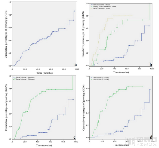
Figure 1. Lobulated sign, initial mean diameter, initial volume, and initial mass are key indicators for predicting whether pGGN will progress
Meanwhile, the paper “A deep network for tissue microstructure estimation using modified LSTM units” (https://doi.org/10.1016/j.media.2019.04.006), co-authored by DeepWise Research Institute and Beijing Institute of Technology, was published in Medical Image Analysis (MIA). As the official journal of the International Conference on Medical Image Computing and Computer-Assisted Intervention (MICCAI), MIA was founded in 1996 and focuses on research advances in applying computer vision, virtual reality, and robotics to medical imaging. With its impact factor rising year by year—reaching 8.88 in 2018—the journal has garnered significant attention from renowned experts and scholars in the field and is regarded as a benchmark for high-quality publications.
This paper proposes MESC-Net, a general-purpose deep network based on improved Long Short-Term Memory (LSTM) units for estimating tissue microstructure, thereby enhancing the quality of Diffusion Magnetic Resonance Imaging (dMRI). dMRI provides a unique tool for non-invasive assessment of tissue microstructure; however, due to the complexity of the models involved, it typically requires scanning sequences with a large number of diffusion gradients to improve image quality, which leads to prolonged scan times. When using fewer diffusion gradients, accurately characterizing tissue microstructure with complex signal models presents significant challenges. Therefore, achieving high-quality dMRI images with fewer diffusion gradients remains a persistent challenge in scientific research.
The MESC-Net proposed in this article can enhance image quality under reduced diffusion gradient conditions, offering clinical value by shortening scan time and improving clinical efficiency. Furthermore, the network architecture presented in this study is a general-purpose framework for estimating microstructural tissue properties, rather than being limited to a specific model. To validate the generalizability of the algorithm, experiments also evaluated microstructural tissue estimates derived from three signal models: NODDI (Neurite Orientation Dispersion and Density Imaging), SMT (Spherical Mean Technique), and EAP (Ensemble Average Propagator). Experimental results demonstrate that MESC-Net can be successfully applied to all three different models, indicating that this method can serve as a general approach for microstructural tissue estimation with broad prospects for clinical application.
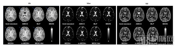
Figure 2. Cross-sectional schematic diagrams of vic/viso/OD calculated using MESC-Net based on the NODDI model,
AMICO/MLP/MEDN/MEDN+/m-MEDN+ are all comparison methods
DeepWise is a company dedicated to exploring cutting-edge technologies. As a leader in the field of AI healthcare, it consistently applies the most advanced artificial intelligence technologies to the medical sector. Centered around Professor Yizhou Yu (ACM Distinguished Scientist/IEEE Fellow), the DeepWise Research Institute has been committed to exploring the application of frontier AI technologies in medicine. Leveraging the Dr.Wise® multimodal research platform, DeepWise continuously deepens its scientific research collaborations with hospitals and universities on clinical issues, enhances research efficiency, produces high-quality academic papers, and promotes the translation of research findings. This approach ensures that cutting-edge AI technologies and research are closely aligned with clinical needs, enabling AI to be effectively implemented in real-world clinical scenarios and contributing to the development of national smart healthcare infrastructure.
In 2019, DeepWise Research Institute had eight papers accepted at CVPR 2019, a top-tier conference in artificial intelligence, achieving innovative breakthroughs in technologies such as image recognition and medical image analysis. This accomplishment positioned DeepWise among the leading technology companies in China in terms of the number of published papers. At MICCAI, the premier international conference on medical image analysis held in October, and ICCV, the top international conference on computer vision held in November, DeepWise Research Institute had an additional ten research papers in the field of medical AI accepted. To date, DeepWise Research Institute has published more than 50 top-tier academic papers, with a cumulative impact factor exceeding 80 and an acceptance rate surpassing 50%, underscoring its robust scientific research capabilities.