GE Healthcare Receives FDA Clearance for First Mobile X-ray System with Embedded AI for Rapid Critical Care Imaging in ICU
GE Healthcare recently announced that the U.S. FDA has approved its 510(k) Critical Care Suite for the Optima XR240amx, the industry’s first mobile X-ray imaging system with embedded artificial intelligence algorithms.
The device’s artificial intelligence algorithm was developed through a collaboration between GE and the University of California, San Francisco (UCSF), and launched via GE Healthcare’s Edison platform. GE Healthcare submitted its application for this product to the FDA last November.
Radiologists today face immense pressure to interpret an ever-increasing volume of medical images while delivering rapid and accurate diagnoses. However, in X-ray examinations, 60% of cases are labeled as STAT (urgent) for delivery. How can radiologists determine which images are truly urgent, and whether delayed interpretation would pose a risk to patients? This presents a thorny challenge for the protracted radiology workflow in the United States.
According to a study published in the paper “Reducing STAT Portable Chest X-Ray Turnaround Time: A Pilot Study” in Current Problems in Diagnostic Radiology, even X-ray images marked as “STAT (emergency test)” require an average wait time of up to 8 hours during the radiologist’s diagnostic process.
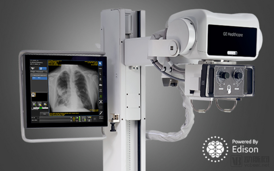
GE’s Optima XR240amx is a mobile, intelligent X-ray device primarily used in intensive care units (ICUs). The integration of the Critical Care Suite is designed to rapidly identify and help physicians prioritize critical conditions such as pneumothorax, thereby reducing radiologists’ interpretation time.
Rapid Intelligent Imaging Detection in the ICU
When patients in the ICU undergo X-ray scans via mobile devices using the GE Critical Care Suite, the AI system automatically analyzes the images in real time. If pneumothorax is suspected, alerts along with the original chest X-rays are sent directly to radiologists for review through the Picture Archiving and Communication System (PACS). The notifications enable radiologists to identify which images require prioritized attention. The AI algorithms can also simultaneously analyze and flag protocol and field-of-view errors, as well as automatically rotate images acquired from mobile devices, thereby standardizing the imaging output and saving physicians’ time.
According to official data from GE Healthcare, the Critical Care Suite demonstrates a sensitivity of 96% for large pneumothorax and a sensitivity of 75% in detecting three out of four types of small pneumothorax.
Furthermore, embedding artificial intelligence algorithms into mobile devices offers numerous benefits to radiologists and technologists. GE Healthcare’s algorithm provides a rapid and reliable method for generating AI results within seconds of image acquisition, without requiring network connectivity for processing. These AI-derived findings are then transmitted to radiologists concurrently with the raw diagnostic images, ensuring no additional processing delays.
Furthermore, automated image quality checks are performed on the device, integrating quality control into the technologists’ standard workflow and standardizing bedside operations, thereby ensuring image quality before transmission to the PACS.
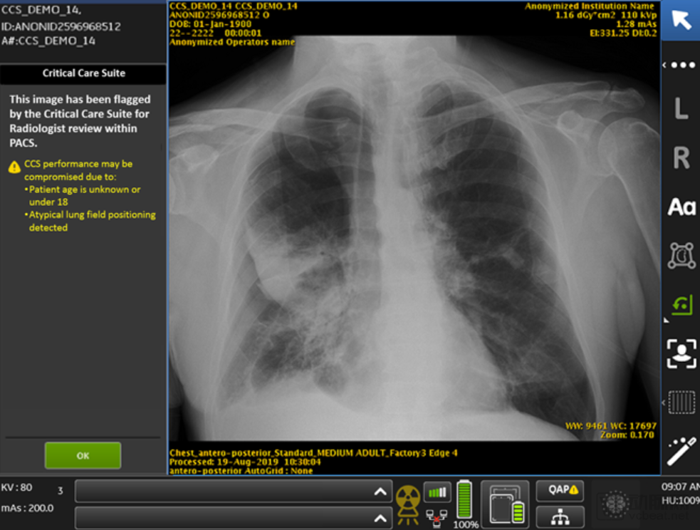
After acquiring the raw images, the system automatically performs protocol checks, quality assessments, and intelligent analysis, flags suspicious images, and sends alerts.
“Currently, 62% of imaging studies are labeled as ‘STAT’ or for urgent reading, yet not all of them are critical. This impacts patients who truly require urgent care and may lead to serious consequences,” added Jie Xue, President and CEO of GE Healthcare’s X-ray business. “The Critical Care Suite not only demonstrates impressive accuracy in flagging images suggestive of pneumothorax, enabling radiologists to immediately prioritize these cases, but it also makes AI more accessible. Our embedded AI algorithms provide hospitals with an opportunity to adopt artificial intelligence without the need to invest in additional IT infrastructure, conduct safety assessments, or implement cybersecurity measures for transmitting images off-site to external platforms.”
The Critical Care Suite and AI algorithms were developed using GE Healthcare’s Edison platform, which facilitates the rapid and secure deployment of AI algorithms.
Edison is the intelligent platform of GE Healthcare, designed to help healthcare professionals improve efficiency, enhance patient treatment outcomes, and provide opportunities for patient care. Edison applications can be embedded into existing workflows, integrate and absorb data from various sources, and apply analytics or advanced algorithms to generate clinical, operational, and financial insights. The Edison platform can be installed directly on smart devices or connected via online methods such as the cloud and Edison HealthLink.
Each patient generates a vast amount of data during the diagnosis and treatment process. The Edison platform leverages machine learning, deep learning, and artificial intelligence to transform this extensive information into directly actionable insights. These insights help healthcare professionals improve work efficiency, prioritize workflows, reduce repetitive tasks, and deliver the most personalized patient care.
At the Radiological Society of North America (RSNA) annual meeting in late 2018, GE Healthcare launched a series of new medical applications and intelligent medical devices based on the Edison platform. GE Healthcare has already provided auxiliary support for more than 200 medical imaging applications worldwide through the Edison platform.
A Collaborative Achievement of GE Healthcare, the University of California, and St. Luke’s University
The approval of this AI-powered pneumothorax diagnostic product marks a milestone in the collaboration between GE Healthcare and the University of California, San Francisco (UCSF). In late 2016, GE (before GE Healthcare became an independent entity) entered into a partnership with UCSF to jointly create a library of deep learning algorithms aimed at improving diagnostic workflows for users of GE’s imaging equipment and cloud platforms.
GE’s direct partner is the Center for Digital Health Innovation, affiliated with the University of California, San Francisco (UCSF). At the time, Dr. Michael Blum of UCSF stated that the key aspect of this collaboration lies in further integrating deep learning algorithms into the analysis of clinical data and medical imaging, thereby enabling clinicians to obtain accurate patient information in the shortest possible time. Moreover, in press releases at the time, GE researchers cited pneumothorax as a representative example when introducing their AI technology.
Dr. Rachael Callcut, Associate Professor of Surgery at the University of California, San Francisco (UCSF), is a surgeon in the UCSF Department of Health and Director of Data Science at the Center for Digital Health Innovation. She collaborated with GE to develop the Critical Care Suite. “The time spent processing and interpreting images when patients undergo X-ray examinations can affect final outcomes. AI provides us with opportunities to accelerate diagnosis and transform patient care, ultimately saving lives and improving prognoses,” said Dr. Rachael Callcut.
In GE Healthcare’s press release, in addition to UCSF, several partner institutions were mentioned: St. Luke’s University, Humber River Hospital, and Mahajan Imaging from India.
St. Luke’s University has long awaited the announcement of these results. In an interview three months ago, they mentioned that over the past year, Dr. Karl Yaeger, a radiologist at St. Luke’s University Health Network (SLUHN), and his colleagues collaborated with an international research team from GE Healthcare to develop the industry’s first X-ray system with embedded AI algorithms. This technology is designed to alert clinical teams to signs of pneumothorax.
During the development of this artificial intelligence product, Dr. Yaeger and his team at SLUHN reviewed hundreds of chest X-rays depicting pneumothoraces of varying sizes and severities. They then employed deep learning software algorithms to process these images and assess their accuracy.
“The purpose of this artificial intelligence product is to improve the detection rate and accuracy of these potentially lethal indicators during the diagnostic process, enabling patients to receive early treatment,” said Dr. Yaeger. “Ultimately, this study aims to focus on improving patient care and saving lives.”