Lingyi Zhihui Partners with Zhongshan Ophthalmic Center to Launch International Angle Closure Glaucoma Evaluation Challenge
On October 17, the MICCAI conference, a top-tier international academic meeting on medical imaging, concluded successfully in Shenzhen, China. Lingyi Zhihui led the organization of one of the conference’s highlight events—the Ophthalmic Medical Image Analysis Workshop (hereinafter referred to as the OMIA Workshop). In collaboration with the Zhongshan Ophthalmic Center of Sun Yat-sen University and the Medical University of Vienna, Lingyi Zhihui hosted the Angle Closure Glaucoma Evaluation Challenge (hereinafter referred to as AGE) during the workshop. The initiative aimed to address clinical needs in angle-closure glaucoma, advance related research, and drive progress in the AI-driven ophthalmology industry.
“I am delighted to see that researchers from industry have also made significant contributions to MICCAI, particularly in initiatives focused on practical implementation, such as OMIA and AGE. Such activities benefit a larger number of young medical imaging researchers and greatly advance research oriented toward real-world applications,” remarked Professor Leo Joskowicz, President of the MICCAI Society.
Centered on angle-closure glaucoma, the AGE competition focused on two major tasks using anterior segment optical coherence tomography (hereinafter referred to as AS-OCT) images: classification of open versus closed angles and localization of the scleral spur. The dataset used in the competition was jointly released by Lingyi Zhihui and Professor Zhang Xiulan’s team from the Zhongshan Ophthalmic Center at Sun Yat-sen University. Comprising 4,800 AS-OCT images, it is the world’s first large-scale public AS-OCT dataset specifically dedicated to angle-closure glaucoma.
Since the launch of the online preliminary round on July 9, AGE has attracted more than 200 competing teams, including renowned medical schools such as Johns Hopkins University School of Medicine, prestigious universities like The Chinese University of Hong Kong, Tsinghua University, and Peking University, as well as AI-industry leaders such as Tencent, Ping An Technology, and IBM. The onsite challenge was held at the OMIA Symposium, with Hangzhou Dianzi University ultimately claiming the overall championship, while South China University of Technology and Shenzhen University secured the runner-up and third-place titles, respectively.
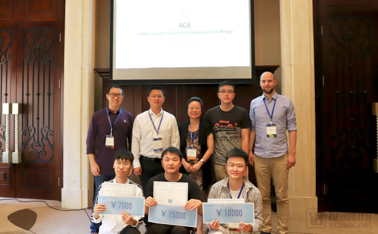
Group Photo of AGE Competition Organizers with the Champion, Runner-up, and Third-Place Teams
As a representative ocular disease, glaucoma is known as the “invisible killer of vision.” It causes continuous, irreversible damage to the optic nerve; if left untreated, it can lead to complete vision loss and blindness. In clinical practice, researchers can utilize various imaging modalities to visualize ocular structures and thereby diagnose glaucoma. Among these, Optical Coherence Tomography (OCT) offers advantages such as being non-contact, rapid, and high-resolution, holding significant value for morphological examination of the anterior segment of the eye.
Anterior Segment Optical Coherence Tomography (AS-OCT) images can clearly visualize ocular structures such as the cornea, sclera, anterior chamber angle, and iris morphology, providing a more objective record of the anterior segment. By assessing whether the anterior chamber angle is open or closed, AS-OCT enables the classification of glaucoma into open-angle or closed-angle types. Furthermore, AS-OCT is well-suited for measuring ocular biometric parameters and evaluating the anterior chamber angle, allowing for non-invasive quantitative analysis and biometric statistical measurements of the anterior chamber. This provides an objective and reliable basis for anterior segment analysis in glaucoma. The primary step in achieving these critical parameter measurements is the localization of the scleral spur.
This AGE model directly addresses the clinical challenges in glaucoma diagnosis by specifically performing two key tasks: classification of open-angle versus closed-angle glaucoma and localization of the scleral spur. It effectively facilitates the measurement of many critical biological parameters, offering significant reference value for rapid qualitative early screening and quantitative subtyping diagnosis of angle-closure glaucoma.
It is reported that AGE is part of the iChallenge competition series, launched by Lingyi Zhihui in 2018. Having evolved into the most influential and widely watched event in the field of AI-based ophthalmic image analysis, iChallenge aims to share large volumes of high-quality, finely annotated ophthalmic imaging data to enhance communication among researchers and promote the development of AI algorithms for diagnosis and image analysis. To date, it has hosted two competitions: the Retinal Fundus Glaucoma Challenge (hereinafter referred to as REFUGE) and the Pathological Myopia detection from retinal images challenge (hereinafter referred to as PALM).
September 2018 REFUGE
Numerous studies have demonstrated that the vertical cup-to-disc ratio (CDR) plays a significant role in clinical practice and glaucoma assessment: generally, a larger vertical CDR is associated with a higher risk of glaucoma. In clinical settings, CDR determination relies heavily on visual estimation by physicians, leading to considerable inter-observer variability. Consequently, many researchers are exploring automated and objective methods for measuring CDR.
However, due to the general lack of high-quality, task-specific datasets within the industry, related research has been difficult to advance. To address this challenge, REFUGE released a dataset of 1,200 color fundus photographs focused on glaucoma. Through three tasks, it evaluated and compared automated algorithms for glaucoma detection, optic disc/cup segmentation, and fovea localization on color fundus photograph datasets, enabling more researchers interested in cup-to-disc ratio (CDR) measurement methods to refine their work using high-quality data. In September 2018, the REFUGE Challenge was successfully held at the 5th MICCAI OMIA Workshop, attracting over 400 participating teams.
April 2019 PALM
Among myopic patients, approximately 35% have high myopia. As the degree of myopic refractive error increases, high myopia can progress to pathologic myopia (hereinafter referred to as PM), causing irreversible visual impairment in patients.
To advance research on pathologic myopia (PM), Lingyi Zhihui partnered with the Zhongshan Ophthalmic Center of Sun Yat-sen University to launch the PALM challenge and released a large volume of high-quality, annotated color fundus photographs of PM. These data are intended for the research and development of algorithms related to PM diagnosis and for achieving segmentation of lesions in fundus images of PM patients. The PALM Onsite Challenge was successfully held in April 2019 at the International Symposium on Biomedical Imaging (ISBI), a top-tier medical imaging conference, attracting more than 400 outstanding teams from China and abroad.
Although the competition has concluded, its value for research “never ends.” At this year’s MICCAI conference, three AI-related ophthalmology papers from Lingyi Zhihui were accepted. Previously, a review article on REFUGE was also published in Medical Image Analysis, the international journal with the highest impact factor in the field of medical imaging (IF=8.88). It is reported that participants have already published a series of papers using the REFUGE dataset in top-tier journals such as Transactions on Medical Imaging (IF=7.816) and at premier conferences like MICCAI 2019. Colleagues from both academia and industry have highly recognized the exceptional annotation quality and significant influence of the dataset. These research papers on intelligent analysis algorithms for ophthalmic images lay a solid technical foundation for exploring cutting-edge technologies and implementing practical products in ophthalmology.
The public data competition organized by Lingyi Zhihui is merely a microcosm of its open-minded approach to empowering the AI ophthalmology industry. This September, Lingyi Zhihui announced that the enterprise standard “Annotation and Quality Control of Color Fundus Photography for Artificial Intelligence,” co-developed with Professor Zhang Xiulan’s team, would be upgraded to a group standard under the auspices of the China Medical Education Association. As the first annotation standard for color fundus photography tailored to artificial intelligence within the industry, this standard significantly addresses the current inconsistency in annotation quality and provides a reference for establishing annotation standards for other types of ocular images in AI applications. Within just two days of announcing the launch of the group standard, more than 50 institutions applied to join, including hospitals, health examination centers, mobile screening companies, and AI algorithm developers, demonstrating strong industry recognition and enthusiastic response to the standard.
With data as the foundation and standards as the source, Lingyi Zhihui has always upheld a professional attitude of being “down-to-earth and meticulous.” We aim to enable more participants in the ophthalmic AI sector to benefit from high-quality data and high-standard development, thereby comprehensively promoting the rapid and healthy growth of ophthalmic AI and contributing to significant progress in the industry.