Over 10 Domestic Companies Enter Imaging-Based FFR Field as China Races to Catch Up with Global Unicorn HeartFlow
The heart was once considered a no-go zone for surgery, making it the last organ to be opened in the field of modern surgery. As the body’s engine, the heart’s constant beating and abundant blood flow had long prevented surgeons from opening it to perform operations.
The renowned Austrian surgeon Theodor Billroth once said, “To operate on the heart is a blasphemy against the art of surgery; anyone who attempts such a procedure is doomed to ruin and disgrace.”
Today’s surgeons can not only open the heart safely but are also striving to treat cardiac diseases through minimally invasive approaches.
Physicians can now visualize the cardiovascular system not only through two-dimensional imaging but also via “4D” imaging, enabling them to view three-dimensional models of blood vessels and assess hemodynamic conditions for more precise diagnosis and treatment.
In the process of achieving "4D" visualization of the heart, medical imaging-based computation of fractional flow reserve (FFR) for coronary arteries in patients with chronic coronary artery disease is currently a relatively mature technology, with CT-FFR and QFR being the primary techniques.
In this field, HeartFlow, a U.S. company, received FDA approval for its CT-FFR product in 2014. To date, HeartFlow has raised $467 million in financing and is valued at $1.5 billion. In China, startups are also beginning to enter this hot sector one after another.
According to VCBeat (WeChat ID: vcbeat), there are approximately 15 companies worldwide whose business portfolios include image-based FFR calculation, with one company’s product already approved in China. In this article, we review the primary application scenarios for image-based FFR diagnostic products, the key players in this field, and their respective core technological principles.
Before understanding image-based FFR estimation, we need to understand FFR, which stands for Fractional Flow Reserve. Its primary function is to assess the extent to which stenosis in coronary artery lesions affects distal blood flow. The main condition it applies to is coronary atherosclerosis, also known as coronary heart disease.
According to data from the "Report on Cardiovascular Diseases in China 2018," there are as many as 290 million patients with cardiovascular disease in China, including 11 million patients with coronary heart disease.
Coronary heart disease is a condition in which atherosclerotic plaque accumulation in the coronary arteries leads to arterial narrowing, thereby reducing blood supply to the myocardium and causing myocardial ischemia.
In simple terms, a layer of deposits composed primarily of lipids, inflammatory cells, smooth muscle cells, connective tissue, thrombi, and calcium deposits forms within the coronary arteries. These deposits can obstruct blood flow, leading to myocardial ischemia, and may also rupture or detach, causing a myocardial infarction.
In the diagnosis of coronary heart disease, diagnostic methods include electrocardiogram (ECG), coronary computed tomography angiography (CCTA), coronary angiography, intravascular ultrasound (IVUS), and fractional flow reserve (FFR) measurement.
Among these diagnostic methods, coronary angiography is a commonly used approach for confirming the diagnosis of coronary heart disease (CHD). For many years, coronary angiography has firmly held its position as the “gold standard” for diagnosing CHD.
Coronary angiography involves puncturing the radial artery at the wrist, advancing a guidewire into the coronary arteries of the heart, and then injecting contrast agent to visually observe its filling in the various coronary vessels. The presence of filling defects indicates stenosis caused by plaque. Based on the degree of vascular stenosis, the physician then determines whether stent implantation is required to dilate the vessel.
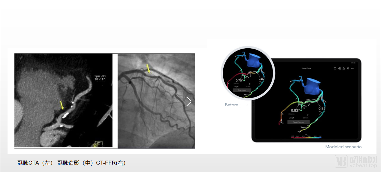
Coronary CTA visualizes the course and degree of stenosis of blood vessels through CT angiography and computer-based three-dimensional reconstruction. Coronary CTA can partially replace coronary angiography, but its accuracy is not high.
Coronary angiography can only visualize the degree of stenosis caused by lesions; however, clinicians have increasingly recognized that the angiographic severity of stenosis does not necessarily determine the clinical severity of the disease.
At times, coronary artery stenosis appears severe on visual inspection by physicians but does not cause myocardial ischemia. Conversely, some coronary lesions appear mild on imaging studies but actually result in significant myocardial ischemia.
The advent of fractional flow reserve (FFR) has effectively addressed this issue.
Fractional Flow Reserve (FFR) refers to the ratio of the maximum blood flow that can be obtained in the myocardial region supplied by a coronary artery with stenotic lesions to the maximum blood flow that would be obtained in the same region under normal conditions.
Simply put, imaging allows physicians to assess the degree of stenosis, whereas FFR can evaluate the actual impact of the stenosis on distal blood flow. Therefore, FFR can accurately assess the relationship between coronary artery lesions and myocardial ischemia, thereby guiding rational treatment decisions and improving patient prognosis.
Currently, the clinical method for measuring FFR involves using a pressure wire. The pressure wire is advanced via the radial artery to the distal end of the coronary lesion. During maximal hyperemia induced by administering adenosine or ATP to the patient, the distal pressure (Pd) and aortic pressure (Pa) are measured, thereby calculating FFR as Pd/Pa.
After 20 years of development, FFR has gained recognition among physicians and is assigned a Class I, Level A recommendation in both the Chinese Guidelines for Percutaneous Coronary Intervention (2016) and the 2014 ESC/EACTS Guidelines on Myocardial Revascularization.
Although guidewire-based FFR measurement allows physicians to assess coronary blood flow, the high cost of pressure guidewards results in a per-test expense for patients of nearly RMB 10,000. Furthermore, FFR technology is monopolized by foreign companies such as Abbott (which acquired St. Jude Medical) and Philips (which acquired Volcano Corporation).
Relevant data indicate that the global patient utilization rate of fractional flow reserve (FFR) is approximately 6% to 8%. In developed countries, FFR has been more widely adopted; in 2014, the utilization rate was 18% in the United Kingdom and reached 30.8% in the United States. By contrast, the utilization rate of FFR in China remains below 1%.
Without the advent of technology for calculating FFR based on medical imaging, the widespread adoption of FFR measurement might have had to wait until the cost of pressure guidewires decreased—a process that could have taken several years or even more than a decade.
The emergence of FFR estimation based on medical imaging has provided physicians with an alternative measurement approach, enabling them to leapfrog traditional methods. Non-invasive FFR diagnosis eliminates the need for expensive pressure guidewires inserted into blood vessels. Techniques such as CT-FFR and QFR can estimate FFR from imaging data by reconstructing the three-dimensional geometry of coronary arteries directly from CT scans, coronary angiography, OCT, and IVUS images, followed by hemodynamic calculations.
Physicians can assess whether a vessel is functionally ischemic while visualizing stenosis, without incurring high medical costs for patients.
The first company to bring this technology into clinical practice was the U.S.-based HeartFlow, with its FFRct (a patented name), which received approval in 2014. Reportedly, HeartFlow’s product demonstrates a sensitivity of 84% and a specificity of 86% per vessel.
HeartFlow’s FFRct calculates FFR values based on CT images, but CT-FFR is not the only method for calculating FFR from imaging data.
Among the methods for measuring fractional flow reserve (FFR), FFR calculation approaches vary based on data sources and computational algorithms, including CT-FFR, quantitative flow ratio (QFR), and intravascular imaging-derived FFR (OFR). These modalities serve distinct roles: acting as a “gatekeeper” in the cardiac catheterization laboratory, assisting in the formulation of coronary intervention plans, and optimizing percutaneous coronary intervention (PCI), respectively.
Lin Xiaojie, Head of the Marketing Department at Bodong Medical Imaging, told VCBeat that the primary difference between CT-FFR and QFR lies in their imaging data sources.
CT-FFR performs three-dimensional reconstruction of vessels based on CT images; QFR calculates FFR through three-dimensional reconstruction from coronary angiography images; while OFR is calculated based on OCT images.
Different products have different application scenarios. For QFR, it is mainly used intraoperatively to help doctors better assess functional stenosis after identifying vascular narrowing, determine which vessel branch is the primary lesion, identify the precise stent implantation site, and assist operators in selecting stent size.
For CT-FFR, the industry benchmark company HeartFlow positions its product as a “gatekeeper” for the catheterization laboratory, assisting physicians in determining whether patients require coronary angiography screening. This helps reduce unnecessary invasive coronary angiography procedures, supports clinicians in formulating optimal treatment plans, and thereby lowers healthcare expenditures.
Wang Xiao, Director of Product Marketing at Shukun Technology, stated, “The assessment of cardiovascular diseases and the formulation of treatment plans constitute a comprehensive evaluation process, encompassing anatomical structure analysis, parameter measurement, functional analysis, and more. When evaluating a patient’s condition, physicians employ multiple diagnostic methods. CT-FFR provides an additional reference value for clinicians during patient assessment. A prerequisite for the technological development of CT-FFR is the precise characterization of anatomical structures.”
Products can also be classified based on differences in their underlying technical principles.
HeartFlow employs finite element analysis to first calculate the blood volume of the myocardium. It then reconstructs all coronary arteries, simulating the blood flow status at every point along the vessels. Consequently, HeartFlow requires a relatively long computation time, currently exceeding three hours.
The one-dimensional model developed by Siemens AG can be directly processed on a post-processing workstation, significantly reducing computation time to approximately 30 minutes.
Another approach employs deep machine learning models based on artificial intelligence algorithms. These models leverage big data to analyze and establish the relationship between coronary anatomical structures and corresponding hemodynamics, but are currently applicable only to specific populations.
From a technical perspective, CT-FFR currently utilizes CT imaging combined with computational fluid dynamics principles to calculate blood flow velocity and pressure, thereby deriving three-dimensional FFR values.
The workflow of CT-FFR technology includes CT modeling, three-dimensional reconstruction of coronary arteries, and hemodynamic analysis. This process involves technologies related to AI-based intelligent image recognition, graphical image processing, and hemodynamic simulation.
Three-dimensional organ reconstruction and accurate extraction of coronary arteries constitute one of the core technological barriers. In CT imaging, coronary arteries are extremely thin, with a diameter of only approximately 2 mm; coupled with various motion artifacts and noise, this makes automated vessel recognition highly challenging.
In image recognition and processing, the application of AI-based image processing can yield more precise and effective medical images.
Secondly, in terms of computational speed, due to the involvement of extensive calculations, the current primary solution is to distribute computational tasks across multiple CPU threads to reduce computation time.
Although CT-FFR can provide physicians with a more rapid reference for disease assessment, it inevitably has its own limitations at present.
CT-FFR demonstrates lower diagnostic accuracy in certain calcified diseases. Coronary artery calcification makes image segmentation on CT scans particularly challenging, thereby complicating FFR calculations.
Furthermore, the diagnostic accuracy of CT-FFR is also relatively low in certain complex cases. Taking diffuse long coronary artery lesions as an example, previous studies have shown that such lesions account for approximately 20% of all patients with coronary heart disease. Patients with diffuse long coronary artery lesions exhibit more extensive atherosclerotic involvement, characterized by a diffuse distribution of lesions and frequent involvement of the left main coronary artery. These cases are often associated with small vessel diameter, angulation, calcification, and tortuosity.
Simply put, diffuse long lesions involve multiple plaques and thrombi within a single vessel segment. For such complex cases, the diagnostic accuracy of general models is significantly reduced. VCBeat has learned that some companies are developing dedicated models specifically for complex cases.
QFR: Products in China have obtained Class III registration certificates
The only domestic company approved for image-based FFR measurement is Pulse Medical Imaging with its QFR product.
QFR technology, short for Quantitative Flow Ratio, was independently developed by Pulse Medical Imaging Technology (Shanghai) Co., Ltd. in collaboration with Shanghai Jiao Tong University. By performing 3D quantitative coronary angiography (QCA) analysis on coronary angiograms and integrating computational fluid dynamics with pressure drop formulas, QFR calculates the pressure drop caused by lesions, thereby enabling rapid computation of fractional flow reserve (FFR) under resting conditions. To date, QFR technology has been validated in over 40 clinical studies, resulting in more than 40 SCI-indexed publications in prominent interventional cardiology journals, which have thoroughly demonstrated its diagnostic accuracy and long-term clinical benefits for patients. This year, QFR was also included in the official European textbook on percutaneous coronary intervention published by the European Society of Cardiology (ESC).
It is reported that Pulse Medical Imaging has also licensed its QFR technology to the Dutch company Medis for product integration.
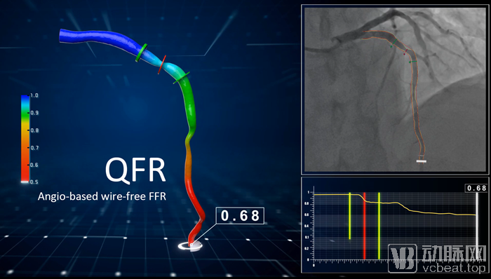
Pulse Medical Imaging QFR Product
It is reported that the FAVOR III China and FAVOR III Europe-Japan studies are currently underway in more than 60 countries across China, Japan, and Europe. The FAVOR III China study, initiated by Fuwai Hospital, plans to enroll 3,830 patients and aims to demonstrate the significant superiority of QFR-guided percutaneous coronary intervention (PCI) over angiography-guided PCI in terms of patient benefits. The FAVOR III Europe-Japan study plans to enroll 2,000 patients and seeks to establish the non-inferiority of QFR-guided PCI to FFR-guided PCI regarding patient outcomes. The results of these two studies are expected to reshape clinical guidelines by incorporating recommendations for QFR-guided PCI.
We can categorize the major market players into three groups: first, AI companies that expand into disease-specific applications based on artificial intelligence technology, such as Arterys, Shukun Technology, and Xingmai Technology; second, companies specializing in CT-FFR, represented by HeartFlow; and third, companies focused on cardiac imaging, exemplified by Circle Cardiovascular Imaging, Pulse Medical Imaging, and PulseFlow Technology.
If only CT-FFR, a single technology, is evaluated, the overall market size is not substantial.Currently, HeartFlow has raised $467 million in financing, with a valuation of $1.5 billion.However, virtually all manufacturers currently possess product expansion capabilities and are developing multiple products targeted at cardiovascular imaging.
Taking Circle Cardiovascular Imaging as an example, this cardiovascular imaging platform provides physicians with cardiovascular MRI and CT image analysis, as well as interventional procedure planning. According to the official website of Circle Cardiovascular Imaging, its related products (excluding CT-FFR products) have received regulatory approval and registration in China. Domestically, PulseFlow Technology serves as another example; it is a medical technology company specializing in general artificial intelligence for cerebrovascular and cardiovascular diseases. Leveraging big data from medical imaging, the company applies computational simulation and artificial intelligence technologies to deliver precise and personalized care for cerebrovascular and cardiovascular conditions, thereby revolutionizing their treatment.
In addition, digital software development companies can also partner with large medical device manufacturers to offer bundled sales solutions. Abroad, Arterys is a renowned medical imaging platform that enables physicians to capture cardiac blood flow data within a visualized three-dimensional space. Arterys has entered into a significant partnership with GE Healthcare to jointly launch the next generation of MRI scanners.
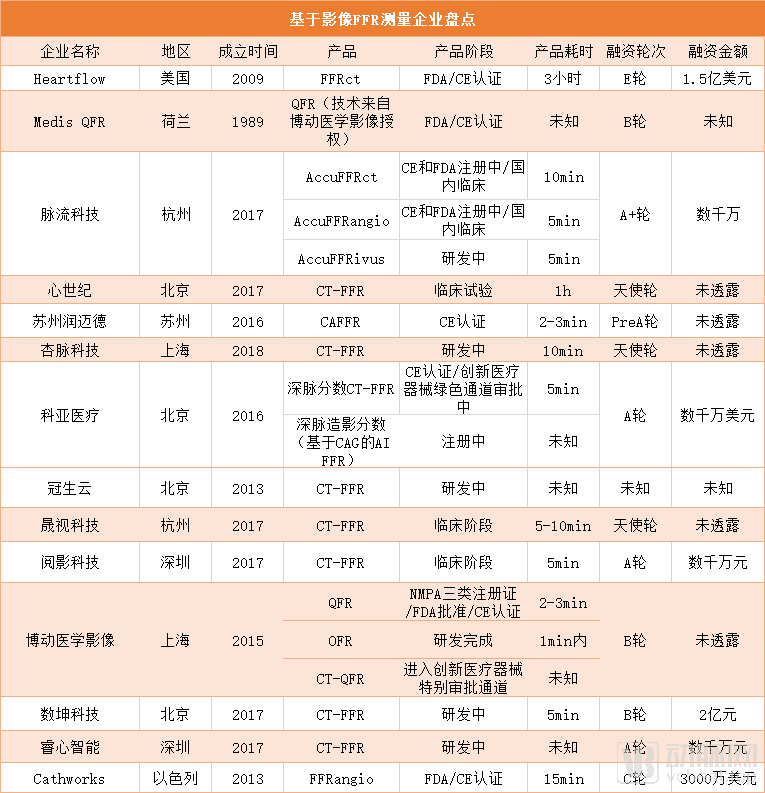
*Companies not included in this table may also contact VCBeat for further discussion.
Data Source: VCBeat Knowledge Base
From an economic perspective, FFR offers patients a cost-effective and precise diagnostic approach. A single CTA scan costs approximately RMB 2,000–3,000, whereas a pressure wire costs nearly RMB 10,000. Image-based FFR can also help reduce unnecessary coronary angiography in patients with chest pain. Data from HeartFlow indicates that more than half of patients undergoing invasive testing show no significant coronary artery obstruction.
In China, there were 915,000 cases of PCI surgery in just one year (2018), and approximately over 3 million people underwent coronary angiography. In China, the mortality rate of cardiovascular diseases is rising year by year. Surgical techniques are continuously evolving, but the threat of cardiovascular diseases to humans has not diminished. Data published by The Lancet shows that cardiovascular disease has always been the leading cause of death in our country. In the United States, since 1995, the mortality rate of cardiovascular diseases has significantly decreased.
In the early 19th century, infectious diseases were the leading cause of death in the United States. During the first half of the 20th century, the U.S. successfully brought infectious diseases under control, becoming a legend in the field of public health. Since the 1970s, the U.S. has also successfully reduced mortality from cardiovascular diseases. A major reason for this decline in cardiovascular mortality is the advent of precision medicine, including the control and treatment of hypertension, the widespread use of statins, and the invention of thrombolytic therapy and stent retrievers for thrombectomy.
If image-based FFR algorithms can be implemented in both tertiary hospitals and primary care institutions, thereby improving the screening of coronary artery disease and the precision of intraoperative diagnosis and treatment, it will help reduce mortality from cardiovascular diseases.