Dr. Huang Xinsheng of Beijing Anzhen Hospital: 11-Year Exploration of 3D Ultrasound Navigation in Ventricular Aneurysm Surgery, Advocating Greater Integration of Technology and Clinical Practice
Looking back, Huang Xinsheng would single out one experience as a pivotal milestone in his scientific research career: a comparative study on the application of two-dimensional/three-dimensional ultrasound navigation in ventricular aneurysm surgery.
From a doctoral student to the Chief Physician of the Department of Cardiac Surgery at Beijing Anzhen Hospital, he has been continuously conducting research on this topic for 11 years and has participated in over 600 ventricular aneurysm surgeries.
“Two-dimensional ultrasound can only display a plane.Three-dimensional dynamic stereoscopic imaging conveys tumor size and morphology more precisely and intuitively,Particularly, it provides valuable reference for clinical surgical procedures involving ventricular aneurysms characterized by wall thinning, ventricular dilation, and compromised cardiac function.“Huang Xinsheng said.
In clinical surgeries for ventricular aneurysms characterized by wall thinning, ventricular dilation, and impaired cardiac function, what specific benefits does three-dimensional technology offer compared to traditional two-dimensional techniques? Recently, VCBeat had the privilege of interviewing Chief Physician Huang Xinsheng, who provided insights on these questions.
Ventricular aneurysm is more like a "ticking time bomb" in the body of patients with myocardial infarction. With even slight negligence, patients may die due to heart failure and other causes.
Huang Xinsheng introduced,Ventricular aneurysm is a mechanical complication following myocardial infarction.. It refers to the condition following myocardial infarction in which the infarcted area undergoes ventricular wall dilation and thinning, with transmural myocardial necrosis. The myocardium becomes thinned due to fibrotic replacement of necrotic tissue, resulting in a weakened region of the ventricular wall that loses contractile function or exhibits paradoxical motion during systole, thereby forming a ventricular aneurysm. Ventricular aneurysms are most commonly found in the left ventricle.
Over 90% of ventricular aneurysms are associated with coronary atherosclerotic heart disease; other rare causes include traumatic ventricular aneurysm, sarcoidosis, congenital ventricular aneurysm, and Chagas disease.
“Ventricular aneurysm has a high incidence rate. Data indicate that,”Currently, the incidence rate of ventricular aneurysm among patients with myocardial infarction in China is approximately 5%.“Over time, the morphology of ventricular aneurysms also changes. Many young patients present with ventricular aneurysm disease, often as their initial manifestation,” introduced Huang Xinsheng.

(Figure caption: Chief Physician Huang Xinsheng, Department of Cardiac Surgery, Beijing Anzhen Hospital)
Ventricular aneurysm formation leads to cardiac structural remodeling, posing a life-threatening risk.Treatment options for ventricular aneurysm include medical therapy and surgical intervention. Among these, surgical resection of the ventricular aneurysm is the most aggressive and effective treatment measure; however, it is technically challenging, carries high risks, and is associated with high rates of mortality and complications, thereby demanding a high level of expertise from the surgeon and their team.
“Traditional open-chest surgical resection requires the patient’s heart to be stopped during the procedure, resulting in significant postoperative trauma and poor prognosis. Many elderly patients cannot tolerate this approach,” introduced Huang Xinsheng.
In addition to surgical intervention, there is another treatment option: minimally invasive left ventricular aneurysm plication. This technique has been widely recognized and adopted by physicians both in China and internationally.
“Minimally Invasive Left Ventricular Aneurysm Plication“It is relatively less invasive, causes minimal trauma, and is suitable for elderly patients; it is currently a highly recommended surgical approach,” said Huang Xinsheng.
He pointed out that whether it is traditional open-heart surgery or minimally invasive left ventricular aneurysm plication, there is a crucial step involved—real-time ultrasound navigation.
“The application of real-time ultrasound navigation plays a pivotal role in intraoperative procedures. In minimally invasive left ventricular wall plication, the ability to more accurately depict the morphology and size of the aneurysm through two-dimensional or three-dimensional ultrasound guidance holds significant instructional value for surgeons.“said Huang Xinsheng.”
Accordingly, Huang Xinsheng conducted a detailed comparative study on the application of three-dimensional ultrasound navigation versus two-dimensional ultrasound navigation in minimally invasive left ventricular aneurysm plication.
Cast your mind back to 2008. That year, the research focus of Huang Xinsheng’s doctoral dissertation was “A Clinical Randomized Controlled Trial on Left Ventricular Remodeling and Function Following Real-Time Three-Dimensional Echocardiography-Guided Non-Extracorporeal Circulation Ventricular Aneurysm Plication.”
“This was my first research project in the field of cardiac surgery at that time,” said Huang Xinsheng. In his view, if a study can clearly demonstrate its significance and value, then it is worth pursuing thoroughly.
“After completing my doctoral degree, I joined the Department of Cardiac Surgery at Beijing Anzhen Hospital. Upon assuming my position, I became a member of the department’s research team. Led by Director Gu Chengxiong, our team conducted a series of studies on cardiac surgical diseases and achieved notable research outcomes. My primary focus was on ventricular aneurysms,” said Huang Xinsheng.
Huang Xinsheng introduced,In minimally invasive left ventricular wall folding procedures, the information provided to surgeons by two-dimensional ultrasound is highly limited. Consequently, surgeons primarily rely on personal experience to assess the size and morphology of ventricular aneurysms for surgical resection, rendering the procedure heavily dependent on subjective human judgment.
“Two-dimensional ultrasound can only identify structures and provide static images; this planar format fails to supply physicians with more accurate data. The application of three-dimensional technology can effectively address this pain point,” said Huang Xinsheng.
“He introduced that their team currently includes both cardiac surgeons and sonographers. ‘We are jointly exploring the application of this technology and validating its advantages,’ pointed out Huang Xinsheng.”
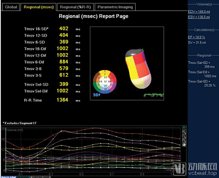
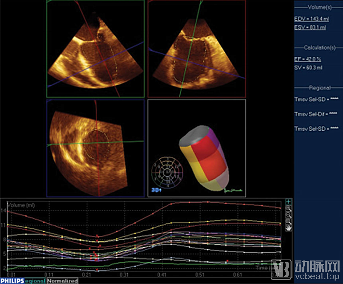
(Figure caption: Intraoperative 3D echocardiographic navigation of changes in ventricular systolic synchrony during ventricular aneurysm repair)
Through hundreds of clinical cases, the team has identified two key advantages of 3D ultrasound navigation technology:Advantages: 1. Greater precision. 2. Pre-assessment of near and far effects.It can display the severity of ventricular aneurysm in real time, providing a reference for physicians’ resection decisions.
“Furthermore, this technology enables quantification, thereby saving physicians time and effort.“When encountering patients with complex conditions, some doctors are uncertain about the extent and volume of tissue to resect. This tool can help them make such judgments in a highly intuitive and effective manner,” said Huang Xinsheng.
Currently, this research has also gained considerable recognition from peers. Clinical practice involving hundreds of cases has further confirmed its advantages, including enhanced safety, higher surgical success rates, and reduced patient pain.
Nowadays, an increasing number of physicians are using 3D ultrasound navigation to perform surgeries. “This is the qualitative change brought about by technology,” said Huang Xinsheng."In the future, he hopes to see more technology-driven products for the treatment of ventricular aneurysms, such as AI-integrated clinical solutions, to help surgeons perform procedures more effectively."