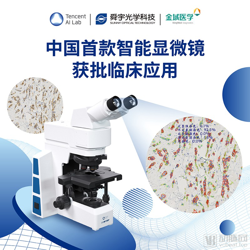
China's First AI-Powered Smart Microscope Receives NMPA Approval for Clinical Use: Jointly Developed by KingMed Diagnostics, Tencent AI Lab, and Sunny Optical
KingMed Diagnostics
Third-Party Medical Testing and Pathological Diagnosis Service Provider
Recently, the intelligent microscope jointly developed by KingMed Diagnostics, Tencent AI Lab, and Sunny Optical Technology has obtained a registration certificate issued by the NMPA, becoming the first intelligent microscope product approved for clinical use in China.
This microscope provides precise quantitative analysis to support pathologists’ microscopic interpretation in immunohistochemistry (IHC) scenarios. Once the field of view is selected, the microscope runs algorithms to rapidly calculate the number of IHC-positive tumor cells in that area within 300 milliseconds, thereby offering real-time assistance to pathologists, saving their time and effort, and enhancing the consistency and accuracy of diagnostic interpretations.
It is reported that this marks another advancement by KingMed Diagnostics in the field of medical AI, following its joint release with Huawei Cloud last June of R&D achievements in AI-assisted cervical cytology screening.
Pathological analysis is the “gold standard” for diagnosis, prognosis prediction, and guidance of cancer treatment. As a critical link in the healthcare chain, pathological diagnosis serves as an essential basis for physicians to determine tumor characteristics and forms the foundation of precision medicine. With advances in diagnostic technologies, precise pathological diagnosis is playing an increasingly vital role in the field of precision oncology.
Currently, pathological diagnosis encompasses routine hematoxylin and eosin (H&E) histomorphology, immunohistochemistry (IHC), fluorescence in situ hybridization (FISH), and molecular pathological testing methods such as genetic testing.
Among these, immunohistochemistry is primarily used for auxiliary disease diagnosis, differential diagnosis, prognosis assessment, and guiding clinical treatment plans, targeted therapies, and immunotherapy. Many of its indicators require precise quantitative analysis; otherwise, this may impact treatment strategies for tumors, including endocrine therapy, targeted therapy, and immunotherapy, and even affect medication choices and prognosis management for patients with malignant tumors.
However, immunohistochemistry (IHC) frequently suffers from poor stability and consistency in subjective interpretation during detection and pathological diagnosis. Additional challenges include image analysis tools being disconnected from routine clinical workflows, the inability to perform precise quantitative analysis, and a lack of standardized criteria for interpreting biomarkers that guide antibody-based therapies. Pathologists are required to examine IHC-stained tissue sections under a microscope to identify and estimate tumor lesions. This process often relies heavily on physician experience, is time-consuming, and yields results that are difficult to standardize with high accuracy.
Meanwhile, the scarcity of pathologists is also constraining the development of precise pathological diagnosis. According to statistics, China currently has only 15,000 pathologists, with a shortfall of nearly 100,000, resulting in a severe imbalance between supply and demand. Furthermore, the training of pathologists faces challenges such as insufficient motivation among the younger generation, due to the lengthy training period and generally low compensation within the industry.
To facilitate more precise pathological diagnosis for patients, improve the accuracy of immunohistochemistry interpretation in pathology, and address the shortage of pathologists, KingMed Diagnostics has collaborated with Tencent and Sunny Optical since 2018 to jointly develop integrated hardware and software solutions for smart microscopes. Through continuous iterations to enhance user experience, they have ultimately developed the first efficient and precise smart microscope product.
Previously, pathologists had to rely on their diagnostic experience to manually select fields of view under the microscope and estimate the number of positive cells. Today, with the approval and clinical application of the first smart microscope, pathologists can quickly calculate the number of tumor-positive cells in a representative tumor lesion area identified on the slide using algorithms embedded in the smart microscope. The entire process is completed directly under the microscope, eliminating the need to switch back and forth between the computer and the microscope.
This integrated hardware-software solution was jointly developed by Tencent AI Lab, Sunny Optical Technology, and KingMed Diagnostics, with the current deployment utilizing an offline computing version. The three parties are also developing an upgraded algorithm based on deep learning methods, which offers higher accuracy and greater potential for future enhancements.
Tencent AI Lab provides leading AI algorithms and software solutions. During training data acquisition, the lab employs active learning and hard example mining to avoid disrupting physicians’ workflows and reduce their burden of manual annotation. By adopting advanced model design strategies, the algorithmic models achieve real-time, full-field-of-view analysis of immunohistochemistry (IHC) slides within 300 milliseconds while maintaining high accuracy. Furthermore, transfer learning and generative adversarial networks (GANs) are leveraged to normalize microscopic images, enabling robust compatibility across different hospitals and slide preparation methods, thereby enhancing the algorithm’s robustness and generalizability.
Sunny Optical Technology has provided customized hardware solutions. For instance, to address inconsistencies in optical imaging environments, condenser lenses and apertures are equipped; to accommodate physicians’ habitual switching of objective lens magnifications, a dedicated magnification memory device has been developed, which adjusts brightness accordingly when a magnification level is selected and directly transmits magnification data to algorithms for analysis. Furthermore, the eyepiece height and illumination design have been optimized specifically for clinical usage scenarios.
KingMed Diagnostics provides expert pathological resources and data assets, ensuring that the microscope system supports interpretation across a variety of disease scenarios, facilitates effective algorithm training, and aligns closely with clinicians’ workflows and practices. For borderline or challenging cases, KingMed Diagnostics assigns full-time pathologists to further review image patches captured from multiple microscopic fields and to guide the annotation team, thereby ensuring data accuracy and laying a solid foundation for precise algorithm training.
KingMed Diagnostics also organized a team of pathologists to conduct field tests on the intelligent microscope, revised the workflow, optimized computational methods, and accelerated algorithm performance.
It is estimated that the intelligent microscope requires only 300 milliseconds on average to analyze a single microscopic field of view, aligning with physicians’ habits and workflow rhythm when switching between fields. Furthermore, by leveraging augmented reality (AR) technology, the analysis results can be directly projected into the microscope’s field of view without interfering with image interpretation or documentation, thereby effectively enhancing physicians’ work efficiency.
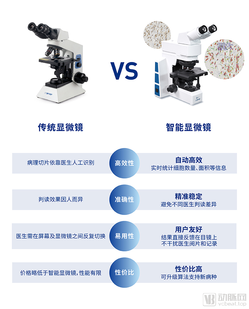
Performance Comparison Between Smart Microscopes and Traditional Microscopes
Validated results demonstrate that the accuracy and real-time performance of the smart microscope meet the practical requirements of pathological diagnosis. It not only assists physicians in reaching diagnostic conclusions more effectively but also significantly enhances their work efficiency. Much like pressing the accelerator while driving, pathologists need only lightly tap a pedal to have the smart microscope rapidly and accurately display results within the microscopic field of view.
Luo Pifu, a pathology expert at KingMed Diagnostics, stated that the practical value of this system is particularly significant in regions and hospitals facing a shortage of pathologists. “The intelligent microscope enables pathologists at primary care hospitals, where diagnostic expertise and capabilities are more limited, to benefit from rapid and accurate interpretations, ultimately benefiting cancer patients.”
Currently, through collaboration with KingMed Diagnostics, the intelligent microscope supports the interpretation of quantitative analysis scenarios for commonly used nuclear and membrane staining in breast cancer immunohistochemistry, including Ki67 (tumor cell proliferation index), ER (estrogen receptor), PR (progesterone receptor), and HER2 (human epidermal growth factor receptor 2).
In the future, KingMed Diagnostics will continue to deepen its collaboration with partners on this project, advancing the research and application of intelligent microscope products in the pathological diagnosis of high-prevalence diseases in China, such as breast cancer, lung cancer, colorectal cancer, and gastric cancer. This initiative aims to enhance the technical standards of pathological diagnosis in China, alleviate the diagnostic pressures stemming from the shortage and limited experience of pathologists, and assist clinicians in achieving precise diagnosis and targeted treatment for malignant diseases such as tumors.