
Percutek Therapeutics
Developer of Minimally Invasive Cardiovascular Treatment Devices

Today's sharing is byProfessor Li Xuan's Team from Peking University International HospitalA High-Risk Thoracic Aortic Aneurysm Surgery Case with a Tricky Anatomical Location of the Lesion. The patient is a 78-year-old male who was admitted to the hospital 8 days after discovering an aortic arch aneurysm during examination. CTA confirmed that the lesion involved the root of the left subclavian artery (LSA), with a severely insufficient proximal anchoring zone. Additionally, there was extensive calcified plaque in the aortic arch, further increasing the difficulty of the surgical procedure. More challenging still, the patient had a history of comorbidities including hypertension, postoperative renal cell carcinoma, and multiple metastases in the pancreas, descending part of the duodenum, and left adrenal gland, leading to compromised multi-organ function. This situation imposed extremely high demands on the safety, minimally invasive nature, and efficacy of the treatment.
Facing multiple challenges such as "insufficient anchoring area requiring LSA reconstruction, and vascular calcification prone to complications," Professor Li Xuan's team, after thorough evaluation and weighing multiple options, innovatively adopted the new process thoracic aortic stent graft system from Percutek Therapeutics, combined with meticulous guidewire puncture techniques, to perform thoracic endovascular aortic repair (TEVAR) combined with in-situ fenestration of the LSA for the patient. This surgical approach ingeniously overcame the limitations of traditional methods, precisely isolating the lesion and reconstructing LSA blood flow while minimizing trauma to the patient’s body. The surgery proceeded smoothly, and the treatment outcome met expectations.

Medical History Introduction
Gender:Male
Age:78 years old
Chief Complaint:Thoracic aortic aneurysm detected 8 days ago.
History of Present Illness:Eight days ago, the patient underwent CTA at another hospital due to fever, which revealed an aortic arch aneurysm with mural thrombus and risk of impending rupture. The patient now comes to our hospital for further surgical treatment.
Past Medical History:Hypertension, postoperative renal cell carcinoma, with metastases to the pancreas, descending portion of the duodenum, and left adrenal gland.
Detailed Explanation of CTA:Thoracic aortic aneurysm, with lesions localized to the anterior wall of the aortic arch, involving the root of the left subclavian artery (LSA), approximately 7mm from the left common carotid artery (LCCA). The aneurysm's long and short diameters are approximately 64.5mm and 40.5mm, respectively. The distance between the LSA and LCCA is about 4mm. The aortic diameter at the root of the LCCA is approximately 40mm. No significant lesions were observed in the ascending and descending segments of the aorta. Large areas of calcified plaque are present in the aortic arch.
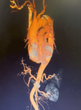
Preoperative Three-dimensional Reconstruction

Preoperative CTA Cross-Section

Condition of the Aortic Arch

Treatment Challenges
The tumor involves the root of the LSA, with a significantly insufficient proximal anchoring zone, necessitating blood flow reconstruction of the LSA to expand the proximal anchoring zone.
Extensive calcification and plaque are present in the aortic arch, which increases the risk of plaque detachment and vascular injury during endovascular manipulation, requiring the operator to be extremely precise and gentle.
The 78-year-old patient, with a history of hypertension, postoperative renal cell carcinoma, and multiple metastases, has relatively poor overall tolerance, placing higher demands on the minimally invasive nature and time efficiency of the surgery.
The patient has multiple underlying diseases, such as old pulmonary tuberculosis, emphysema, bullae, coronary heart disease, and hypertension, which increase the risks during the perioperative period of surgery and the difficulty of postoperative recovery, demanding higher safety and tolerance for the surgical plan.

Surgical Plan Strategy
Endovascular Repair of Thoracic Aortic Stent Graft + Left Subclavian Artery Chimney Technique:The surgical procedure is relatively simple, but there is a higher risk of endoleaks and occlusions, with suboptimal mid- to long-term outcomes.
Endovascular Repair of Thoracic Aortic Stent Graft with In Vitro Fenestration:The lesion closure effect is good, which can preserve the original hemodynamic characteristics, but the operation is complex. The stent needs to be modified before surgery according to the measurement results, which takes a long time; during the operation, precise positioning of the ultra-selection window is required, posing higher risks.
Single-branched Stent Thoracic Endovascular Aortic Repair:The lesion was effectively sealed, but the left subclavian artery forms an acute angle with the aortic arch, increasing the risk of long-term occlusion of the branch stent.
Endovascular Repair of Thoracic Aortic Stent Graft + In-situ Fenestration:The lesion sealing effect is good, and there is no need to modify the stent before surgery. However, the in-situ fenestration of traditional aortic covered stents has higher requirements for interventional devices, requiring special membrane-breaking instruments such as in-situ fenestration needles, lasers, and biopsy needles.
Percutek Therapeutics' Thoracic Aortic Stent Graft can perform in-situ fenestration using the soft tip of a CTO guidewire. Considering the mid- to long-term treatment outcomes and the simplicity of intraoperative manipulation, Professor Li Xuan's team made a comprehensive assessment and choseHuamao・Tianyi®Thoracic Aortic Stent GraftPerform endovascular repair and achieve blood flow reconstruction of the left subclavian artery through in-situ fenestration technology.

01.After the patient was successfully anesthetized, they were placed in the supine position. Following routine sterilization and draping, bilateral femoral arteries and the left brachial artery were punctured under ultrasound guidance, and vascular sheaths were placed.
02.A pigtail catheter was introduced through the left femoral artery to the ascending aorta. Angiography revealed localized dilation of the thoracic aorta, consistent with an aortic aneurysm, and focal stenosis at the origin of the left subclavian artery (LSA). A vascular closure device was pre-placed at the right femoral artery puncture site, and the vascular sheath was replaced. After systemic heparinization, a guidewire and catheter were advanced through the left brachial artery, followed by replacement with a long vascular sheath.

Preoperative Angiography
03.A stiff guidewire was inserted through the right femoral artery, and the Percutek Therapeutics thoracic aortic stent graft PTBS3232080 was introduced as a distal restrictive stent and deployed in the descending aorta.

Placement of Distal Restrictive Stent

Angiography after distal restrictive stent deployment
04.Implant Percutek Therapeutics thoracic aortic stent graft PTBS4440180 in the aortic arch, with the proximal end of the graft positioned distal to the LCCA opening and the distal end positioned for release within the restrictive stent.
05.A V-18 guidewire was introduced through the left brachial artery sheath, and the guidewire, in conjunction with the MPA catheter, successfully performed an in-situ fenestration. Subsequently, a 4mm balloon and an 8mm balloon were sequentially placed at the fenestration site for gradual dilation.

V-18 Guidewire Successfully Penetrates Membrane for Fenestration

4mm Balloon Dilation Window Opening

8mm Balloon Dilation Opening
06.A 9mm-29mm balloon-expandable covered stent was implanted through the LSA window and deployed to complete the blood flow reconstruction of the LSA. Postoperative angiography showed: satisfactory position and morphology of each stent, no imaging of the aortic aneurysm, no endoleak, and smooth blood flow in the aorta and each branch of the aortic arch.

LSA Stent Placement and Release

Postoperative Angiography

Postoperative Angiography
07.Withdraw all guidewires, catheters, and sheaths; close the puncture site with pressure dressing to ensure complete hemostasis. Close the incision layer by layer. End of procedure.

Summary of Case Experience
This case involves an elderly male patient who was admitted to the hospital due to "thoracic aortic aneurysm discovered 8 days ago during examination." The condition presents multiple high-risk features: hypertension, history of nephrectomy and multiple metastases, poor physical tolerance; the aortic aneurysm involves the root of the left subclavian artery (LSA), with a large aneurysm body, insufficient length of the proximal anchoring zone, and unfavorable aortic arch conditions, making the overall treatment extremely challenging.
In response to the patient's complex condition, Professor Li Xuan's team conducted a comprehensive evaluation and innovatively selectedHuamao・Tianyi®Thoracic Aortic Stent Graft, whose core advantage lies in the ability to directly complete in-situ membrane perforation and fenestration using a standard CTO guidewire soft tip, balancing operational simplicity with medium- and long-term treatment outcomes.Postoperative angiography showed that the lesion area was completely isolated, with no endoleak, and the blood flow of the aorta and branches above the arch was unobstructed. The stent position and morphology were satisfactory.
This case fully validatesHuamao・Tianyi®Thoracic Aortic Stent GraftThe excellent performance in complex anatomical conditions and high-risk patients also reflects Professor Li Xuan's team precise clinical decision-making ability and exquisite interventional operation skills, providing valuable clinical reference for the treatment of similar high-risk and complex thoracic aortic aneurysms.

Department Features
Interventional Vascular Surgery Department, International Hospital of Peking University
Peripheral Vascular Disease
1. Aneurysm:Including surgical treatment of intracranial aneurysms, thoracoabdominal aortic aneurysms, and peripheral aneurysms.
2. Aortic Dissection:Aortic stent angioplasty for the treatment of acute, chronic, and complex aortic dissections, avoiding major surgeries such as thoracotomy and laparotomy.
3. Peripheral Arterial Sclerosis Stenosis, Occlusion:Including surgical treatment for diseases such as stenosis or occlusion of the carotid artery, vertebral artery, subclavian artery, renal artery, and lower limb arteries.
4. Deep Vein Thrombosis of the Lower Limbs, Pulmonary Embolism:Combined treatment with medication, interventional thrombectomy and thrombolysis, and inferior vena cava filter implantation reduces the severe consequences caused by acute pulmonary embolism, decreases the long-term sequelae of lower limb venous insufficiency, and improves quality of life.
5. Varicose Veins in the Lower Limbs:CHIVA, radiofrequency, and sclerotherapy for the treatment of varicose veins in the lower extremities feature minimal trauma, quick recovery, and same-day discharge after surgery.
6. Budd-Chiari Syndrome:Interventional therapy can avoid combined thoracotomy and laparotomy, shorten hospital stays, and improve quality of life.
7. Portal Hypertension:TIPSS and selective embolization surgery for the treatment of portal hypertension and its complications, prevention and treatment of massive hemorrhage caused by rupture of gastric and esophageal varices.
8. Arteriovenous malformations, hemangiomas, arteriovenous fistulas:Endovascular treatment, avoiding open surgery.
Interventional Treatment of Benign and Malignant Tumors
1.Interventional Treatment of Liver Cancer:Including hepatic artery chemoembolization, continuous infusion chemotherapy, etc., with minimal surgical trauma, improving quality of life and prolonging survival.
2. Interventional Treatment of Systemic Malignant Tumors:Embolization chemotherapy for malignant tumors in various parts, such as gynecological tumors, bone tumors, and retroperitoneal tumors, reduces perioperative bleeding, lowers surgical risks, and improves surgical outcomes.
3. Esophageal, duodenal, and biliary obstruction caused by various malignant tumors:Minimally invasive surgery for catheter placement and stent implantation to relieve obstruction, alleviate symptoms, and improve quality of life.
Complex Mid- to Long-Term Venous Access
Establishment, Maintenance, and Complication Management of PICC, CVC, Infusion Ports, and Renal Dialysis Access.


END
Copyright Statement: This platform aims to help medical and health professionals better understand the latest developments in relevant disease areas. The information published on this platform does not imply agreement with its descriptions or viewpoints, but is merely intended to provide more information. If there are any copyright issues, we kindly request the rights holders to contact us, and we will address them as soon as possible. The information is solely for medical and health professionals to stay informed, and such information cannot replace professional medical guidance in any way, nor should it be regarded as medical advice. If such information is used for purposes other than staying informed, this platform and the author assume no related responsibility.Contact email for cooperation:vascular@edoctor.work。