Radiologists Beyond the Reading Room: Embracing Patient-Centered Care and AI Integration – Insights from RSNA 2019
Text and Photos by Dr. Yang Jun
The 2019 RSNA annual meeting feels as if it were just yesterday. In this information era, characterized by rapid technological advancements, RSNA has persisted for many years as a premier international academic conference focusing on technology and diagnostic imaging. This longevity has prompted me to reflect on my past conference experiences with new perspectives.
“What has enabled RSNA to consistently maintain high-level academic and medical vitality for 105 consecutive years?”
The answer is undoubtedly the integration of innovation and humanism, a union of body and soul where neither can be dispensed with. This integration is reflected not only in medical imaging but also in the increasing proximity of intelligent technologies and wearable devices to people. However, the primary driving force behind this progress stems from the interaction and advancement among “technology, physicians, and patients.”
Each time I attend the conference, I return with unique insights and knowledge—reconnecting with old friends, meeting new ones, engaging in academic debates on clinical issues, and exploring future technologies. I believe this is the very vitality that defines RSNA. Although Chicago may not be the most exciting city, every early December, we gather here to reflect on the past and look toward the future.
This year’s conference theme and values have been clearly articulated by Chair Dr. Valerie P. Jackson. Drawing on the overarching themes of previous editions, I summarize my personal reflections as follows:
Beyond image reading room,
Embrace a patient-centered care;
Endeavor to be an interactive radiologist with both your patients and clinical colleagues (See Possibilities Together).
The patient-centered integration of imaging workflows and technological innovation, proposed by former Chair Dr. Rao, has been carried forward in spirit during the current term. Hot topics continue to be discussed, now supplemented by more rational reflection and practical application from physicians.
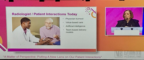
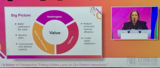
Encouraging physicians to step out of the reading room, engage more closely with patients, and collaborate with multidisciplinary teams—thereby enhancing observation, interaction, and research to effectively address clinical challenges, improve overall value, and maximize patient benefits—represents a return to best practices centered on patients or diseases.
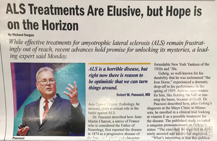
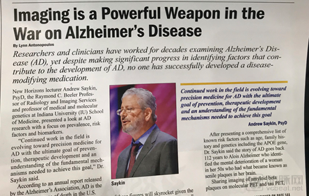
One particularly strong start was the keynote address on the second day of the RSNA annual meeting, which drew a packed house at the Arie Crown Theater that afternoon. Dr. Jackson invited clinical neurologist Dr. Robert Pascuzzi (specializing in amyotrophic lateral sclerosis, ALS) and molecular genetics and imaging expert Dr. Andrew Saykin (specializing in Alzheimer’s disease, AD) to discuss how neurologists and molecular imaging specialists understand and research these two major neurodegenerative diseases beyond the reading room, as well as the ongoing demand for and innovation in medical imaging.
This enables more radiologists to genuinely focus on the disease itself and re-engage in patient interactions, thereby enhancing the value contributed by radiologists within multidisciplinary teams oriented toward disease treatment. Drawing from my own research experience in molecular imaging of Alzheimer’s disease (AD) and multiple sclerosis (MS), I recognize that for neurological disorders lacking effective treatments, medical imaging indeed plays a critical role in exploring mechanisms during the prodromal phase and identifying biomarkers during the treatment phase. Furthermore, imaging serves as a key component and assessment tool in integrated diagnostics, bridging fields such as epigenetics, bioassays, radiomics, and machine learning.
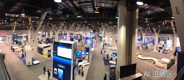
In the realm of artificial intelligence and related research, equipment exploration, major manufacturers showcased AI-driven hardware devices and platform applications this year. Examples include GE Healthcare’s Edison platform and its deep learning-based CT reconstruction algorithms, which were highlighted again; Siemens’ AI-powered Somatom CT; and Neusoft Medical’s neuAI 256- and 512-slice CT and MR systems from China. Additionally, the AI Showcase (sponsored by Zebra) featured a rich lineup of presentations and introduced an AI Deep Learning Lab and workshops, allowing physicians to engage in hands-on, interactive learning. Chip and internet companies such as NVIDIA, Google, and Zebra also actively participated.
Artificial intelligence remained a focal point for research and product development at the conference. Non-diagnostic decision-support deep learning applications were primarily concentrated on CT and MR image reconstruction and optimization, rapid post-processing of imaging data, as well as PET image synthesis and optimization under low-dose tracer conditions. These areas are largely dominated by major vendors or pursued through collaborations with startups. Diagnostic decision-support applications focused mainly on the classification and detection of intracranial hemorrhage in CT scans, detection of vascular lesions in CT, and lesion segmentation in cardiothoracic, pulmonary, and breast imaging. However, there were fewer deep learning-based auxiliary diagnostic tools for MRI, and less than 30% of such products had officially received FDA clearance via the 510(k) or De Novo pathways.
Manufacturers from South Korea, China, and Israel occupied approximately 50% of the main AI exhibition area. Google Cloud has engaged in deep collaboration with the Mayo Clinic on segmentation of abdominal organ composition and the extraction of knowledge summaries from multi-parameter, interdisciplinary charts. Neusoft Medical’s intelligent cloud platform, along with its associated post-processing algorithms, has begun to gain traction in automated stroke assessment products. Meanwhile, Philips Healthcare is leveraging computational power to facilitate big data analytics and intelligent optimization of emergency care workflows.
Unlike the previous two editions, doctors and enterprises are now approaching AI with greater rationality, with data volume and computing power no longer being the primary focus. Key considerations and challenges now center on: 1) healthcare institutions recognizing that while consensus-based annotations are scarce, the workload is substantial and quality varies; 2) interdisciplinary communication barriers between clinical radiology and technical/computer engineering fields, coupled with a scarcity of multidisciplinary talent; 3) the high cost of quality validation due to biases in training datasets across different institutions; and 4) issues related to regulatory registration processes, quality control, and commercialization pathways. Only after overcoming most of these challenges can products truly alleviate the burden on radiologists, improve workflow efficiency, and free up their time to actively engage in patient interactions and contribute value within multidisciplinary treatment teams.
Final Thoughts:
Beyond imaging and AI, radiologists face a broader range of responsibilities. To uncover greater opportunities within the patient care ecosystem, they must proactively assume their professional role as interactive caregivers.