Behind the Surge: The Unsung Heroes of Wuhan’s Radiology Departments Amid a 10-Fold Workload Increase | Tech in the Fight Against Pandemic

Infervision
Artificial Intelligence Product Developer
On February 13, Hubei Province included clinically diagnosed cases in the newly reported cases of COVID-19 for the first time, with the majority of the daily new cases being clinically diagnosed. (From 0:00 to 24:00 on February 12, Hubei Province reported 14,840 new COVID-19 cases, including 13,332 clinically diagnosed cases.)
In the diagnostic criteria outlined in the latest “Trial Version 5 of the Diagnosis and Treatment Protocol for Novel Coronavirus Pneumonia” issued by the National Health Commission, suspected cases within Hubei Province exhibiting characteristic imaging findings are classified as “clinically diagnosed cases,” thereby affirming the critical role of medical imaging in the diagnosis of COVID-19. Behind these figures lie the immense efforts and hardships endured by countless radiologists in Hubei Province.
What is the current status of radiologists on the front lines in Wuhan, Hubei? Recently, VCBeat interviewed Professor Xia Liming, Director of the Department of Radiology at Tongji Hospital Affiliated to Tongji Medical College of Huazhong University of Science and Technology (hereinafter referred to as Wuhan Tongji Hospital).
“The situation has improved significantly compared to New Year’s Eve. At the very least, doctors’ working hours have begun to follow a regular schedule,” stated Director Xia Liming. “We now operate with two teams of physicians and two teams of technicians working in shifts, each team covering 12 hours to ensure uninterrupted 24-hour service. The hotels surrounding the hospital have been reserved by our institution; technicians off duty proceed directly from the hospital to these hotels for rest. This arrangement is the best option for both them and their families.”
Director Xia Liming attempted to adopt a more relaxed tone during the conversation, yet reporters could still sense the unmaskable gravity of the situation from the other end of the line. It is understood that since the onset of the epidemic, Wuhan Tongji Hospital has been conducting hundreds of CT scans daily at a single campus, requiring physicians to interpret images and draft reports. When aggregating the CT examinations across all three campuses of the hospital, the daily volume ranges from several hundred to over one thousand cases. Consequently, radiologists and technicians in the hospital’s Department of Radiology often find themselves with no time for meals or rest.
“The radiologic technologists endure the greatest hardship. They must be ‘fully geared up,’ wearing masks, goggles, and airtight protective suits as they enter the examination rooms to help position patients, coming into close contact with them. Working nearly 24 hours without interruption, they face both physical exhaustion and intense stress; their bodies are soaked in sweat within half an hour. Meanwhile, the interpreting radiologists contend not only with fatigue but also with immense pressure,” said Dr. Xia Liming. “Every minute we save in delivering imaging reports to the clinical team allows them to initiate patient management, isolation, and treatment one minute earlier. The demand for both speed and absolute accuracy presents a tremendous challenge for our radiologists, who work 12-hour shifts every day.”
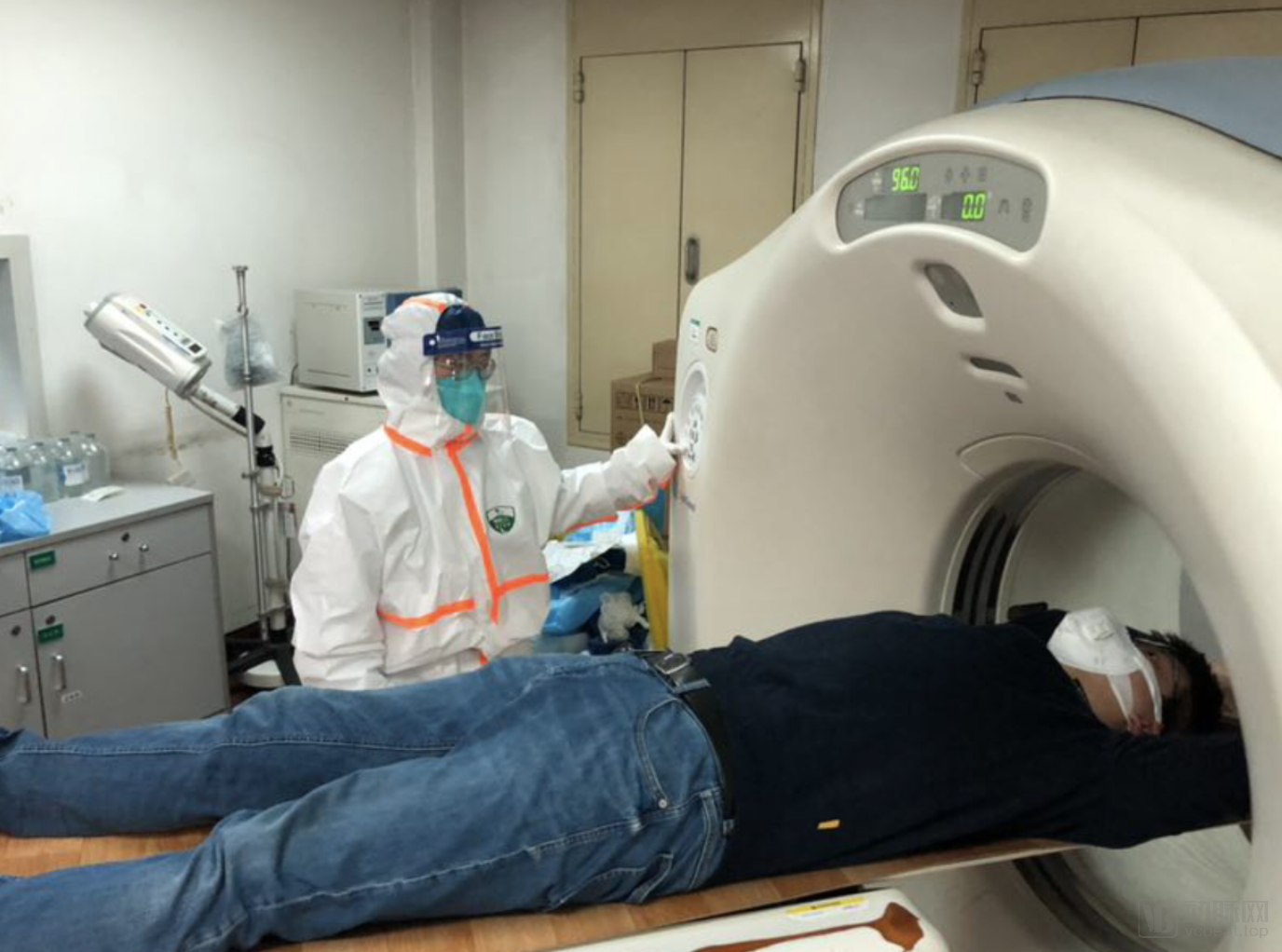
Doctor in Protective Gear Positions Patient
The pandemic placed immense pressure on the Department of Radiology at Tongji Hospital in Wuhan. However, instead of retreating, Director Xia Liming’s team leveraged artificial intelligence to alleviate this burden and accelerate workflow. In collaboration with Infervision, they integrated clinical features to study the CT imaging manifestations of COVID-19 and train AI models. During the intense battle against the epidemic, when workloads surged dramatically, this tireless AI assistant—requiring neither food nor rest—supported physicians in making accurate distinctions and diagnoses.
While most people were still immersed in the Spring Festival holiday, Infervision had already taken the lead in launching the “AI Special Edition for Pneumonia.” This product, born from the front lines of the epidemic, was also among the first to be deployed on those very front lines. Wuhan Tongji Hospital, of course, was among its first users.
“This special AI edition for pneumonia, developed by Infervision, was designed specifically for the current epidemic. The qualitative capabilities of Infervision’s AI have facilitated the tracking of patient disease progression and enabled retrospective studies. On one hand, Infervision’s AI assists physicians in performing quantitative measurement and analysis of pulmonary lesions; thus, during a patient’s follow-up examination, we can rapidly assess changes in the lesions by comparing them with prior data, which significantly aids clinical diagnosis. On the other hand, these quantitative data allow us to conduct more comprehensive correlation studies between imaging findings and clinical manifestations in subsequent research,” introduced Director Xia Liming.
“Secondly, our three hospital campuses have long been overcrowded, with patients spending considerable time queuing for examinations and waiting for results, thereby facing a substantial risk of cross-infection. The efficiency gains brought by AI can help minimize the time patients spend waiting for results. While the examinations themselves are quick, patients devote much of their time to awaiting reports. With the integration of AI, report generation time has been accelerated from tens of minutes to just a few minutes.”
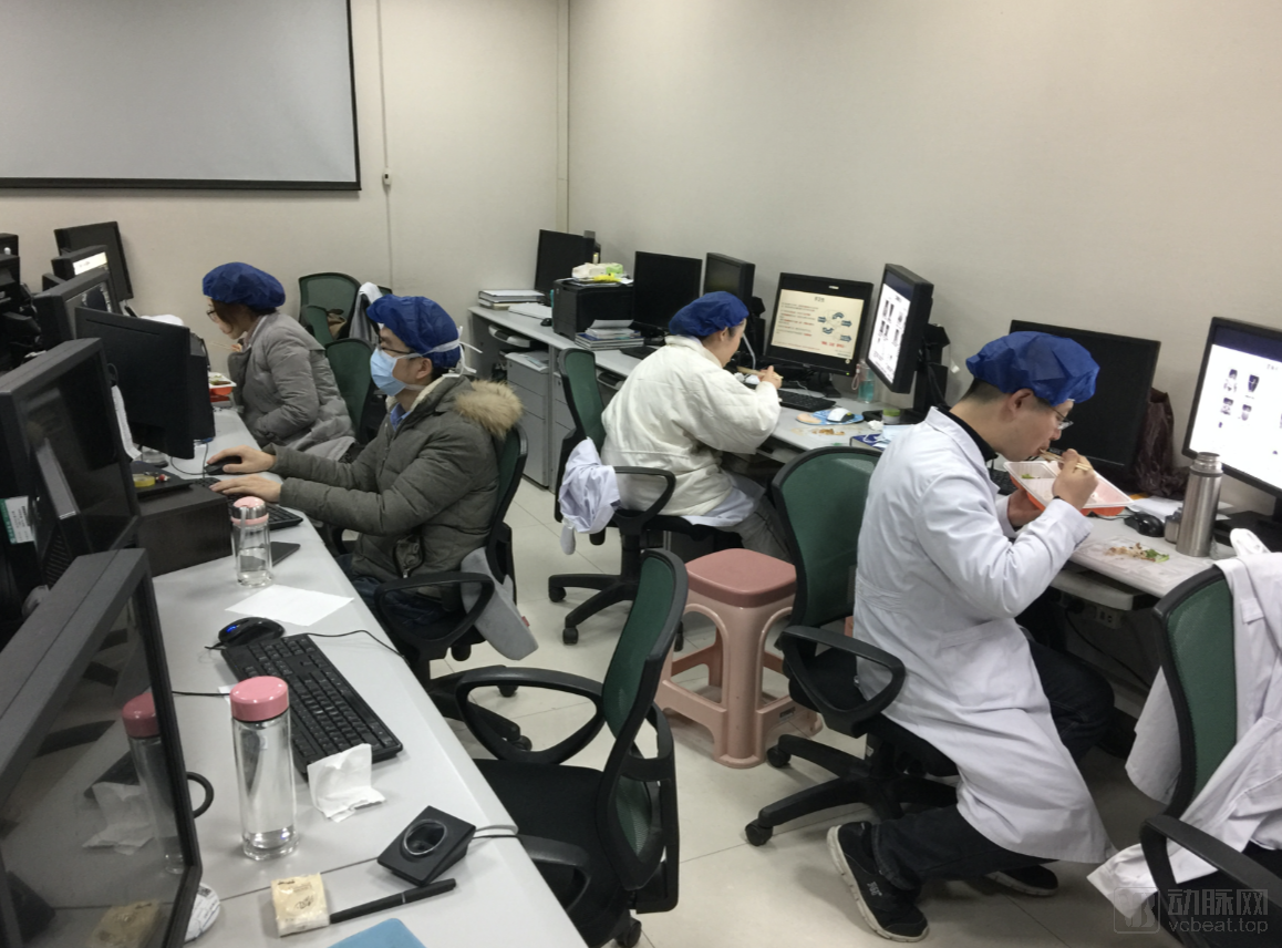
Radiologists at Wuhan Tongji Hospital Holding the Line
Regarding the heated recent debate over “whether imaging diagnosis or nucleic acid testing should serve as the diagnostic standard,” Director Xia believes that there is no definitive answer; different testing modalities will play distinct roles across different regions and scenarios.
“Every diagnostic method has its own advantages and disadvantages. CT images offer high clarity and can detect early, subtle lesions; however, when the disease is confined to the larynx, trachea, or bronchi without significant involvement of the lung parenchyma or interstitium, chest CT findings may be negative. Furthermore, COVID-19 is a type of viral pneumonia, and during this season, other viral pneumonias—such as those caused by influenza viruses or adenoviruses—are also common, making differential diagnosis based on imaging challenging. Although nucleic acid testing is the gold standard for diagnosing COVID-19, throat swab tests typically yield a positive rate of only 30–50%, often requiring repeated testing. Additionally, the quality of test kits varies among manufacturers, and there was initially an insufficient supply of kits.”
“Furthermore, the current level of medical practice also limits the effectiveness of nucleic acid testing to some extent. Due to healthcare workers’ limited experience in using test kits, it is often difficult to collect saliva samples from the upper respiratory tract, leading to false-negative results in patients. If a patient has just contracted the disease, the viral load in the sample may be too low, resulting in false negatives (missed diagnoses). In addition, PCR testing imposes stringent requirements on laboratory facilities, testing equipment, and operational personnel. In many primary-care hospitals, even if test kits are available, healthcare workers may still fail to collect valid samples.”
Therefore, each technology has its own advantages and limitations; only by making the right decisions based on specific scenarios can optimal testing performance be achieved.
In summary, the role of artificial intelligence in this epidemic prevention and control campaign can be summarized in the following five points:
I. Providing More Effective Diagnostic Assistance to Key Epidemic Areas Such as Hubei ProvinceAs previously mentioned, in accordance with the Diagnosis and Treatment Protocol for Novel Coronavirus Pneumonia (Trial Version 5), AI combined with CT serves as an effective tool for determining clinically diagnosed cases. This approach will significantly accelerate the efficiency of case diagnosis in Hubei Province, eliminating concerns over false-negative results from nucleic acid tests and slow testing speeds. This development is beneficial for both patient treatment and epidemic prevention and control in Hubei Province.
II. It can accelerate imaging diagnosis and reduce the risk of infection for a large number of patients waiting for test results within the hospital. Designated fever clinics for COVID-19 in Wuhan and other regions are operating beyond capacity, with a significant number of suspected patients queuing for chest CT scans, posing a substantial risk of cross-infection. AI enables physicians to provide faster and more accurate recommendations even under excessive workloads, thereby reducing patient wait times and lowering the risk of cross-infection among patients and between patients and medical staff within the hospital.
III. AI-Integrated CT Can Serve as a Primary Force in Epidemic Prevention and Control at the Grassroots Level. While nucleic acid testing is the gold standard for confirming COVID-19, its application is constrained not only by limited throughput but also by stringent laboratory environmental requirements. Most grassroots hospitals lack the necessary conditions for such testing; the entire process, including sample transport and result retrieval, can take several days, thereby delaying epidemic containment efforts. In contrast, AI-integrated CT can be rapidly deployed to grassroots hospitals, enabling swift diagnosis and buying valuable time for epidemic prevention and control.
4. It helps identify asymptomatic carriers and early-stage patients, thereby supporting the treatment and prognosis of COVID-19. The long incubation period of COVID-19, coupled with mild early symptoms and the presence of asymptomatic cases who may still be infectious, poses significant challenges to epidemic control. However, even in patients with mild or no symptoms, radiological abnormalities are often present. Therefore, timely identification of infected individuals through imaging is of great significance for epidemic prevention and control. Furthermore, AI is not merely an auxiliary tool for image analysis; it offers robust capabilities in disease course management and therapeutic efficacy assessment. In this fight against COVID-19, AI can assist physicians in early detection, monitoring disease progression and outcomes, and guiding clinical treatment.
In addition to enhancing diagnostic and treatment efficiency through artificial intelligence, the substantial investments previously made in health informatics also played a significant role during this pandemic.
Recalling the SARS outbreak 17 years ago, many hospitals did not even have PACS systems; misfiled or incorrectly retrieved films were common occurrences, and the retrieval, review, and comparison of images were highly time-consuming. Acquiring clinical data for subsequent analysis was straightforward.
Today, Tongji Hospital in Wuhan stores all patient imaging records within the hospital, which not only ensures the accuracy of clinical workflows but also makes archived data readily available for research by relevant authorities. Furthermore, the hospital’s teleconsultation program has significantly addressed diagnostic challenges for patients at primary healthcare institutions.
“The medical informatics infrastructure built in recent years has played a crucial role in this incident. If there are frequent situations where the workload at a certain campus surges, we can transmit patient imaging data from other hospitals to our hospital remotely. Once we have completed the imaging reports, we can quickly send them back to the requesting hospital.”
“Through our medical consortium, many complex and rare cases at the primary care level can be examined locally, with images then transmitted to our hospital for interpretation. During the pandemic, every outing posed increased risks for patients; telemedicine inherently eliminated the potential for cross-village and cross-county transmission.”
Third-party medical institutions have also leveraged telemedicine to help alleviate the shortage of radiologists in epidemic-affected areas. Specifically, the 365 Hospital website and Yimai Yangguang launched free online remote consultation services to support these regions; Meinian Onehealth collaborated with companies such as Daxiang Doctor and Wanli Cloud to provide physicians and patients with remote image interpretation, psychological counseling, and other services; Ping An Health (Testing) Center has also utilized local and cloud-based PACS systems to offer hospitals and patients across various regions services including remote image interpretation, imaging data hosting, remote consultations, and AI-assisted imaging diagnosis.
“Urgently hiring truck drivers: 18-meter-long vehicle to drive a box truck carrying ‘Mobile CT’ to Wuhan Union Hospital for COVID-19 screening.” On January 23, upon learning of the severity of the outbreak in Wuhan, Chen Shengchao, General Manager of Zhejiang MinFound Medical Equipment Co., Ltd., posted this message on his WeChat Moments.
Telemedicine is not the only approach to stratify patient populations and alleviate pressure on core hospitals. Consequently, many enterprises have begun to expand the scope of CT utilization by improving CT imaging equipment, with MinFound being one such example.
Director Xia told VCBeat, “From a practical standpoint, the novel coronavirus does not impose high hardware requirements on CT imaging; 16-slice CT scanners used at the primary care level are sufficient to detect lesions.” Therefore, enhancing the mobility of CT scanners could potentially provide more CT imaging services to primary healthcare facilities and epidemic-affected areas with strained medical resources.
MinFound’s mobile CT units have been adapted to meet the needs of epidemic-affected areas. Equipped with a 16-slice CT scanner, these units feature independent isolation and a mobile design, enabling their deployment in hospital epidemic prevention and control, makeshift hospitals, communities, and other outdoor (emergency) settings. They address clinical demands such as the shortage of fixed CT scanners, the inability to rapidly establish protected CT rooms in emergency locations, and the need for isolated usage. In practice, each mobile CT unit can diagnose nearly 200 patients per day at full capacity.
However, mobile CT units impose significant demands on medical personnel, requiring each unit to be staffed with engineers, clinical training specialists, on-site CT operators for equipment installation, and hospital training personnel. In response, MinFound deployed its own experts and medical staff to Wuhan for on-site operations.
Ping An Health (Testing) Center has also adopted mobile CT units to expand the scope of CT screening. Previously, these mobile CT units were primarily used to address the lack of medical infrastructure in remote areas. On January 29, a 64-slice mobile CT imaging vehicle developed based on 5G technology was deployed to Hubei Provincial People's Hospital, and on February 7, it was reassigned to a "Fangcang Shelter Hospital," achieving mobile deployment with a single device.
By enhancing the mobility of CT equipment and leveraging the remote intelligent image interpretation capabilities of imaging platforms, we were able to rapidly establish a temporary hospital with diagnostic and treatment capabilities equivalent to those of a Grade 3A hospital in epidemic areas, thereby alleviating the shortage of medical resources and radiology specialists in Wuhan.
Compared with radiology departments during the SARS era, today’s physicians are better equipped, possessing more precise diagnostic capabilities and more efficient image interpretation skills; however, there is still room for improvement in the application of artificial intelligence technology in the medical field.
Director Xia stated, “Although AI-assisted diagnostic products have already provided substantial support to physicians, there remains room for improvement in terms of precision and depth. For instance, from an imaging perspective, I hope to obtain more intelligent big data analytics to help us promptly identify patterns in lesion changes, which would also facilitate the development of related pharmaceuticals. Of course, for routine clinical use, their sensitivity is sufficient to address the current epidemic, achieving effects such as improved efficiency and enhanced triage.”
We have now entered the critical phase of the fight against the epidemic. Empowered by technology, radiologists continue to work tirelessly day and night. Although crowds outside remain bustling, we can already see the faint light of hope kindled by our collective efforts, signaling the approaching end of the outbreak.
Gratitude to the doctors and colleagues who stepped forward in the fight against the epidemic.