Seven Wuhan Radiology Experts on COVID-19: A Comprehensive Overview of the Pandemic and Related Medical Technologies
As the medical community’s understanding of viruses continues to deepen, it has become evident that COVID-19 features a prolonged incubation period, mild early symptoms, and even asymptomatic cases that may still be infectious. These factors pose significant challenges to epidemic prevention and control. Since individuals with mild or no symptoms often exhibit radiological abnormalities, timely screening of infected populations through imaging techniques holds substantial significance for effective epidemic control.
On February 13, Hubei Province included clinically diagnosed cases in the newly reported COVID-19 cases for the first time, with the majority of daily new cases being clinically diagnosed. The radiological progression of the novel coronavirus has garnered widespread attention. In response to frontline needs, medical imaging companies donated a large number of CT and DR devices to address equipment shortages at hospitals such as Huoshenshan and Leishenshan. While accelerating production, some companies also promptly initiated the research and development of box-type CT scanners, remote diagnosis systems, and AI-assisted diagnostic technologies to support epidemic control efforts.
On January 29, Neusoft Medical donated high-end CT equipment and software valued at RMB 27 million; on February 3, Mindray successfully completed the installation of approximately 1,800 medical devices at Wuhan Huoshenshan Hospital; on February 8, Mingfeng Medical’s Ark CT arrived at the Wuhan Fangcang Shelter Hospital and underwent power-on commissioning; on February 10, United Imaging Healthcare’s artificial intelligence solutions provided comprehensive support for the fight against the epidemic...
From the hospital perspective, designated fever clinics for novel coronavirus in cities such as Wuhan have seen overwhelming numbers of suspected patients queuing for chest CT scans, posing a substantial risk of cross-infection. How physicians can provide recommendations more rapidly and accurately to reduce patient wait times and mitigate the risk of cross-infection among patients and between patients and healthcare workers has become an urgent challenge for hospitals. In Wuhan, at the epicenter of the epidemic response, how can hospital management and medical imaging diagnosis be effectively maintained amid strained medical resources and high-load operations of the hospital system?
On March 20, 2020, the online course “Medical Imaging Practices in the Fight Against the Epidemic” successfully concluded. The event was hosted by the Radiology Branch of the Hubei Medical Association, co-organized by MinFound Medical Systems Co., Ltd., and undertaken by VCBeat. Following the conference, VCBeat compiled the professors’ presentations into four key themes: understanding COVID-19, the value of CT in epidemic control, infection prevention and control in radiology departments, and the construction of Wuhan hospitals and makeshift cabin hospitals.
Since the discovery of the novel coronavirus, our understanding of this uninvited guest has gradually deepened. In an online lecture, Dr. Li Zhen, Director of the Department of Radiology at Tongji Hospital Affiliated to Tongji Medical College of Huazhong University of Science and Technology, and Dr. Zha Yunfei, Director of the Department of Radiology at Renmin Hospital of Wuhan University, provided a detailed account of research progress on SARS-CoV-2 by reviewing recent papers published in top-tier medical journals. They covered five aspects: clinical features, imaging characteristics, prognostic analysis, studies related to mortality, and damage to other organs.
Clinical Features
The first article on COVID-19, published in the NEJM, conducted an epidemiological analysis of the earliest 425 confirmed cases of novel coronavirus pneumonia and described the general characteristics of the virus. The sample data showed that 58.1% of the patients were male and 41.9% were female, with a median age of 59 years.
Before January 1, the majority of confirmed cases had exposure to the Huanan Seafood Market, accounting for 55%; however, after January 1, this proportion dropped sharply to 8.6%, with a mean incubation period of 5.2 days. In the early phase of the epidemic, the number of patients doubled every 7.4 days, and the reproduction number (R) was estimated at 2.2, indicating that COVID-19 is highly contagious.
A subsequent study published in the NEJM yielded results similar to those described above. Furthermore, the article’s statistical analysis showed that critically ill patients accounted for 6.1% of the sample, including 5.0% admitted to the ICU, 2.3% who received invasive mechanical ventilation, and 1.4% who died. Among these patients, only 1.9% had a history of direct contact with wildlife.
So, what are the clinical manifestations of these patients? The most common symptom is fever (present in 43.5% of patients upon admission and 88.7% during hospitalization), followed by cough (67.8%), while diarrhea is relatively rare (3.8%).
Ground-glass opacities in the lungs were the most common finding on chest CT scans (56.4%). Among 887 patients with mild disease, 157 (17.9%) showed no radiological or CT abnormalities; among 173 patients with severe disease, 5 (2.9%) showed no radiological or CT abnormalities, indicating that even CT scans carry a risk of missed diagnosis. Additionally, 83.2% of patients exhibited lymphopenia.
If we delve deeper into the research and examine outpatients, critically ill patients, and ICU patients separately, the data reveal further differences.
A study published in The Lancet investigated the outpatient characteristics of COVID-19. Among the 628 patients in the sample, the mean age of nucleic acid-positive individuals was 53 years, significantly higher than the mean age of 49 years observed in nucleic acid-negative individuals. The rate of positive chest CT findings was higher in nucleic acid-positive patients (96.3%) than in nucleic acid-negative patients (88.5%). Of these patients, 553 (88.1%) had mild cases and 75 (11.9%) had severe cases; 78.2% of patients presented with fever, and 80.0% reported myalgia.
Another article in The Lancet focused on critically ill patients, 67% of whom were male; 40% had chronic diseases, and 98% experienced fever during the critical phase.
The disease severity in critically ill patients progresses rapidly, with 61.5% of these patients dying within 28 days. The mean age of the deceased patients was 64.6 years, which is higher than that of the survivors (51.9 years).
Most critically ill patients experienced varying degrees of organ dysfunction following disease onset, with 67% developing acute respiratory distress syndrome (ARDS), 29% experiencing acute kidney injury, 23% exhibiting abnormal liver function, and 2% presenting with pneumothorax. Overall, the characteristics of SARS-CoV-2 were more pronounced in critically ill patients.
Characteristics in the ICU share similarities with severe cases, and a JAMA article has identified some interesting phenomena. In ICU patients, white blood cell, lymphocyte, and neutrophil counts differ significantly from those in non-ICU patients. Meanwhile, D-dimer, procalcitonin, and troponin levels are markedly elevated, as are indicators of hepatic and renal impairment, including ALT, AST, BUN, creatinine, and urea nitrogen.
Imaging Findings
Given the distinct radiological manifestations of COVID-19 on CT scans and the widespread availability of this modality, CT has played a pivotal role during the current outbreak.
Some retrospective studies have indicated that the most prominent imaging features of COVID-19 include ground-glass opacities (GGOs), GGOs combined with focal consolidation or multiple irregular areas of consolidation along the bronchovascular bundles, diffuse GGOs, and the crazy-paving pattern. Other findings observed in some patients include diffuse bilateral lung involvement, air bronchograms, reticular or linear opacities, solid nodules, thickening of the bronchovascular bundles, and vascular enlargement.
Children are not merely miniature adults. In the article “Clinical and CT features in pediatric patients with COVID-19 infection: Different points from adults,” it is reported that 65% of pediatric patients had a history of close contact with family members infected with COVID-19, 60% presented with fever, and 65% exhibited cough. A notable observation is that 80% of children showed elevated procalcitonin levels, and 40% had co-infections, which are uncommon in adults. Furthermore, chest CT scans were normal in 20% of pediatric cases, a proportion slightly higher than that observed in adults.
Prognostic Analysis
Based on early imaging and clinical features, physicians can to some extent predict patient prognosis.
A study published in The Lancet, which analyzed 56 cases from fever clinics, found that 85.7% of patients exhibited ground-glass opacities on CT scans, and 78.6% showed vascular dilation; both factors were significantly associated with patient prognosis. Meanwhile, no significant prognostic correlation was observed for white blood cells, neutrophils, lymphocytes, platelets, or C-reactive protein. In simple terms, the study found that patients with lower CT values—characterized by prominent ground-glass opacities and predominant exudation—were more likely to experience disease progression.
Analysis of Factors Contributing to Death
The article “Clinical course and risk factors for mortality of adult inpatients with COVID-19 in Wuhan, China: a retrospective cohort study” analyzed factors associated with mortality in COVID-19. The sample included a total of 191 patients, of whom 137 were discharged and 54 died. Among the deceased patients, 30% had hypertension, 19% had diabetes mellitus, and 8% had coronary heart disease.
Based on data analysis, the article concludes that older patients have a higher risk of mortality, and D-dimer levels greater than 1 μg/L are associated with increased likelihood of death. Among survivors, the median time to viral clearance was 20 days, with the longest duration being 37 days. This suggests that many patients may remain potentially infectious for up to a month after clinical recovery, as they have not yet tested negative for the virus.
Other researchers conducted a multivariate regression analysis of factors associated with mortality and identified the following characteristics in this population:
1. Predominantly male, with more than half of the patients aged over 60 years.
2. Most patients have comorbidities, especially hypertension, heart disease, and diabetes.
3. Respiratory failure remains the leading cause of death, followed by sepsis, heart failure, hemorrhage, and renal failure.
4. Lymphopenia, neutropenia, and thrombocytopenia are commonly observed in patients upon admission; the neutrophil-to-lymphocyte ratio is elevated in most patients; systemic immune-inflammation index is high; C-reactive protein levels are increased; and lactate dehydrogenase and D-dimer levels are elevated.
5. The median time from initial symptom onset to death was 15 days.
Other Organ Injuries
“Acute Myocardial Injury of Patients with Coronavirus Disease 2019” investigated other organ injuries caused by COVID-19. Among the 53 patients, 79.25% were found to have cardiac complications, including six cases of acute myocardial infarction, all of whom were over 60 years of age.
Another study on liver dysfunction investigated 148 patients, 75 of whom presented with abnormal liver function upon admission. Furthermore, patients with elevated liver function indices were more likely to experience moderate high fever.
In studies on renal impairment, researchers investigated 85 cases of COVID-19, with 27.06% of patients exhibiting acute kidney failure. H&E staining revealed severe acute tubular necrosis in postmortem liver tissue samples from six cases, but no glomerular lesions or lymphocytic infiltration in the tubulointerstitium was observed.
Finally, Professor Li Zhen summarized the characteristics of the novel coronavirus into five points:
1. Most patients present with mild symptoms and are susceptible to infection; the primary clinical risk factors include advanced age, male predominance, and underlying conditions such as hypertension, which increase susceptibility to infection.
2. Patients often present with high fever, an elevated neutrophil-to-lymphocyte ratio, a high systemic immune-inflammation index, increased C-reactive protein levels, and elevated lactate dehydrogenase and D-dimer levels; the CT severity score is a high-risk factor.
3. Ground-glass opacities (GGOs) and GGOs with interstitial network changes are the primary CT features; the distribution, extent, and characteristics of imaging findings such as GGOs can help determine prognosis.
4. In children, 80% present with elevated procalcitonin levels and are prone to co-infections; some early cases show negative CT findings.
5. Evidence of multi-organ dysfunction involving the heart, liver, and kidneys.
As explained by Director Zha Yunfei and Director Li Zhen, the imaging manifestations of COVID-19 can reflect the patient’s infection status to a certain extent. In his course, Dr. Hu Daoyu, Director of the Department of Radiology at Tongji Hospital in Wuhan, further elucidated the role of multi-slice spiral CT (MSCT).
Regarding the value of CT, Hu Daoyu stated, “CT has a high detection rate for COVID-19. Although its imaging findings are not specific, they do exhibit certain characteristic features, such as multifocal involvement in both lungs. Furthermore, CT can visualize the extent and nature of the lesions, facilitating physicians in stratifying patients for appropriate management.”
Therefore, CT scans hold exceptional clinical significance. For patients with negative nucleic acid test results, CT imaging serves as the basis for intervention in cases of false negatives; in other words, CT examinations can advance the therapeutic window, thereby securing critical time for patient treatment.
Furthermore, CT imaging can be used to assess the severity and progression of lesions. Since changes in pulmonary imaging manifestations progress by more than 50% within 24–48 hours, this typically indicates that the patient’s condition is deteriorating into severe or critical illness. This enables physicians to take appropriate interventions based on these findings, thereby preventing the patient’s condition from becoming critical.
With the assistance of AI, computer-aided diagnosis, and radiomics, CT enables rapid detection of lesions and their locations, as well as precise quantitative assessment of lesion extent, disease progression, and pulmonary function.
Finally, CT results can assist physicians in evaluating the efficacy of clinical treatment and guiding adjustments to therapeutic regimens; furthermore, CT can serve as one of the bases for assessing disease progression and determining recovery.
However, CT scans also have a certain false-negative rate. During image interpretation, physicians must differentiate COVID-19 from pneumonia caused by influenza viruses, parainfluenza viruses, adenoviruses, and other viral pneumonias such as SARS and MERS; it is also necessary to distinguish it from Mycoplasma pneumonia, Chlamydia pneumonia, and bacterial pneumonia; in some cases, differentiation from non-infectious diseases (such as vasculitis and organizing pneumonia) is required.
In addition to accelerating the understanding and research of COVID-19, hospital and physician management is equally important. From the perspective of infection prevention and control, Xu Haibo, Director of the Department of Radiology at Zhongnan Hospital of Wuhan University, and Zheng Chuansheng, Director of the Department of Interventional Medicine at Union Hospital affiliated with Tongji Medical College of Huazhong University of Science and Technology, shared their management experiences during the pandemic in an online lecture series.
Zhongnan Hospital of Wuhan University, where Director Xu Haibo serves, shouldered significant responsibilities during the epidemic prevention and control efforts. In addition to treating patients at its own facility, Zhongnan Hospital was entrusted with the management of Leishenshan Hospital, the Living Room Fangcang Shelter Hospital, and the Seventh Hospital, assuming responsibility for a total of 5,400 patient beds. However, the Department of Radiology at Zhongnan Hospital had fewer than 100 medical staff members, comprising only 38 physicians, 48 radiologic technologists, and 13 nurses. Despite immense pressure, the doctors and nurses ultimately rose to the occasion, shouldering this heavy burden and completing an enormous volume of radiological tasks.
Given the variations in patient conditions, hospital scales, and local environments across different institutions, hospitals need to adopt adaptive management models, such as integrating centralized management, in-depth management, and leading management.
In terms of specific implementation, to win the battle against the epidemic, hospitals must first build consensus among healthcare workers and ensure they study relevant guidelines and prevention and control measures. Secondly, staff should be familiar with applicable regulations and guidelines, such as the Law on the Prevention and Treatment of Infectious Diseases, the Diagnostic and Treatment Guidelines for Novel Coronavirus Pneumonia (NCP), and the Imaging Guidelines for NCP, so that they can firmly remember the appropriate response measures for different scenarios.
In addition, Zhongnan Hospital established a prevention and control management group, with the department director assuming overall responsibility. Each team was assigned corresponding functions to enable unified coordination of personnel, finances, and materials.
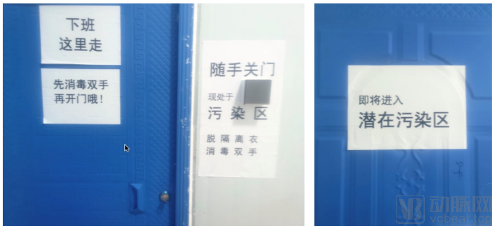
During the pandemic, infection prevention and control measures in radiology departments must be rigorously implemented. The Department of Radiology at Zhongnan Hospital has consistently adhered to regulations governing personal protective equipment management, zonal barrier protection, area-specific containment protocols, humane shift scheduling, comprehensive disinfection practices, strict waste management, and evidence-based emergency response.
In terms of equipment layout, the Radiology Department has designated one CT scanner exclusively for patients from the fever clinic, suspected cases, or confirmed cases, while keeping another CT scanner on standby. Regarding environmental modifications, the central air conditioning supply system was shut down and air outlets were sealed to prevent nosocomial cross-infection; human-machine coexistent air disinfectors were installed in both the operator control rooms and examination rooms. For cleaning and disinfection, the Radiology Department implemented a scheduled and assigned personnel management system for equipment and environment, requiring documented signatures, and specified the types, concentrations, and application methods of disinfectants.
Furthermore, the Department of Radiology standardized entry and exit routes, with clearly marked pathways for physicians and patients, and mandated strict adherence to these designated routes. These measures collectively contributed to Zhongnan Hospital’s achievement of “zero infections.”
The Department of Radiology at Wuhan Union Hospital also shouldered significant responsibilities, managing operations across six fronts: the main campus, Xiyuan Campus, Jianghan Fangcang Hospital, the Cancer Center, and the Jianghan Development Zone Fangcang Hospital, overseeing nearly 4,000 beds. According to Professor Zheng Chuansheng, Head of the Department, during peak periods, the department conducted 600–800 imaging examinations per day, completing over 30,000 CT and DR scans within two months.
Upon detection of the outbreak, the Department of Radiology at Wuhan Union Hospital promptly established a Leading Group for Combating Novel Coronavirus Pneumonia (COVID-19), headed by the department director. The group comprised seven subcommittees—Infection Prevention and Control, Logistics Support, Medical Diagnosis, Medical Technology, Scientific Research, Publicity and Media Relations, and Education—which collaborated through clear division of labor and coordinated efforts to jointly combat the epidemic.
Furthermore, the radiology department has refined staff scheduling management by implementing a unified roster for all physicians, technicians, and nurses, regardless of their subspecialty groups. Technicians adhere to a daily rotation system to prevent fatigue and alleviate psychological stress. Work areas and equipment are managed through zoning, with CT and DR equipment areas designated as either clean or contaminated zones, each governed by specific regulations. Separate pathways have been designed for patients and healthcare personnel, and rigorous disinfection protocols are strictly enforced.
Meanwhile, the Department of Radiology at Wuhan Union Hospital also emphasized strengthening clinical summary research, taking the lead globally in publishing “Preliminary Study on Imaging Features of 19-nCov Pneumonia in Wuhan,” thereby paving the way for research on COVID-19.
The development of a radiology department falls under departmental construction, whereas hospital-level development is a more formidable task. During the online lecture series, Zhu Wenzhen, Vice President of Tongji Hospital Affiliated to Huazhong University of Science and Technology, and Liu Yulin, Vice President of Hubei Cancer Hospital, shared their insights on hospital-level development, respectively.
Vice President Zhu Wenzhen stated: In early January 2020, Tongji Hospital convened a special meeting on medical operations, established leadership and working groups, activated mechanisms for fever clinics and infectious disease prevention and control, and set up the temporary Party branch of the nation’s first fever clinic. The hospital made every effort to manage public opinion. It held meetings of the Standing Committee of the Party Committee to strengthen Party-led decision-making under emergency conditions, with the president providing overall command and leadership team members assuming divided responsibilities.
The fever clinic at Tongji Hospital expanded from an initial 110 square meters to over 5,000 square meters, growing from one ward to three wards—a 50-fold increase. Within 15 days, the entire hospital campus renovated and put into use 77,300 square meters of space, opening 2,025 critical care beds. Nearly 4,000 staff members worked tirelessly for almost two months. The number of available beds, the volume of critically ill patients, the patient visits to the fever clinic, and the volume of online consultations provided by the virtual fever clinic all ranked highest in Wuhan.
How to Improve the Quality and Safety of Critical Care for Severe Cardiopulmonary Cases? How to Unite 35 National Medical Teams into a Cohesive Clinical Force? How to Ensure Logistical Support? How to Effectively Prevent and Control Hospital-Acquired Infections to Protect Healthcare Workers from Infection? These are the four major challenges facing Tongji Hospital. Tongji Hospital has adopted three strategies: First, integrated management of homogeneous medical care across multiple campuses and medical teams. Second, close integration of logistical support with clinical operations. Third, strengthened prevention and control of hospital-acquired infections to ensure the safety of healthcare workers.
Through the collective efforts of the entire hospital, Tongji Hospital has significantly improved its critical care survival rate, with the mortality rate decreasing from an initial 5% to 3%, and currently falling below 2%. This experience can be summarized in five key points: 1. Efficient and robust organizational management. 2. Scientifically rigorous system development. 3. Meticulous and comprehensive patient monitoring. 4. Fully coordinated multidisciplinary collaboration. 5. Standardized holistic nursing care. These critical care management experiences serve as the best testament and reflection of Tongji Hospital’s commitment to applying its research and expertise for the benefit of the nation.
Regarding the Fangcang shelter hospitals, Vice President Liu Yulin provided a comprehensive introduction.
After entering February, Wuhan's medical demand surged into an explosive phase, with more than 1,000 new cases reported daily. The increase in available beds at designated medical institutions in Wuhan fell far short of keeping pace with the rapid growth in patient numbers.
As the pandemic progressed, the bed capacity of designated hospitals could not be rapidly expanded; at this point, the construction of makeshift hospitals began to compensate for the shortage of beds in designated medical institutions.
The advantage of mobile cabin hospitals lies in their rapid construction cycle; a large-capacity facility can be built within days. Furthermore, concentrating patients with the same infectious disease reduces the risk of cross-infection.
After the construction of a cabin hospital is completed, a critical step involves designing the imaging workflow. This process must take into account factors such as equipment room location and access routes, as well as the design of RIS and PACS systems. Regarding equipment configuration, a CT scanner with at least 16 slices is generally required.
Overall, throughout the prevention and control of COVID-19, Fangcang shelter hospitals served as a vessel of life, safeguarding the health and lives of many patients. Within this system, CT equipment and information technology solutions acted as the engine, enabling us to better navigate this life-saving vessel and leading us to victory in the fight against COVID-19.
As stated by Vice President Liu Yulin, the fight against the epidemic in makeshift hospitals relies heavily on technological power. It is precisely with the support of technologies such as artificial intelligence and mobile CT scanners that doctors are able to perform their duties more accurately and systematically under heavy pressure.
During the pandemic, MinFound Medical was among the first teams to provide emergency support in Wuhan. Before the Lantern Festival, the first domestically produced Ark CT scanner had already arrived at the Wuhan Fangcang Shelter Hospital.
Yuan Jianhua, Director of the Clinical Application Center at MinFound Medical Systems, stated in the Cloud Classroom that MinFound’s mobile CT units have been adapted to meet the specific conditions of epidemic areas. Equipped with a 16-slice CT scanner and featuring independent isolation and a mobile design, these units can be deployed for outdoor (emergency) applications in hospital epidemic prevention and control, makeshift hospitals, and communities. They address clinical needs such as the shortage of fixed CT scanners, the inability to rapidly establish protected CT rooms in emergency settings, and the requirement for isolated use. In practice, each mobile CT unit can diagnose nearly 200 patients per day at full capacity.
By enhancing the mobility of CT equipment and leveraging the remote intelligent image interpretation capabilities of its imaging platform, MinFound Medical Systems can help Wuhan rapidly establish a temporary hospital with diagnostic and treatment capabilities equivalent to those of Grade A tertiary hospitals in epidemic areas, thereby alleviating the shortage of medical resources and radiology specialists in Wuhan.
Now, the battle against the epidemic has finally seen a glimmer of hope. As the roads leading to Wuhan gradually reopen, this heroic city, which stood at the epicenter of the outbreak, will slowly regain its former glory. Although the shadow of the pandemic will eventually dissipate, we must always remember the doctors and their colleagues who stepped forward to fight the disease throughout this journey.