How Pocket-Sized Ultrasound Is Empowering Interventional Radiology: Insights from a Leading Chinese Hospital
The Cancer Hospital of the University of Chinese Academy of Sciences (Zhejiang Cancer Hospital), founded in 1963, is one of the first four cancer hospitals established after the founding of the People's Republic of China. Nestled at the foot of mountains, the hospital is flanked by lush greenery on both sides.
As the academic leader of the Interventional Radiology Department at the Cancer Hospital Affiliated to the University of Chinese Academy of Sciences, Professor Shao Guoliang, who also serves as Vice President of the hospital, Chief Physician, and holds a Medical Doctorate, is a renowned expert in the field of interventional medicine across China. He currently chairs the Interventional Medicine Branch of the Zhejiang Medical Association and spearheads the development and capacity building of the interventional medicine community in Zhejiang Province.
Imaging data is lifeless, yet it can save lives through the expertise of physicians. Interventional radiology is a diagnostic and therapeutic system closely integrated with imaging diagnostics. Professor Shao Guoliang, who has long kept abreast of advancements in imaging technologies both domestically and internationally, recognized the potential of handheld ultrasound devices at an early stage.
At an academic conference, Professor Shao Guoliang visited the handheld ultrasound booth of Chengdu Stork Healthcare Co., Ltd. This handheld ultrasound device, capable of delivering image quality comparable to that of mid- to low-end large-scale ultrasound systems, caught his attention.
Subsequently, this handheld ultrasound product entered a trial phase in the Department of Interventional Radiology at the Cancer Hospital of the University of Chinese Academy of Sciences. Previously, ultrasound equipment was mostly confined to dedicated ultrasound departments; now, it can be carried in clinicians’ white coats for on-demand use.
Handheld ultrasound devices, with their compact size, wireless connectivity, and high-quality imaging, are ideally suited for scenarios such as guided puncture, interventional procedures, and point-of-care diagnostics. Serving as a handheld “GPS” for physicians, they have gained widespread recognition and favor among medical staff since their introduction into clinical departments.
Why Did the Department of Interventional Radiology at the Cancer Hospital of the University of Chinese Academy of Sciences Dare to Be the First Mover? What Role Can Handheld Ultrasound Truly Play in Clinical Practice? VCBeat Interviewed Professor Shao Guoliang.
At its inception, handheld ultrasound aimed to equip more primary care clinics with low-cost diagnostic tools through miniaturized devices. In clinical settings, handheld ultrasound holds significant potential across departments such as interventional medicine, nursing, anesthesiology, and cardiology. For procedures like interventions and venipuncture in various clinical departments, ultrasound guidance with handheld devices can improve success rates. Additionally, it enables clinicians to rapidly assess conditions such as pleural and ascitic fluid, lung pathology, blood volume status, and pericardial effusion, facilitating swift clinical decision-making.
How Can Handheld Ultrasound Move Beyond the Ultrasound Department and Be Truly Implemented in More Scenarios? The Interventional Radiology Department Serves as an Ideal Entry Point.
Interventional radiology is a diagnostic and therapeutic system closely integrated with imaging diagnosis, characterized by its distinct disciplinary feature of combining imaging diagnosis with minimally invasive treatment.
Interventional radiology refers to the science of diagnosing and treating diseases using interventional devices such as puncture needles, guidewires, and catheters under the guidance of medical imaging equipment. It is recognized as the third major diagnostic and therapeutic system in modern clinical medicine, following internal medicine and surgery. Interventional radiology offers advantages such as minimal invasiveness, high efficiency, convenience, safety, and strong repeatability, thereby transforming traditional internal medicine and surgical treatment models in many aspects. Compared with other clinical specialties, interventional radiologists possess greater expertise in imaging diagnosis.
As a leading center for the development of interventional medicine in Zhejiang Province, the Department of Interventional Radiology at the Cancer Hospital of the University of Chinese Academy of Sciences has consistently championed the concept of "comprehensive intervention" under the leadership of Professor Sha Guoliang.
The so-called "Grand Interventional" concept refers to the rapid proliferation of various interventional technologies, driven by advancements in modern technology and the transformation of healthcare service models. Beyond traditional medical imaging, interventional techniques across multiple disciplines—including digestive endoscopy, neurology, emergency medicine, and ultrasonology—have witnessed swift development across diverse categories and fields. By integrating multiple imaging modalities and fostering multidisciplinary collaboration, this approach delivers advanced, minimally invasive interventional therapies to a broad patient population. This constitutes the "Grand Interventional" philosophy long advocated by Professor Guoliang Shao.
The introduction of handheld ultrasound devices, in combination with angiography equipment such as DSA, has enhanced the independent operational capabilities of interventional radiologists.
Professor Shao Guoliang used an analogy: “Handheld ultrasound devices are like handguns—simple yet practical. For us, having an extra handgun would solve many problems.”
In point-of-care diagnostic scenarios, Professor Guoliang Shao stated, “Handheld ultrasound devices are compact enough to fit easily into a white coat pocket. I carry one with me during ward rounds. For patients complaining of abdominal distension, I can immediately perform a quick ultrasound examination to check for ascites. For patients with bilateral foot swelling, I can use the handheld ultrasound to determine on the spot whether deep vein thrombosis in the lower extremities is present. In patients with liver cancer, I can use it to roughly assess the size and location of lesions. With handheld ultrasound, I can make judgments about some common changes in clinical conditions without needing to send patients immediately to queue up in the ultrasound department.”
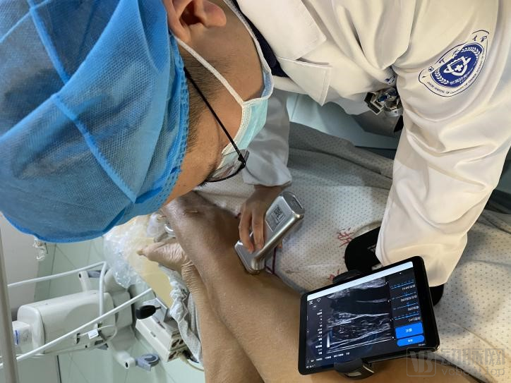
Deep Vein Examination of the Patient
During interventional puncture guidance, physicians can utilize handheld ultrasound to guide procedures such as aspiration, puncture, and catheterization. Blind puncture requires extensive clinical experience to perform successfully and carries inherent risks. Precise puncture under handheld ultrasound guidance helps avoid multiple needle insertions. Compared with X-ray-guided puncture, ultrasound-guided puncture eliminates radiation exposure risks for both physicians and patients.
After multiple clinical trials, Professor Shao Guoliang also found that the quality issues he was most concerned about, such as the image resolution and penetration of handheld ultrasound devices, did not occur.
Stork’s handheld ultrasound system, powered by GPU architecture and harmonic imaging, delivers smooth, high-resolution images with virtually no latency or lag. During needle-guided procedures, the ultrasound visualization of the needle is nearly synchronous, significantly improving procedural success rates.
"Meanwhile, equipped with specialized chips and algorithms, it not only enables imaging on tablets or smartphones but also allows for arbitrary image zooming while maintaining clear image quality. It even supports screen mirroring, ensuring real-time, distortion-free image transmission, thereby eliminating the need for an assistant to hold the tablet at all times."
Whenever a new technology is introduced into a clinical department, it inevitably faces initial skepticism. For physicians, who already contend with heavy workloads, an excessively long learning curve for new technologies can lead to resistance. However, the adaptation period for medical staff in the interventional radiology department to integrate handheld ultrasound devices is relatively short.
“I mastered the operation of the handheld ultrasound device within a single day. Additionally, the device features a built-in battery that supports up to 4.5 hours of continuous use, and it comes with a wireless charging dock for on-demand power, eliminating the risk of battery depletion during procedures. The handheld ultrasound connects wirelessly, freeing users from the constraints of cables and facilitating surgical operations; it can be used for scanning immediately after being covered with a sterile sheath.” The head nurse of the operating room expressed particular satisfaction with this feature.
“With so many advantages of handheld ultrasound, do the large machines in the ultrasound department seem redundant? Certainly not. As a convenient tool for initial screening, handheld ultrasound is efficient and saves time for both patients and physicians. However, for complex or challenging cases, further examination with the advanced equipment in the ultrasound department is still necessary.”
For primary healthcare institutions, there is a shortage not only of senior physicians but also of efficient and convenient medical equipment. Professor Shao stated, “To my knowledge, handheld ultrasound devices can share high-definition images in real time remotely, facilitating expert guidance. Some doctors and nurses without a background in imaging can transmit images to me in real time via their mobile phones when working at the grassroots level, allowing me to guide them in performing certain interventional procedures.”
In the near future, Stork’s handheld ultrasound devices can leverage 5G remote technology to establish a telemedicine platform, regularly inviting relevant experts to conduct teaching and training sessions. This initiative will deliver valuable expertise to primary-care hospitals, thereby maximizing the value of handheld ultrasound technology.