Deepwise AI-Powered CT Analysis Links Upper Lung Consolidation to Adverse Outcomes in COVID-19 Patients, Study Published in Theranostics

DeepWise
Developer of Artificial Intelligence Medical Imaging Diagnosis System
Due to the distinct radiological manifestations of COVID-19 and the wide availability of imaging, medical imaging modalities such as CT and X-ray have played a pivotal role during the pandemic. To date, major hospitals worldwide have accumulated extensive datasets of COVID-19 CT images.
Relevant retrospective studies are also ongoing. Many artificial intelligence companies have developed algorithms with a certain degree of interpretability, which can assess the probability of patients contracting COVID-19 by analyzing medical images and even highlight the specific image regions influencing the algorithm’s decisions. These studies have, to some extent, advanced the prevention and treatment of COVID-19.
However, the factors associated with adverse clinical outcomes remain unclear. For patients confirmed to be infected with COVID-19, how should physicians make decisions regarding subsequent treatment based on the numerous observational indicators?
Recently, a paper titled “Multicenter cohort study demonstrates more consolidation in upper lungs on initial CT increases the risk of adverse clinical outcome in COVID-19 patients,” which explores the relationship between the composition, quantity, distribution of pneumonia and adverse clinical outcomes, was accepted by Theranostics (Impact Factor: 8.063). This study was jointly conducted by the team led by Director Ju Shenghong from the Department of Radiology at Zhongda Hospital Southeast University and DeepWise Research Institute.
This study employs deep learning to identify ground-glass opacities and consolidations in CT images of COVID-19 patients, calculates various quantitative metrics, and investigates the association between these metrics and clinical outcomes.
The results indicate that older patients and those with extensive consolidation in the upper lungs have a higher probability of adverse clinical outcomes. Therefore, healthcare providers should pay closer attention to patients with these characteristics and initiate treatment more promptly.
Given the rapid progression of severe COVID-19 cases, a retrospective study published in *The Lancet* reported that 61.5% of critically ill patients died within 28 days. Therefore, for patients with COVID-19, if we can predict clinical outcomes in advance based on pulmonary imaging and implement corresponding interventions, this would significantly improve survival rates and prognosis.
Professor Ju Shenghong, a renowned radiologist, recipient of the National Science Fund for Distinguished Young Scholars, and a leading talent in technological innovation under the National “Ten Thousand Talents Program,” who serves as the corresponding author of the paper, stated, “This study provides a scientifically grounded risk assessment for the clinical outcomes of COVID-19 patients. It holds significant clinical value for guiding treatment planning, optimizing the allocation of medical resources, and issuing early warnings of treatment risks for high-risk patients during subsequent care.”
This study collected data from 625 patients with laboratory-confirmed COVID-19 across 24 hospitals in Jiangsu Province. After screening, patients who did not undergo CT scans and those under 18 years of age were excluded, resulting in a final sample size of n=421. Specifically, the median age of the sample was 48 years, with males accounting for 53% of the cohort. Among these patients, 64 cases reached the clinical composite endpoint (including admission to the intensive care unit, acute respiratory failure, and shock during hospitalization).
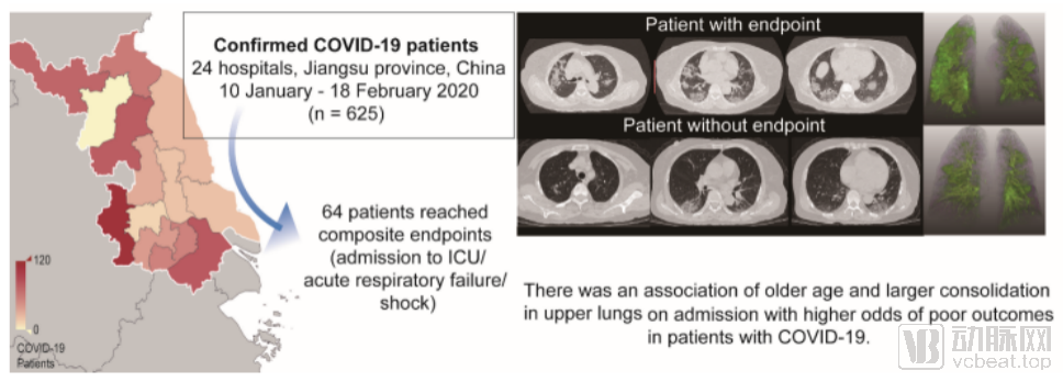
Figure 1. Inclusion criteria and conclusions of this study
Researchers first processed the CT images of each case using DeepWise’s Intelligent Solution for Lung Diseases (COVID-19 Enhanced Version) to detect and segment pneumonia lesions as well as perform lung lobe segmentation. The AI-generated detection and segmentation results were then submitted to senior experts for review. Throughout this process, researchers could quantitatively extract lesion features such as volume, density, and location.
Subsequently, using a multivariable logistic regression model, researchers can identify which variables are risk factors for the composite clinical endpoint. Experimental validation confirmed that age, the volume of consolidation in the upper lungs, and the presence of lesions in the lower lungs near the pleura were all associated with the clinical endpoint. Specifically, older age and a greater volume of consolidation in the upper lungs were more likely to lead to adverse clinical outcomes.
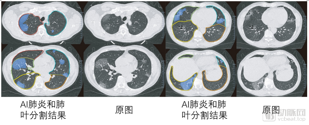
Figure 2. Schematic diagram of deep learning-based pneumonia segmentation and lung lobe segmentation results
Overall, the significance of this study can be summarized in three points: First, it moves beyond the qualitative descriptions of previous studies by quantitatively elucidating the relationship between the composition, volume, and distribution of pneumonia and adverse clinical outcomes. Second, it is the first study to quantitatively demonstrate the correlation between upper lobe consolidation and adverse clinical outcomes in patients with COVID-19. Third, it highlights the positive role of precision AI in pneumonia-related research.
“As one of the authors of this paper, Professor Yu Yizhou, Chief Scientist at DeepWise, stated: ‘This study demonstrates the potential of artificial intelligence. This technology can not only identify lesions and perform qualitative and quantitative analyses but also reveal the correlation between analytical results and clinical outcomes, holding practical value in areas such as COVID-19 risk assessment and treatment decision-making.’”
As of April 30, confirmed cases had been reported in more than 200 countries and regions worldwide, with the total number of infections exceeding 3.19 million. Over 60 countries had declared a state of emergency, and Asia, Europe, and the Americas had successively become hard-hit areas. The outbreak in the United States was particularly severe, with cumulative confirmed cases surpassing 1 million and deaths exceeding 50,000.
Against the backdrop of global collaborative efforts to combat the pandemic, China’s experience in epidemic control offers valuable lessons. In response to the severe epidemic situation in China, the National Health Commission issued the *Diagnosis and Treatment Protocol for Novel Coronavirus Pneumonia (Trial Version 5)* as early as the end of the Spring Festival holiday, adjusting its strategy by incorporating CT imaging findings into the diagnostic criteria for clinically diagnosed cases.
At different stages of the pandemic, the role of CT has been evolving. In the sixth and seventh editions of the diagnosis and treatment guidelines, in addition to listing CT imaging as a key basis for differential diagnosis of COVID-19, it also provides support for staging pulmonary inflammation, assessing disease severity, evaluating treatment efficacy, and guiding decisions on hospital admission, entry into makeshift hospitals, discharge, and exit from such facilities.
The empowering role of AI in CT equipment has also been confirmed by frontline hospitals during the pandemic. This technology can automatically and accurately detect pulmonary opacities that are not easily visible to the naked eye in the early stages of the disease. After admission, it enables rapid quantitative analysis of lesions through multiple follow-up scans, helping to determine the nature and progression of the lesions, assess disease severity, and effectively improve diagnostic efficiency and accuracy.
Nowadays, Fellows of the American Society for Radiation Oncology have also publicly pointed out that it is urgent to use CT as the diagnostic standard for novel coronavirus pneumonia. Undoubtedly, “CT + AI” will continue to play an important role in the ongoing global fight against the epidemic. Therefore, as the global battle against the pandemic enters a critical and intense phase, such clinically significant scientific achievements are undoubtedly a valuable technological force for global epidemic prevention and control.
Taking DeepWise’s enhanced COVID-19 solution as an example, in addition to screening suspected cases, physicians can also use the software for follow-up monitoring of severe and critically ill patients. By leveraging follow-up data to observe disease progression, assess the stage and severity of the condition, and accumulate clinical data, this information can be utilized for future scientific research on infectious diseases.
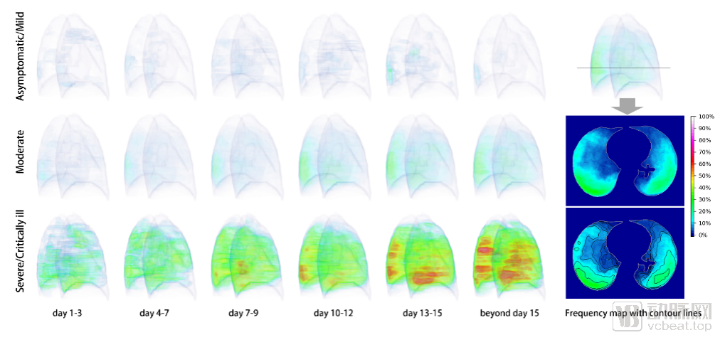
Figure 3. Dynamic progression of pneumonia on CT images in COVID-19 patients from symptom onset to beyond day 15, as revealed in the European Radiology article
In addition to this paper, another collaborative achievement by Professor Ju Shenghong’s team and DeepWise, titled “Dynamic evolution of COVID-19 on chest computed tomography: experience from Jiangsu province of China,” has also been accepted by the prestigious European journal European Radiology (Impact Factor: 3.962). This study elucidates the evolutionary patterns of lesion volume, density, and location in COVID-19 pneumonia, providing significant value for clinical dynamic follow-up.
As of press time, DeepWise had an additional eight recent research achievements accepted by the IEEE/CVF Conference on Computer Vision and Pattern Recognition (CVPR 2020) and the IEEE International Symposium on Biomedical Imaging (ISBI 2020), four of which were selected for oral presentations. These papers represent cutting-edge international research progress in the field of computer vision, particularly in medical imaging. Furthermore, as a leading AI healthcare company in China, DeepWise will continue to collaborate with various medical institutions to contribute to the global prevention and control of COVID-19.