Global Medical Imaging Patent Analysis Report: The Midfield Battle of the GPS Triad
According to statistics, over 90% of current medical data originates from medical imaging, making it an indispensable piece of “evidence” for clinical diagnosis. This includes common imaging modalities such as X-ray, CT, MRI, PET, ultrasound, and pathology images.
In recent years, with the rapid advancement of AI technology, artificial intelligence has increasingly enriched its role in assisting diagnostic processes within radiological imaging. For instance, techniques such as image detection, image recognition, model clustering, and model aggregation in AI algorithms have demonstrated greater accuracy and speed in lesion identification and diagnostic decision-making.
To explore the evolving trends and competitive landscape of global medical imaging technology, VCBeat’s VBInsight, in collaboration with Chofan Intellectual Property, has jointly produced the “Global Medical Imaging Patent Analysis Report.” This report focuses on the field of medical imaging, conducting a global search of relevant patents and analyzing patent application distributions as well as key inventors.
Key Points:
1. The United States and China are the two countries with the highest number of medical imaging patent applications, accounting for over 50% of the total;
2. Traditional medical device giants such as Siemens, GE HealthCare, and Philips remain in the first tier in the field of medical imaging;
3. Samsung Group has seen a rapid increase in medical imaging patents since 2010, aiming to create a second semiconductor miracle in the healthcare sector;
4. Over 80% of GE HealthCare’s medical imaging patent applications are filed in the United States;
5. Samsung’s technological distribution is concentrated in the field of image processing, while GE Healthcare focuses on data acquisition and image reconstruction.
1. Global Distribution of Medical Imaging Patents
1.1. IPC Classification of Related Patent Technologies
1.2 Global Patent Application Trends in Medical Imaging Over the Past 20 Years
1.3 National Rankings of Medical Imaging Patent Application Volume
1.4 Patent Application Status of the Top 8 Applicants
2. Interpretation and Analysis of Core Patents Held by Key Applicants
2.1. Samsung Group: Creating a Second Semiconductor Miracle in the Biopharmaceutical Sector
2.2. GE Healthcare: One of the Three Global Giants in Medical Devices
3. Patent operations, litigation, reexamination, and invalidation information for key applicants
3.1. Patent Litigation Status
3.2 Patent Reexamination Status
Statistical analysis of the distribution of technical fields in global patent applications reveals the key areas of focus for domestic and international applicants, as indicated by IPC classification codes (see Table 1). Among the top 10 patents in each technical direction within medical imaging, the main distributions are in G06T7 (image analysis), G06K9 (pattern recognition), A61B6, and A61B5 (diagnostics), accounting for 18.95%, 16.59%, 15.74%, and 14.31%, respectively.
Table 1: Distribution of the Number of Technical Branches
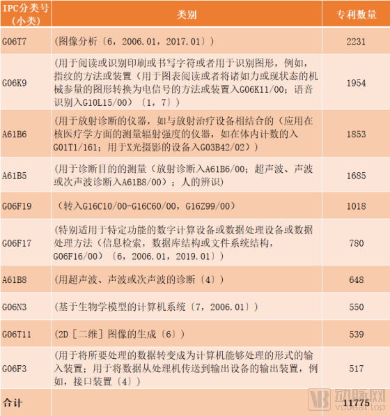
Source: Chofan IP, VCBeat
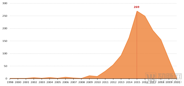
Prior to 2000, the field of medical imaging was in its nascent technological stage, with a relatively small number of patents. Since 2000, driven by technological breakthroughs, research in medical imaging has continuously expanded, leading to a significant increase in annual patent applications. With the rapid development of AI technology in 2014, the field entered a period of accelerated growth, characterized by a surge in related patent filings.
Figure 1: Global Trends in Medical Imaging Patent Applications
(Due to publication delays, 2019 patent data are incomplete.)
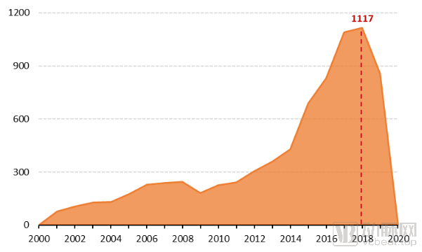
Source: Chofan Intellectual Property, VCBeat
From 2000 to the present, the United States has consistently ranked first in the number of medical imaging patent applications, with a cumulative total of over 3,300 filings as of 2019.
China’s patent filings saw a rapid increase after 2012, rising annually from 50 per year and surpassing the United States for the first time in 2018. This growth was driven by strong government support for the development of hospital radiology departments and policies encouraging the substitution of imported imaging equipment with domestically produced alternatives. United Imaging Healthcare was established in 2011 and gradually emerged as the leading domestic enterprise in high-end medical imaging equipment. In 2013, the Ministry of Health issued the “Notice on Printing and Distributing the Scoring Criteria (Trial) for National Clinical Key Specialty Construction Projects, Including Medical Imaging Departments,” to promote the development of hospital radiology departments across China.
Figure 2: Regional Ranking of the Number of Medical Imaging Patent Applications
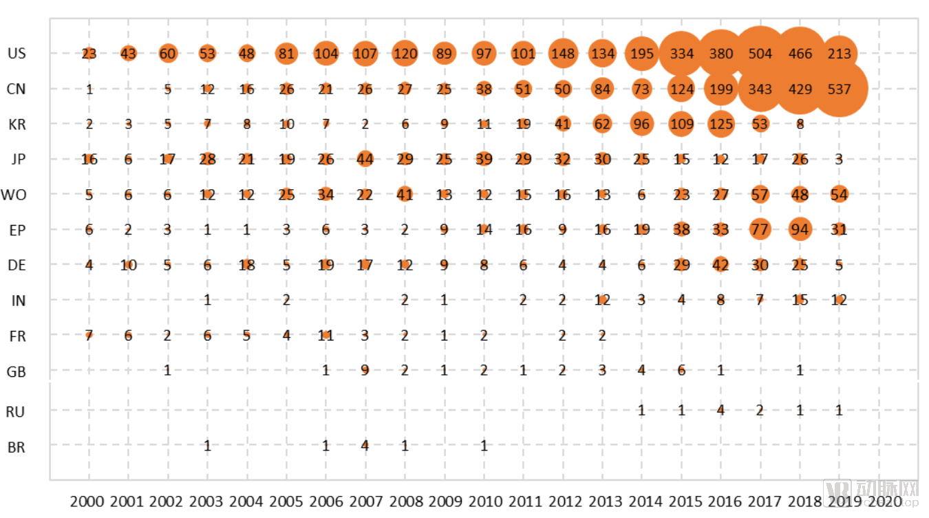
Source: ChaoFan Intellectual Property, VCBeat
In stark contrast to China, South Korea saw an increase in patent applications after 2010, but experienced a precipitous decline in 2017/2018.
As one of South Korea’s largest multinational conglomerates, Samsung is a primary source of medical imaging patent applications in the country. In February 2017, following the arrest of its de facto leader, Lee Jae-yong, by South Korean prosecutors on charges including bribery, Samsung underwent significant management restructuring and strategic shifts. The company refocused on its electronics business while reducing investment in its healthcare sector. This likely constitutes one of the main reasons for the decline in medical imaging patent applications in recent years.
Furthermore, Japan does not hold a significant advantage in the field of medical imaging, with its patent application volume remaining stable over the long term.
Comparisons of innovation strength or influence are often ultimately reflected in the patent application volumes and technological distributions of specific applicants. To this end, an analysis of the distribution of the top eight applicants in this field is presented below.
The top-ranked GPS (G for GE, P for Philips, and S for Siemens Healthineers) are the three giants in the international medical device industry.
Among the "GPS" trio, Philips is the most healthcare-focused company. In 2016, it streamlined its operations by divesting its lighting business to concentrate fully on health technology. Siemens Healthineers went public independently in Germany in March 2018, with Siemens AG retaining an 85% controlling stake. In June 2018, GE announced the spin-off of its healthcare division, establishing GE Healthcare as an independent company.
Siemens was founded earliest among the three companies (in 1847), has accumulated the most expertise in the field of imaging equipment, and ranks first in patents. However, after Samsung acquired Medison, a medical device company, in 2010, it gradually surpassed the two giants, GE Healthcare and Philips, following the transfer of patent rights.
Figure 3: Ranking of Patent Applications by the Top 8 Applicants
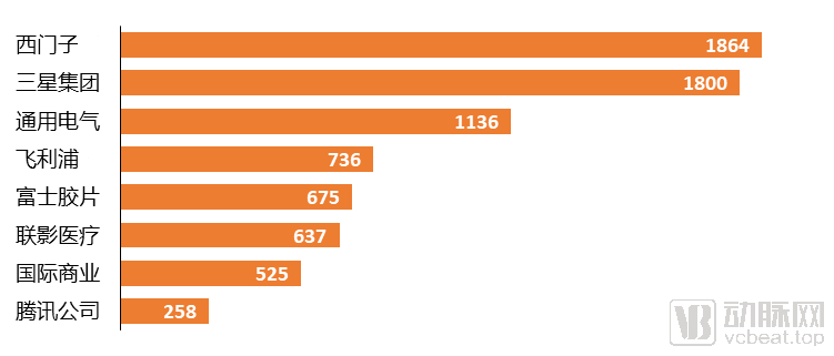
Source: Chofan IP, VCBeat
In addition, two Chinese enterprises, United Imaging Healthcare and Tencent, are also among the top eight in terms of patent application volume. Benefiting from the major trend of import substitution for high-end medical devices and government support, a number of outstanding domestic device companies, such as United Imaging Healthcare, Anke, and Wandong, have emerged in China.
As an internet company, Tencent entered the field of artificial intelligence software and launched Tencent Miying in August 2017, focusing on AI-based early screening for esophageal cancer, lung cancer, and other conditions in medical imaging. Unlike the patent distribution patterns of traditional medical device manufacturers, Tencent Miying’s patents are more concentrated in areas such as image data processing and intelligent decision-making.
As a global leader in consumer electronics, Samsung Group has been increasingly expanding its footprint in the healthcare sector. By partnering with multiple medical institutions and adeptly transferring its construction and operational expertise from the electronics industry to healthcare, Samsung has ultimately developed a unique “Samsung Transformation Logic.”
As early as 2010, South Korea’s Samsung Group announced a $210 billion investment over the following decade in renewable energy and healthcare. It subsequently identified five key growth drivers for its future, including biopharmaceuticals and medical devices. In 2010, Samsung acquired Medison, a South Korean medical device company, to facilitate its entry into the healthcare sector and advance its strategy of diversifying beyond consumer electronics. By integrating mobile devices with mobile services, Samsung has been building a globally leading mobile health company, with continuous business growth in areas such as medical imaging and diagnostics, and medical cloud solutions.
In 2017, NeuroLogica, a medical imaging company under Samsung, partnered with the Israeli artificial intelligence firm MedyMatch to develop an AI platform for the “Mobile Stroke Unit.” By integrating the capabilities of Samsung’s CereTom CT scanner into this device, emergency physicians can rapidly determine whether a patient is suffering from intracranial hemorrhage or thrombosis when using this mobile scanning tool.
As a leader in medical imaging technology, Samsung Group has pursued innovation and in-depth research in ultrasound imaging, digital radiography, computed tomography, and magnetic resonance imaging (MRI). At the Radiological Society of North America 2018 annual meeting (RSNA 2018), it showcased various types of diagnostic imaging software that integrate AI algorithms into imaging equipment to assist physicians in diagnosis. S-Detect™ is an AI-based technology that analyzes breast lesions on ultrasound images, facilitating standardized analysis and classification of suspicious breast lesions. Its AI-powered digital radiography software, SimGrid™, clearly visualizes lung tissue obscured by bones on chest X-rays. The ALND solution is an AI-based computer-aided detection (CAD) solution for automated detection of pulmonary nodules. In the field of MRI, Samsung has leveraged artificial intelligence to develop new software that displays information such as knee cartilage thickness, providing images of affected areas for patients with knee osteoarthritis.
Samsung Group’s Medical Imaging Patents Saw a Rapid Increase After 2010
A global search was conducted on patents in the field of medical imaging held by Samsung Group over the past 20 years. By consolidating patent application numbers, a detailed analysis is presented as follows:
Figure 4: Patent Application Trends of Samsung Group in the Field of Medical Imaging
Source: Chofan Intellectual Property, VCBeat
Samsung’s patent applications in the field of medical imaging have shown an overall upward trend over the past two decades. Prior to 2010, the number of patent applications grew slowly. As attention to the healthcare sector increased, the Samsung Group saw a significant rise in its annual average patent filings compared to the pre-2010 period. Notably, since 2013, the volume of patent applications has experienced rapid, explosive growth.
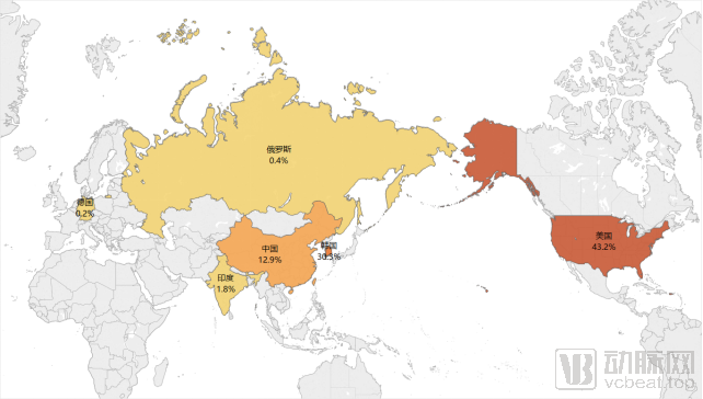
Patent distribution in East Asia and the United States accounts for 86.4%
Figure 5 presents an analysis of Samsung Group’s patent layout in the field of medical imaging, where the intensity of colors in different regions indicates the number of patents related to smart bracelet technology themes in each country/region.
Figure 5: Global Distribution of Samsung Group’s Patents in the Medical Imaging Field
Source: ChaoFan Intellectual Property, VCBeat
It can be seen that patent applications are primarily filed in Asian, European, and North American countries. Among these, the United States has the largest number of patents, accounting for 43.2% of global applications, followed by South Korea and China, with shares of 30.3% and 12.9%, respectively. Furthermore, Samsung Group has filed patents with the European Patent Office, the World Intellectual Property Organization, and in countries such as India, Japan, and Russia. This to some extent reflects Samsung Group’s emphasis on the field of medical imaging.
Samsung Holds the Largest Number of Patents in Image Processing
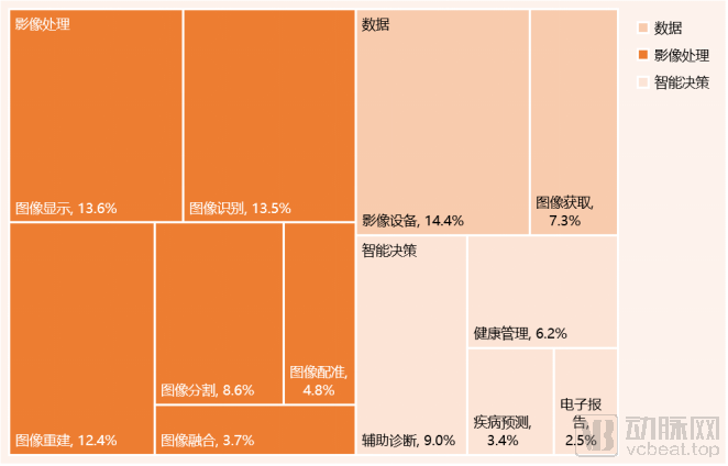
A statistical analysis of the distribution of technical fields in global patent applications reveals the key areas of focus for domestic and foreign applicants. The specific analysis of technological layout is shown in Figure 6.
Figure 6: Samsung Group’s Technological Layout in the Field of Medical Imaging
Source: ChaoFan Intellectual Property, VCBeat
Samsung Group holds the largest number of patents in image processing technology, accounting for 56.8%, covering six technical branches: image segmentation, image registration, image recognition, image fusion, image reconstruction, and image display. Among these, image display, image recognition, and image reconstruction account for a relatively larger proportion.
Underlying data technologies primarily involve image acquisition and imaging equipment technologies, accounting for 7.3% and 14.4%, respectively. In the realm of intelligent decision-making, there are four technological branches: electronic reporting, computer-aided diagnosis, disease prediction, and health management, among which computer-aided diagnosis accounts for the largest share of patents (9%).
Analysis of Core Patents in Samsung Medical Imaging
Core patents refer to patents that hold a pivotal position in a specific technical field, make outstanding contributions to technological development, exert significant influence on other patents or technologies, and possess substantial economic value.
Table 2: Core Patents of Samsung Group in the Field of Medical Imaging
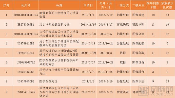
Source: Chofan IP, VCBeat
The following provides an interpretation and analysis of selected core patents from dimensions such as technical problems and technical effects.
1. Title: Method and Apparatus for Measuring Biometric Characteristics of a Subject |
Publication (Announcement) No.: KR1020130080312A |
Claim 1: A method for measuring biometric features of a subject, the method comprising: receiving an image of the subject; simulating the subject to identify at least a portion of the subject; and measuring the biometric features of the subject based on a result of the simulation.
|
Technical Issue: Biometric parameters such as crown-rump length (CRL), nuchal translucency (NT), and intracranial translucency (IT) are measured and reported individually. However, the relative discrepancy between NT or IT and CRL—specifically, values derived from at least two biometric parameters—is utilized to assess fetal status. Therefore, it is necessary to automatically provide users with values calculated based on a comprehensive analysis of biometric parameters like CRL, NT, and IT, along with diagnostic results regarding fetal status derived from these calculated values, thereby enabling early diagnosis and determination of fetal condition.
|
Technical Effect: A method and apparatus for measuring biometric features of a subject are provided, enabling automatic measurement of the subject's biometric features using ultrasound images of the subject.
|
Brief Analysis: This patent enables automated biometric measurement of a subject by receiving image simulations of the subject and processing the simulation results. The patent is currently in force and demonstrates strong technical stability. It has been cited 13 times worldwide, including its patent family members, indicating its technological advancement. With 22 claims and filings in 10 countries and regions—including the United States, China, the European Patent Office, South Korea, and India—the patent offers robust protection scope, reflecting the company’s significant emphasis on this intellectual property asset.
|
2. Title: Devices and Methods for Diagnosis |
Publication (Announcement) Number: EP2733669B1 |
Claim 1: An apparatus, comprising: an analysis unit (110) configured to detect lesion regions from medical images and generate a set of candidate lesion regions relative to the detected lesion regions; and an interface unit (130) configured to arrange and display, in a first area (20) of an interface, the group of candidate lesion regions (21) with brief analytical information based on a priority determined according to predetermined criteria including class information about each candidate lesion region, and to display detailed analytical information about one of the selected candidate lesion regions in a second area (30) of the interface if a user selects one of the candidate lesion regions displayed in the first area (20), wherein the class information is used to indicate the degree of benignity or malignancy of the candidate lesion regions, wherein candidate lesion regions with higher priority in the group of candidate lesion regions are displayed in the first area (20) with at least one aspect relative to the group, the group comprising: being in a more central position; having a larger size; and having a color that is more noticeable than candidate lesion regions with lower priority in the group of candidate lesion regions.
|
Technical Problem: Computer-aided diagnosis (CAD) is a technology used in medicine to assist physicians in interpreting medical images by detecting suspected abnormal regions within the images and analyzing these regions to provide a preliminary diagnosis of lesions. In existing technologies, to analyze the diagnostic results of a CAD system, users must evaluate various images of the abnormal regions from different perspectives. However, such evaluation is constrained by temporal and spatial limitations, making it difficult to analyze and modify the diagnostic results provided by the CAD system through the user interface offered by such systems.
|
Technical Effect: A diagnostic method and apparatus are provided, enabling the diagnosis and analysis of lesion regions.
|
Brief Analysis: This patent detects lesion regions in medical images, generates a set of candidate lesion regions along with their respective priorities relative to the detected lesion areas, and displays the group of candidate lesion regions with concise analytical information to achieve diagnostic analysis of pathological regions. The patent is currently in force and demonstrates good technical stability. It has been cited 19 times globally, including its patent family, indicating strong technological advancement. The patent has been filed in five countries and regions—the United States, China, the European Patent Office, Japan, and South Korea—providing robust scope of protection.
|
3. Title: Method for Extracting Regions of Interest from Tongue Images, and Corresponding Health Monitoring Method and Device |
Publication (Announcement) No.: KR1020040059312A |
Claim 1: A method for extracting a region of interest from a tongue image, the method comprising: constructing a database storing template images, wherein each template image corresponds to personal information and displays a region of interest; segmenting a tongue region from a tongue image obtained from a subject whose health status is to be determined; matching the tongue region with the template images stored in the database; and extracting the region of interest from the matched template image.
|
Technical Issue: In the prior art, devices for detecting the early stages of disease onset and progression based on tongue conditions—such as tongue coating, color, or appearance—are prohibitively expensive and are predominantly available only in hospital settings. Similarly, conventional devices are unsuitable for personal health monitoring by individuals.
|
Technical Effect: Achieves health monitoring by extracting a region of interest from tongue images and analyzing at least one characteristic factor from the extracted region of interest.
|
Brief Analysis: This patent enables health monitoring by extracting regions of interest from tongue images and analyzing at least one characteristic factor from these extracted regions. The patent is currently in force and demonstrates strong technical stability. It, along with its patent family, has been cited 87 times worldwide, indicating a high level of technological advancement. The patent has been filed in five countries and regions—the United States, China, the European Patent Office, Japan, and South Korea—providing robust scope of protection.
|
4. Title: Apparatus and Method for Automatic Landmark Registration in Three-Dimensional Medical Images |
Publication (Announcement) Number: US9799115B2 |
Claim 1: A method for automatically registering landmarks in a three-dimensional (3D) medical image of an object, the method comprising: obtaining the 3D image; determining a set of search points based on a statistical atlas attached to a bounding box corresponding to a portion of the 3D image, wherein the statistical atlas includes information indicating a statistical distribution of previously identified landmarks in the object; extracting features from the determined set of search points; forming a set of landmark candidates based on the extracted features; filtering the candidates and outputting remaining candidates from among the candidates based on the filtering; and outputting a final position of the landmark based on one of the remaining candidates.
|
Technical Challenges: Most registration algorithms are designed to work exclusively with two-dimensional medical images, limiting their applicability to specific domains. Some methods can only process high-resolution, high-quality medical images, which require prolonged acquisition times. In practice, feature extraction algorithms in these approaches are typically employed to address classification tasks. Generally, these feature extraction algorithms are manually tuned based on empirical knowledge. In certain scenarios, generic feature extraction models are trained without accounting for the specific characteristics of the data being used, thereby failing to fully leverage the potential of machine learning methods. Furthermore, most approaches construct only a single level of features rather than establishing a multi-level hierarchical feature representation. In some methods, candidate landmarks are filtered using corresponding thresholds; however, these thresholds are also manually adjusted as rule-based parameters.
|
Technical Effect: Provided is an improved method for automatically registering landmarks in three-dimensional (3D) medical images, which allows for the detection of the positions of unique points within the constraints specific to each type of such points, as well as the positions of multiple points for each of these types.
|
Brief Analysis: This patent is currently in force and exhibits strong technical stability. It and its patent family have been cited five times globally, indicating a high level of technological advancement. The patent has been filed in five jurisdictions—the United States, the World Intellectual Property Organization (WIPO), the European Patent Office (EPO), Russia, and South Korea—providing robust scope of protection.
|
5. Title: Ultrasound Imaging Method and Device Based on Pulse Compression Technology Using Improved Golay Codes |
Publication (Announcement) Number: FR2804516A1 |
Claim 1: An ultrasound imaging method for forming an image of an object using signals reflected from the object after transmitting ultrasound pulses to the object, the method comprising the steps of: (a) transmitting a first set of ultrasound pulses to the object by applying voltages to one or more transducers (1) according to a first code of a pair of modified first Golay codes; (b) performing pulse compression on a first set of reflected signals of the first set of ultrasound pulses reflected from the object; (c) transmitting a second set of ultrasound pulses toward the object to the one or more transducers (1) by means of applied voltages according to a second code of the pair of modified Golay codes; (d) performing pulse compression on a second set of reflected signals of the second set of ultrasound pulses reflected from the object; (e) summing the compressed pulses and signals of the first and second sets of reflected signals; (f) generating a signal after receiving focusing using the summed signal to form an image of the object; and (g) displaying the image.
|
Technical Issue: Due to the sidelobe suppression characteristics of Golay codes, efforts have been made to apply them in long-pulse ultrasound imaging systems. However, one undesirable frequency characteristic of Golay codes is their broader spectrum compared to conventional ultrasound transducers. This results in some power loss of the Golay code on the ultrasound transducer, preventing the system's signal-to-noise ratio (SNR) from reaching the desired level.
|
Technical Effect: To provide codes whose frequency characteristics match those of an ultrasonic transducer, and an ultrasonic imaging method based on pulse compression technology using these codes.
|
Brief Analysis: This patent and its patent family have been cited 57 times worldwide, indicating strong technological advancement. The patent has been filed in eight countries and regions, including the United States, Germany, France, the European Patent Office, Japan, and South Korea, demonstrating broad scope of protection and reflecting the company’s high level of attention to this patent.
|
General Electric (GE), headquartered in Boston, United States, is the world’s largest manufacturer of electrical and electronic equipment and the world’s largest diversified services company. Its operations span a wide range of sectors, from aircraft engines and power generation equipment to financial services, and from medical imaging and television programming to plastics.
GE Healthcare, a subsidiary of General Electric Company, is a global leader in medical imaging, patient monitoring, biomanufacturing, and gene therapy technologies. By providing intelligent devices, data analytics, software applications, and services, it enables comprehensive precision healthcare spanning disease diagnosis, treatment, and monitoring, delivering innovative medical technologies and services worldwide.
Medical Imaging Patents Began to Experience Explosive Growth in 2017
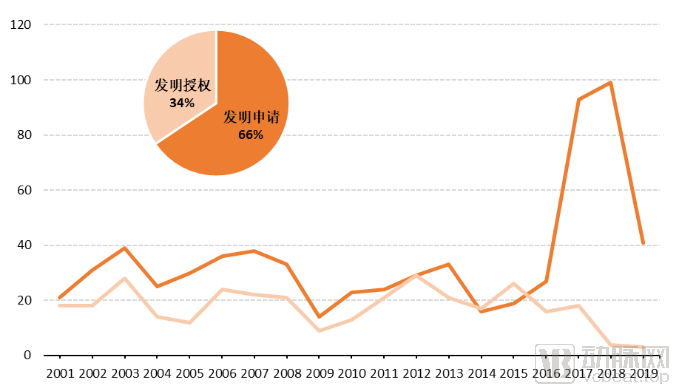
Figure 7 illustrates the global patent application trends and distribution of patent types for GE Healthcare in the field of medical imaging since 2000. In terms of application trends, GE Healthcare has filed a total of 1,050 patents worldwide since 2000, including 689 published invention applications and 361 granted inventions.
Figure 7: Global Medical Imaging Patent Application Trends of GE Healthcare (Unit: Number of Applications)
Source: Chofan Intellectual Property, VCBeat
Since 2000, its patent application volume has gradually increased, surging exponentially in 2017. Due to the 18-month lag in patent publication, the 2019 patent disclosure data is incomplete. Overall, GE Healthcare’s patent applications in the field of medical imaging are on the rise.
Over 80% of GE Healthcare’s Patent Applications Are Distributed in the United States
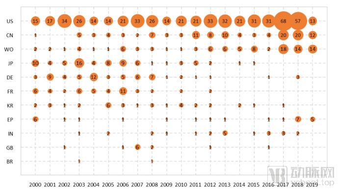
Figure 8 presents an analysis of GE Healthcare’s patent filings by country since 2000. As shown in the figure, GE Healthcare’s patents in the field of medical imaging are primarily filed in the United States, China, and under the Patent Cooperation Treaty (PCT/WO), with filing counts of 542, 123, and 101, respectively.
Figure 8: Analysis of Patent Layout by Country
Source: ChaoFan Intellectual Property, VCBeat
GE Healthcare began its operations in China in 1979 and established its first office in Beijing in 1986. In 1991, Hangwei GE Medical Systems Co., Ltd. was founded in Beijing, becoming GE’s first joint venture in China; thus, the company places significant emphasis on its strategic presence in the Chinese market. Additionally, GE Healthcare has secured patent protections in eight other countries and regions, including Japan, Germany, France, and South Korea.
Key areas of technological layout are distributed across data acquisition and image reconstruction.
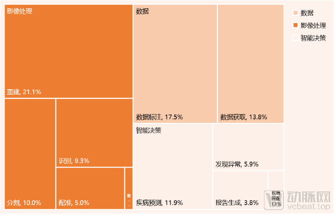
Figure 2-6 illustrates GE Healthcare’s technological distribution in the field of medical imaging, primarily covering data, image processing, and intelligent decision-making. Among these, image processing has the largest patent portfolio, accounting for 46.1%, while data processing and intelligent decision-making account for 31.3% and 22.6%, respectively.
Figure 9: Distribution of GE Healthcare’s Medical Imaging Technologies
Source: Chofan IP, VCBeat
In the field of image processing, GE Healthcare has the most extensive patent portfolio in image reconstruction, accounting for 21.13%. This is followed by segmentation at 10.04%, recognition at 9.31%, registration at 5.02%, and fusion at 0.63%.
In the field of data processing, data annotation and data acquisition account for 17.47% and 13.81%, respectively.
In the field of intelligent decision-making, its overall proportion is lower than that in the fields of image processing and data processing, indicating that its technological capabilities in intelligent decision-making are relatively weak. Within the intelligent decision-making sector, disease prediction accounts for the largest share of patent layouts at 11.92%, while anomaly detection, report generation, and disease screening account for 5.86%, 3.77%, and 1.05%, respectively.
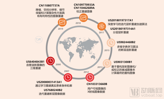
Figure 10: Technical Roadmap for Image Reconstruction Directions
Source: ChaoFan Intellectual Property, VCBeat
As shown in Figure 10, the technology roadmap for image reconstruction represents the field with the largest number of patent filings. In 2000, three-dimensional reconstruction based on angiographic images was proposed, utilizing iterative tomographic images and contrast-enhanced images for image reconstruction. After 2010, the focus shifted primarily to correction methods for reconstructed images. In 2019, image reconstruction techniques were introduced that offer advantageous characteristics in terms of noise reduction, spatial resolution, detail preservation, and computational complexity.
Analysis of Core Patents in Medical Imaging at GE Healthcare
Core patents are those that hold a pivotal position in a specific technical field, make outstanding contributions to technological advancement, exert significant influence on other patents or technologies, and possess substantial economic value. This section identifies GE Healthcare’s core patents in the field of medical imaging based on criteria such as patent citation frequency, family size, and technological importance.
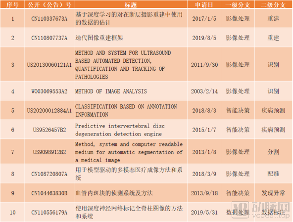
Table 3: Core Patents of GE Healthcare in the Field of Medical Imaging
Source: ChaoFan Intellectual Property, VCBeat
The following section provides an interpretation and analysis of selected core patents from dimensions such as technical problems and technical effects.
1. Title: Deep Learning-Based Estimation of Data Used in Tomographic Reconstruction |
Publication (Announcement) No.: CN110337673A |
Technical Branch: Image Processing Technology Field, Reconstruction Technology Direction
|
Technical Issue: Due to various reasons, a portion of the projection data for a given scan may be corrupted or missing (relative to an ideal or mathematically complete projection dataset), which can consequently lead to image artifacts. Traditional interpolation, extrapolation, or iterative estimation techniques are not always capable of adequately addressing such instances of missing or incomplete data, and in some cases, computational speed may be slow.
|
Technical Solution: A method for estimating missing data used in tomographic reconstruction, the method comprising: acquiring or accessing a set of scan data, wherein the set of scan data has one or more regions of incomplete or unsuitable data; processing the set of scan data using one or more trained neural networks; generating, using the one or more trained neural networks, an estimated dataset for each region of incomplete or unsuitable data, wherein the set of scan data together with the estimated dataset corresponds to a corrected set of scan data; and performing tomographic reconstruction on the corrected set of scan data to generate one or more reconstructed images.
|
Technical Effects: The method involves using deep learning techniques that can be implemented with a trained neural network (50) to estimate various types of missing projection data or other non-reconstructed data. Similarly, as opposed to estimating missing projection data, the method may also be used to replace or correct corrupted or erroneous projection data.
|
Brief Analysis: This patent is currently under examination and demonstrates strong technical stability. It contains 30 claims and has six INPADOC family members, with patent applications filed at the European Patent Office, in Japan, South Korea, the United States, and through the World Intellectual Property Organization. The broad scope of patent protection reflects the company’s significant emphasis on this patent.
|
2. Title: Iterative Image Reconstruction Framework |
Publication (Announcement) No.: CN110807737A |
Technical Branch: Image Processing Technology Field, Reconstruction Technology Direction
|
Technical Issue: Computed tomography (CT) images may be affected by image noise due to the quantum nature of X-rays and detector readout noise. Such images are limited in spatial resolution by several factors, including finite focal spot size, detector element size, and the reconstruction process. Image noise increases when the radiation dose is increased or when spatial resolution is improved. Therefore, it is desirable to minimize image noise and maximize spatial resolution for a given radiation dose. It is also important that the reconstruction process preserves low-contrast details and is computationally efficient.
|
Technical Solution: A method for reconstructing an image, comprising: receiving a sinogram input; generating one or more intermediate sinograms based on the sinogram input or based on one or more intermediate images generated from the sinogram input; iteratively processing the intermediate sinograms, wherein the iterative processing includes at least performing an iterative data fitting operation to compare an output of an iterative loop with an input to the iterative loop; in addition to the data fitting operation, performing a denoising operation, wherein the denoising operation is performed using a trained artificial neural network; and after completing the iterative processing, reconstructing a final image and outputting the final image for viewing, storage, or further processing.
|
Technical Effects: This method combines the simplicity of direct reconstruction with the denoising capabilities of deep learning, while also leveraging the statistical benefits of data fidelity terms. Furthermore, in various specific implementations, the present disclosure can be readily integrated into different CT platforms, for example, by incorporating a first-pass reconstruction step.
|
Brief Analysis: This patent is currently under examination and demonstrates good technical stability. It contains 10 claims and has been filed in four countries and regions, including the United States, China, the European Patent Office, and Japan. The broad scope of patent protection reflects the company’s high level of attention to this patent. |
3. Title: A Method and System for Ultrasound-Based Automatic Detection, Quantification, and Tracking of Pathology |
Publication (Announcement) No.: US20130060121A1 |
Technical Branch: Image Processing Technology Field, Recognition Technology Direction |
Technical Issue: Currently, imaging techniques such as computed tomography (CT), magnetic resonance (MR) imaging, and X-ray are used for the diagnosis of arthritis. However, modalities like X-ray rely on 2D projections of anatomical structures and cannot generate accurate images of the underlying 3D anatomy. Moreover, other imaging methods such as CT and MR are relatively expensive and contraindicated for certain patient groups. Therefore, there is a need to design and develop specialized methods and systems that provide faster and more accurate pathological diagnosis and assessment of treatment response for conditions such as musculoskeletal pathologies. In particular, there is a desire to develop systems and methods for the detection, quantification, and/or tracking of pathologies that allow for easier use, shorter learning curves, faster examination times, and reduced operator dependence.
|
Technical Solution: An automated method for detecting disease status, the method comprising: identifying bone surfaces in one or more image datasets, wherein the one or more datasets correspond to regions of interest in an object of interest; segmenting joint capsule regions corresponding to the one or more image datasets based on the identified bone surfaces; and analyzing the segmented joint capsule regions to identify the disease status.
|
Technical Effects: The described methods and systems facilitate early diagnosis, quantification (scoring), and enhanced longitudinal tracking of pathology, while reducing operator dependency in pathological assessment. Furthermore, an objective method for evaluating pathology is proposed, thereby improving the efficiency of pathological diagnosis. |
Brief Analysis: The patent is currently in force and exhibits strong technical stability. It and its patent family have been cited 18 times globally, indicating a high level of technological advancement. With 21 claims and filings in four jurisdictions—the United States, China, Japan, and Germany—the patent offers robust protection scope, reflecting the company’s significant emphasis on this asset. |
4. Title: METHOD OF IMAGE ANALYSIS (Image Analysis Method) |
Publication (Announcement) Number: WO03069553A2 |
Technical Branch: Image Processing Technology Field, Recognition Technology Direction
|
Technical Issue: MR imaging equipment is used to generate images of known tumors as a "training set." The same equipment is also employed to provide a set of test samples from the scanned body region for detecting secondary tumors. Similarity data are then provided to indicate the degree of similarity between the test samples and training samples, preferably by determining the Euclidean distance between each member of the training set and the test set. A disadvantage of this method is that the training set is defined based on known tumors in the images; in most cases, this training set is inherently heterogeneous, causing adjacent pixels in tumor images to exhibit markedly different behaviors. Another drawback is the lack of appropriate validation for the constructed classes and the absence of physiological information in the processed datasets.
|
Technical Solution: A method for analyzing image data, comprising: a) generating data in image space by acquiring multi-channel data in MR tomography of a human or non-human animal body, wherein at least one subset of the channels describes the dynamic behavior of an MR contrast agent previously administered to said body; b) defining at least one region of interest (ROI) in the image space; c) converting the image data generated in step a) or data corresponding to the ROI into score map data using multivariate image analysis, thereby generating data in score map space; d) determining a region of interest in the score map space corresponding to the ROI; e) selecting relevant data points associated with the ROI; and f) mapping the data points selected in step e) back to the image space, thereby identifying image data having properties similar to those of the ROI.
|
Technical Effect: The present invention relates to a method for analyzing image data obtained in magnetic resonance tomography, and to the use of said method in the identification of pathological tissue, preferably tumor tissue.
|
Brief Analysis: This patent and its patent family have been cited 27 times worldwide, indicating strong technological advancement. The patent contains 12 claims and has been filed in five countries and regions, including the United States, Australia, the European Patent Office, the World Intellectual Property Organization, and Norway, demonstrating broad scope of protection. This reflects the company’s high level of attention to this patent.
|
5. Title: CLASSIFICATION BASED ON ANNOTATION INFORMATION (Classification Based on Annotation Information) |
Publication (Announcement) No.: US20200012884A1 |
Technical Branch: Intelligent Decision-Making Technology Field, Disease Prediction Technology Direction
|
Technical Problem: Artificial intelligence (AI) can be used for the classification and/or analysis of digital images. For example, AI can be employed for image recognition. In certain technical applications, AI can be utilized to enhance imaging analysis. In one example, a region-of-interest-based deep neural network can be used to localize features within digital images. However, it is often difficult to achieve the accuracy and/or efficiency of classifying and/or analyzing digital images using conventional manual techniques. Furthermore, traditional manual techniques for the classification and/or analysis of digital images typically require labor-intensive processes such as pixel annotation and voxel-level annotation. Therefore, there is room for improvement in traditional manual techniques for the classification and/or analysis of digital images.
|
Technical Solution: A machine learning system, comprising: a memory storing computer-executable components; and a processor executing the computer-executable components stored in the memory, wherein the computer-executable components comprise: a training component that trains a convolutional neural network based on training data and a plurality of images; wherein the training data is associated with multiple patients from at least one imaging device, and wherein the plurality of images is associated with a plurality of masks from multiple objects; a first loss function component that generates a first loss function based on the plurality of masks; a second loss function component that generates a second loss function based on a plurality of image-level labels associated with the plurality of images; a third loss function component that generates a third loss function based on the first loss function and the second loss function, wherein the third loss function is iteratively backpropagated to tune parameters of the convolutional neural network; and a classification component that predicts a classification label for an input image based on the convolutional neural network.
|
Technical Effects: By providing richer annotation information (e.g., masks), classification accuracy can be improved, and the convolutional neural network can also output refined localization maps. This can be achieved using the same underlying prediction model for both tasks. Furthermore, the system can apply classification and/or localization to disease detection (e.g., medical condition detection) in medical imaging data (e.g., X-ray images) and/or other digital images.
|
Brief Analysis: This patent has 20 claims and has been filed in two jurisdictions, namely the United States and the World Intellectual Property Organization (WIPO), offering robust patent protection scope.
|
Patent litigation, as a key strategy for companies to build technological and competitive barriers, reflects to a certain extent the high level of importance they attach to specific technical fields.
General Electric has been involved in three patent lawsuits in the field of medical imaging:
(1) Application No.: US09448940
Title of Invention: Image Data Compression Employing Multiple Code Tables
Filing Date: 1999/11/24
Plaintiff: Max Sound Corporation (Matthew D. Davis, Walkup Melodia Kelly Echeverria)
Defendants: Google, Inc., ON2 Technologies, Inc., YouTube, LLC
(2) Application No.: US09448950
Title of Invention: Picture Archiving and Communication System Employing Improved Data Compression
Filing Date: 1999/11/24
Plaintiff: General Electric Company
Defendant: DR Systems, Inc.
(3) Application No.: US09300876
Title of Invention: Method and apparatus for sending ultrasound image data to remotely located device
Filing Date: 1999/4/28
Plaintiff: General Electric Company et al.
Defendant: Sonosite,
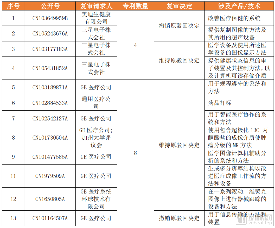
The table below shows the patent reexamination requests filed by Samsung Group and General Electric Company.
Table 4: Patent Reexamination Requests by Key Applicants
Source: ChaoFan Intellectual Property, VCBeat
Among Samsung Group’s patents in the field of medical imaging, four have been subject to requests for reexamination, namely: CN103649959B, CN105243676A, CN103177183A, and CN105431852A, all filed by Samsung itself. According to the reexamination decisions, two of these patents had the original rejection decisions upheld, while the other two had the original rejection decisions overturned.
Among General Electric’s patents in the field of medical imaging, eight have undergone requests for reexamination, namely CN103189871A, CN102884533A, CN102542127A, CN101730504A, CN101477585A, CN1979509A, CN1650805A, and CN101164507A, all filed by GE itself. According to the reexamination decisions, seven maintained the original rejection, while one overturned it. This to some extent reflects General Electric’s emphasis on its medical imaging patent technologies.
In China’s medical imaging sector, although domestic players such as United Imaging and Anke have frequently disrupted the market in the past two years, the position of the overseas “Big Three” GPS (G for GE, P for Philips, and S for Siemens Healthineers) remains solid. Data show that these three companies account for approximately 70% of the market share.
As China’s high-end medical equipment market becomes saturated and healthcare reforms advance, GE, Philips, and Siemens Healthineers—three “century-old” companies—are increasingly exploring a shift from single-product models to comprehensive solutions.
The difference lies in the fact that GE has spun off its healthcare business to focus on digital health applications and solutions; Philips leverages medical AI as a breakthrough point, building its own ecosystem platform, acquiring startups, and collaborating with ecosystem partners; Siemens Healthineers is building its AI capabilities from the ground up, aiming to establish a “App store” for digital healthcare.
The Midfield Battle Among the GPS Trio in Medical Imaging Will Continue.
This article is an excerpt from the report. Scan the QR code below to download the full version for free.
