After 15 Years in Cardiac Imaging, Dr. Li Yu Launches the Pangu Rare Case Museum to Reveal Life-and-Death Moments in Cardiovascular Disease
“If one must grant surgery a certain authority, it should be the power to interpret life.”
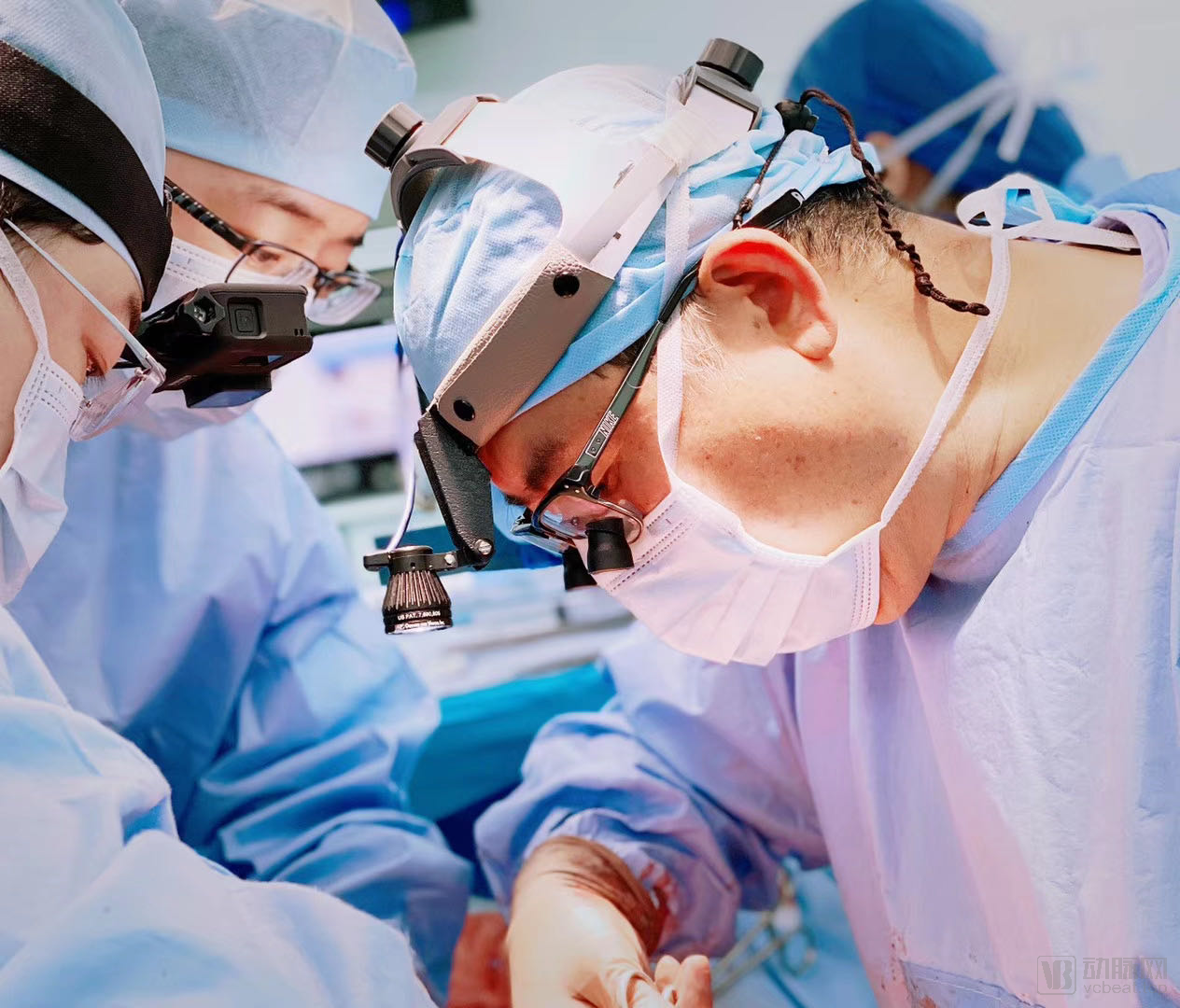
With the advancement of cardiac surgical techniques, modern physicians can now confidently address a wide range of cardiac challenges; nevertheless, heart disease remains the most formidable threat to human life. Data indicate that even in today’s era of highly advanced medicine, cardiovascular patients account for 30% of global deaths. In the face of life and death, cardiothoracic surgeons continue their unwavering efforts.
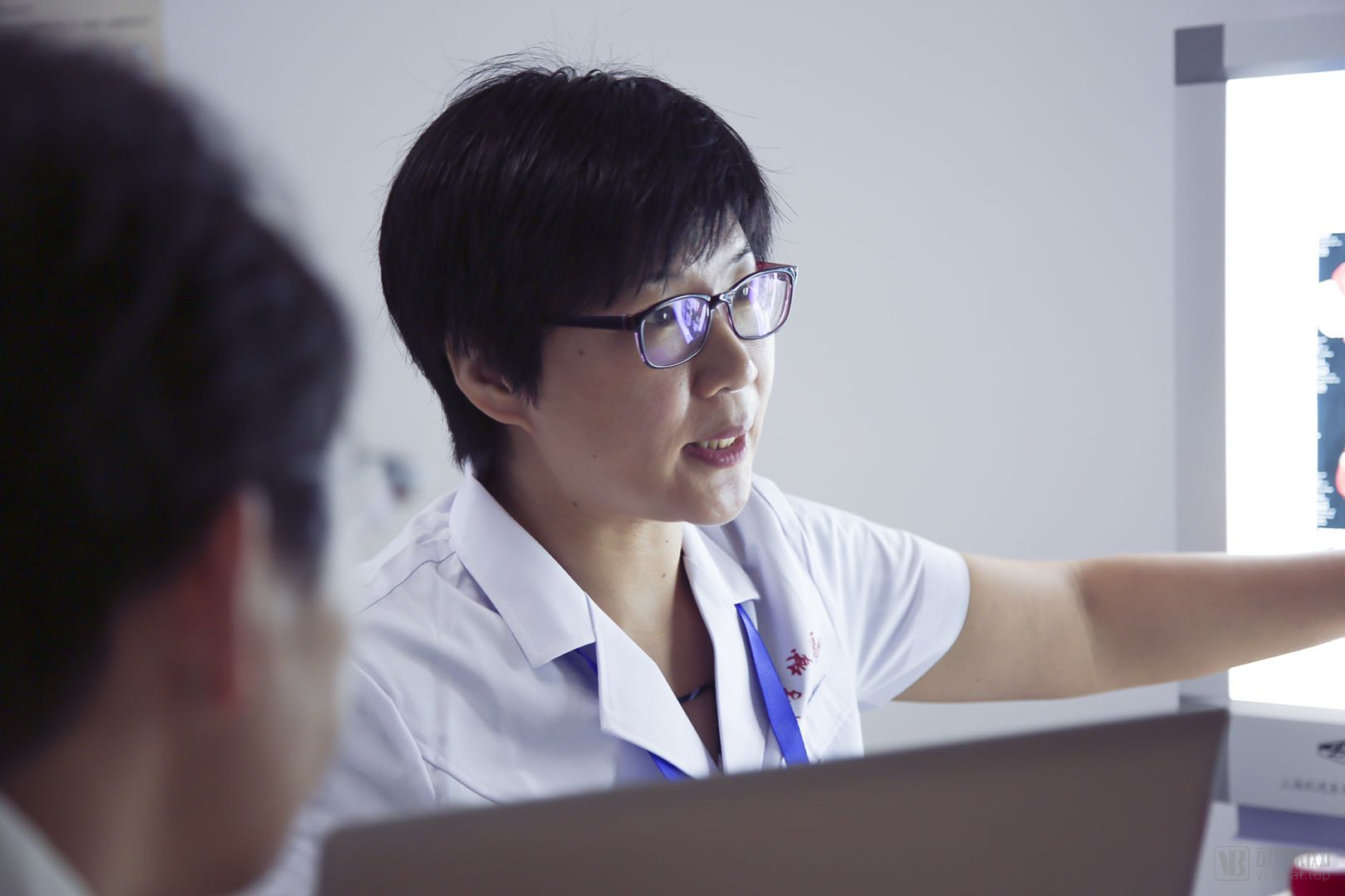
Li Yu
Chief Physician, Department of Radiology, Beijing Anzhen Hospital, Capital Medical University
Who is Li Yu?
Chief Physician, Department of Radiology, Beijing Anzhen Hospital, Capital Medical University,
Founder and Director of the Pangu Rare Case Museum.
Li Yu
Chief Physician, Department of Radiology, Beijing Anzhen Hospital, Capital Medical University
Practicing since 2005,
15 Years of Medical Imaging Data Collection Experience,
Over 1,000 imaging records,
Thirty selected rare cases are featured as exhibits in the Museum of Rare Cases at the inaugural Pangu Conference.
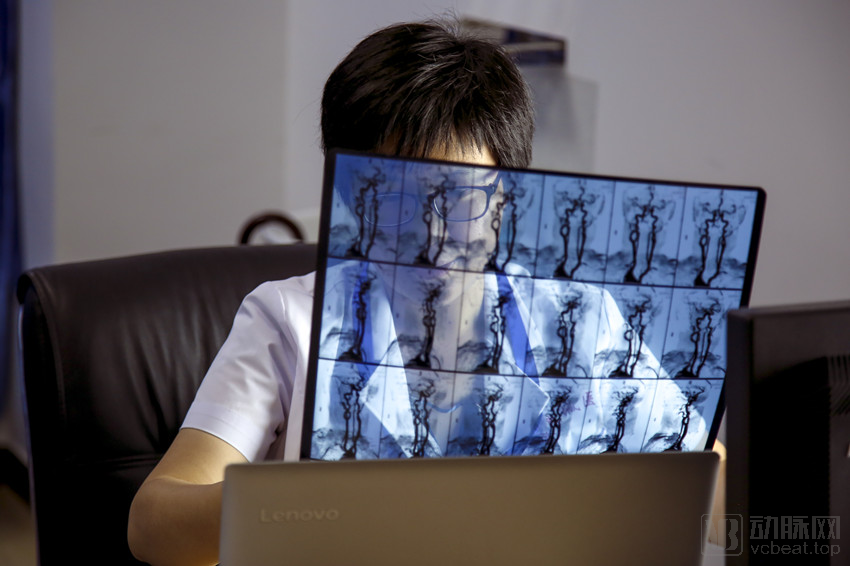
Li Yu Built China’s First Museum of Rare Cardiac Imaging with 5,475 Days of Accumulation.
In her view, the existence of these cases, particularly the rare imaging data, could have a lifelong impact on patients.
Rare, Uncommon, Life, Vision, Diagnosis, Future...
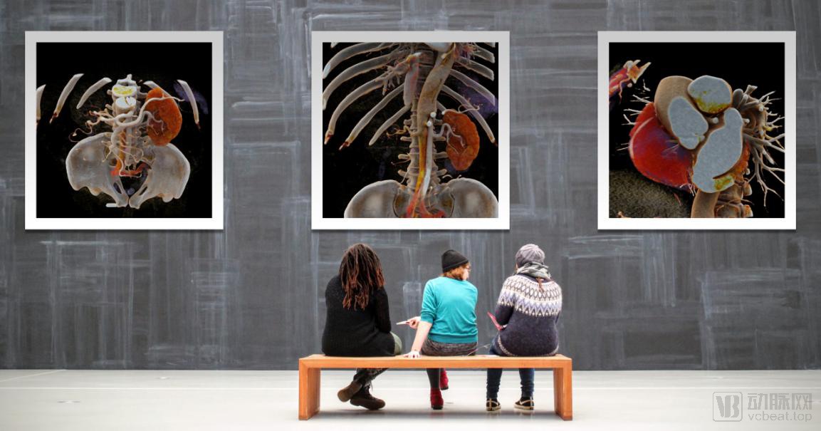
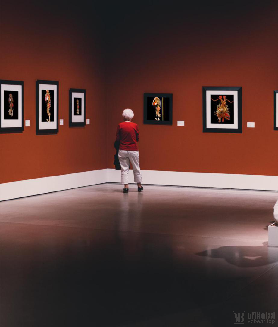
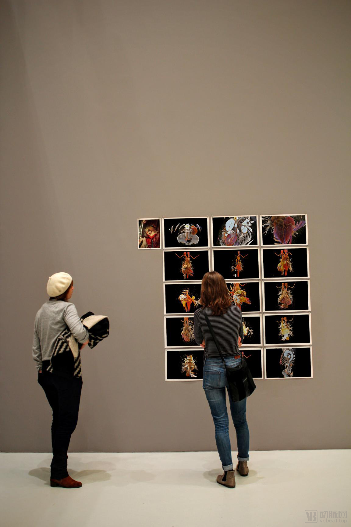
Centralized display of imaging data across four major categories: common cases prone to missed or misdiagnosis, conditions with identical signs but different etiologies, rare cases with distinct features, and complications related to medical procedures.
The Pangu Rare Case Museum Call for Submissions is now fully underway. Its official launch marks a pioneering initiative in the field of cardiac surgery, leveraging bold, boundary-pushing efforts to advance the life sciences. The ultimate aim is to enable the possibility of high-quality extension of human life.
After conceiving the idea of establishing such a museum, Li Yu countless times imagined what it should look like. Should it be large? Should it be luxurious or artistic? Should it be shrouded in mystery?
After envisioning it countless times in her mind, she remained true to the core ethos of a medical professional: “Pragmatic and rigorous, restoring the essence of life.” In her view, every exhibit in the Pangu Rare Case Museum must narrate the story of life and recreate each battle fought in the field of cardiovascular surgery.
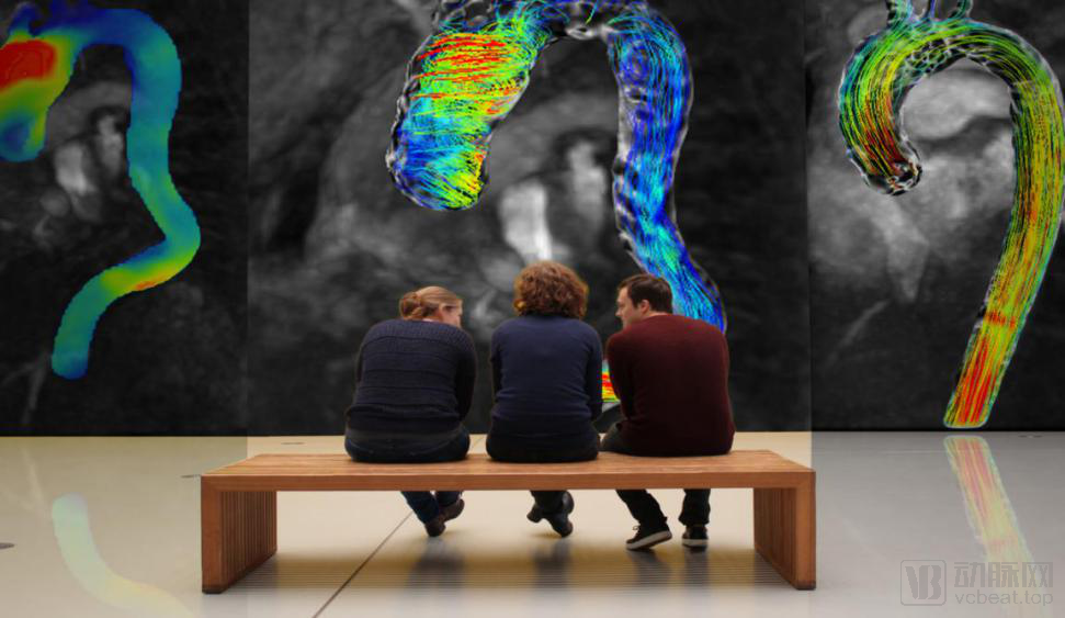
At this point, the overarching theme of the Pangu Rare Case Museum has been established. “We hope to engage professionals from cardiac surgery, cardiology, radiology, and other clinical specialties, inviting submissions of imaging data for any rare cases. The case presentations in the (Rare Case Museum) will be intuitive and three-dimensional, offering a visual feast. Furthermore, the morphological features must create a strong visual impact on visitors to achieve two objectives: first, contemplation, enabling visitors to reflect on and assess each frame; second, validation, allowing us to share the background and diagnostic insights of these cases with both submitters and visitors.”
Impacted by the pandemic, although the Pangu Rare Case Museum could not be physically inaugurated in 2020, it leveraged cutting-edge cloud technology to engage audiences through an immersive virtual exhibition. In Li Yu’s design, the Rare Case Museum is not a one-time event but a permanent institution.
“The Pangu Rare Case Museum will endure, as the life sciences continue to evolve. We aspire to infinite breadth in this field, engaging professionals from cardiac surgery, cardiology, other clinical specialties, radiology, and beyond. This multidisciplinary participation will greatly aid in the diagnosis of complex and rare cases in the future.”
In clinical practice, numerous rare cases play an immeasurable role in the advancement of medicine. Imaging data of aortic rupture, the diagnosis of complex and refractory diseases, and classic cases encountered in routine consultations are all of immense value. Moreover, the accurate interpretation of such imaging data can significantly determine a patient’s prognosis.
“We once treated a patient in whom an aortic lesion was incidentally discovered during evaluation for cerebral infarction. At an outside hospital, CT, MRI, and PET imaging raised suspicion for primary aortic malignancy. The lesion was extensive, involving approximately 15 cm of the descending aorta from the arch, with near-complete occlusion of the aortic lumen. The patient was transferred to our hospital for further management. After reviewing his imaging studies together with Director Sun, I concluded that the lesion was not malignant but rather a thrombus. These two distinct diagnoses warrant fundamentally different surgical approaches: treatment for malignancy would require replacement of the entire diseased segment of the descending aorta, a procedure associated with high technical difficulty and risk, including the potential for spinal cord ischemia leading to paraplegia. In contrast, if the lesion were a thrombus, Director Sun planned to establish a bypass between the ascending and abdominal aorta, excluding the diseased segment while addressing the primary problem. This approach avoids circulatory arrest intraoperatively and is markedly superior in terms of surgical complexity, risk profile, and postoperative quality of life, while also providing psychological reassurance to the patient and his family. Ultimately, our team designed the surgical plan based on the diagnosis of thrombus. At one- and two-year follow-ups, the patient demonstrated good recovery without disease progression. Although histopathological confirmation was not obtained, the clinical outcomes further validated our decision to exclude malignancy and diagnose ‘thrombus.’”
Such imaging data will be continuously archived in the Zhenqi Museum, which embodies the enduring significance of its existence.
Li Yu joined the Department of Radiology at Beijing Anzhen Hospital, Capital Medical University, in 2008, following seven years of experience as a surgeon. Specializing in cardiovascular hemodynamics, he has engaged extensively with various clinical departments, including operating rooms and intensive care units, dedicated to enhancing cardiovascular imaging diagnostic capabilities through a multidimensional approach.
She has participated in research projects funded by the National Natural Science Foundation of China, the Capital Development Fund, and the Beijing Outstanding Talent Fund. At Beijing Anzhen Hospital, where she works, there is a formidable team in the field of large-vessel diseases: the Sun Lizhong Heart and Great Vessel Team, led by Sun Lizhong.
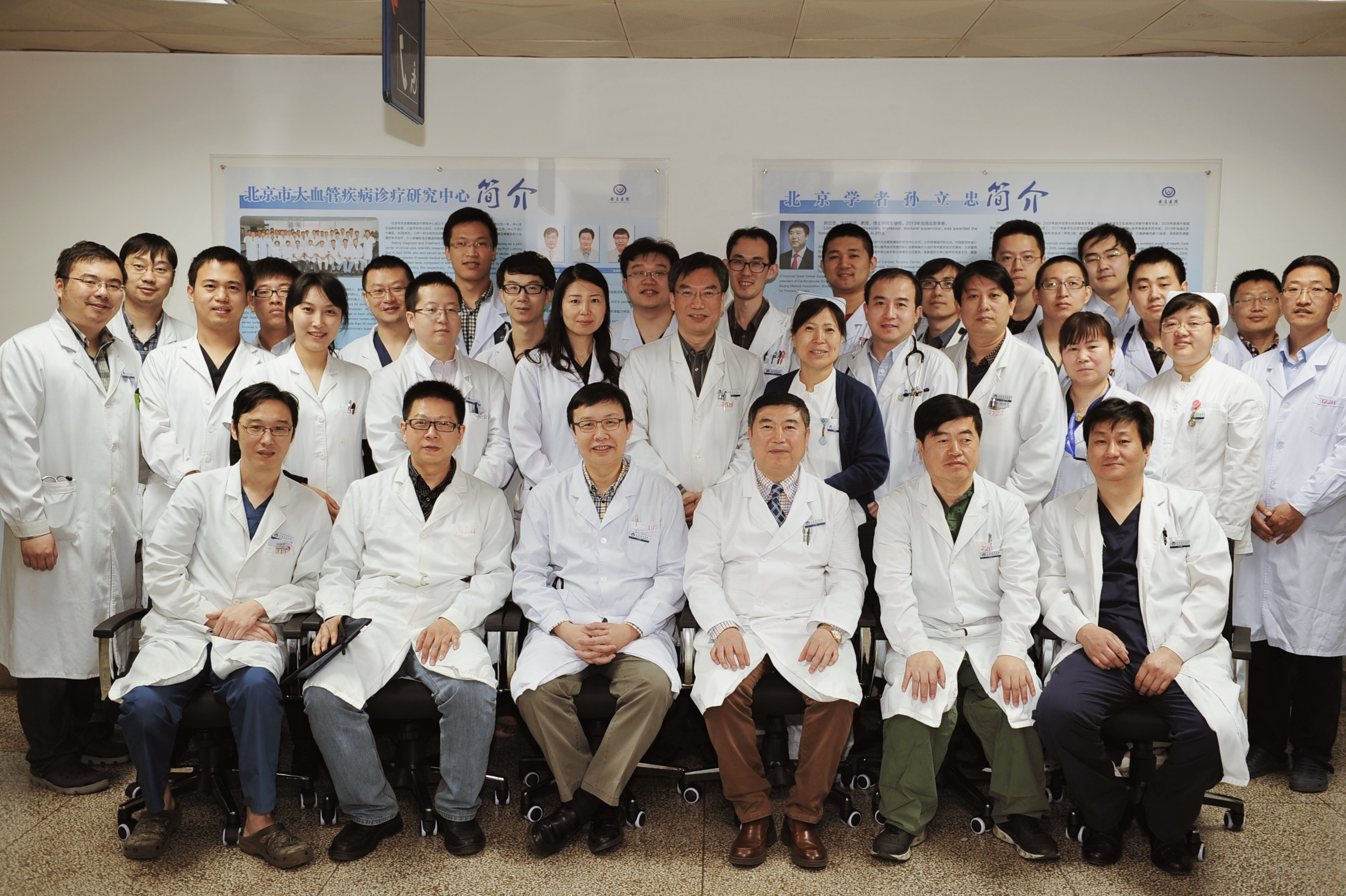
What kind of team is Sun Lizhong’s Cardiac and Great Vessel Team? Perhaps we can describe it in one sentence: the team with the highest volume of aortic surgeries worldwide.
The team consists of 28 members, including 8 chief physicians, 11 associate chief physicians, 5 attending physicians, and 4 resident physicians, with 90% holding intermediate or senior professional titles.
Team leader Sun Lizhong has been engaged in cardiovascular surgery for 37 years, having independently performed or supervised junior physicians in completing more than 10,000 cardiovascular surgical procedures. Based on the morphological characteristics of aortic diseases in China, he developed and applied a self-designed stented graft prosthesis to pioneer a new technique for aortic arch replacement combined with stented elephant trunk implantation. This procedure was formally named the “Sun’s Procedure” by international scholars at the American Association for Thoracic Surgery (AATS) Annual Meeting held in Toronto in April 2014.
To facilitate the exchange and sharing of experiences among researchers across all disciplines and to continuously advance the field of large-vessel diseases, a group of dedicated professionals led by Professor Sun Lizhong’s team has been organizing the “Pangu Large-Vessel Disease Forum” since 2009.
In 2020, due to the impact of the pandemic, the 9th Pangu Great Vessel Disease Forum was moved online and held on August 8–9. As a national-level academic conference in the field of great vessel diseases, the 9th Pangu Great Vessel Disease Forum not only continued its tradition of “professional disciplinary spirit and frontier exploratory attitude,” but also added exciting new segments, including the Pangu Rare Case Museum and a call for paper submissions.
The entanglement of the body with disease, and the race between life and time, have shifted the power determining human lifespan from divine forces on the altar to the surgeon’s scalpel under the shadowless lamp. The advancement of surgery has transformed human life from an unknown brevity into a knowable longevity. Over the millennia, the accumulation of individual case studies has become the cornerstone of modern medical progress. These precious cases serve as powerful weapons in humanity’s fight against disease; particularly in the field of cardiovascular medicine, each technological leap has become a milestone in the progress of life. Yet, how can such things be deemed truly precious?
Guidelines for Case Submissions to the Pangu Rare Cases Museum
Time Description:Submission Period: June 1–July 20; Screening to Begin on July 21
Target Audience:Targeting cardiologists, radiologists, and surgeons across China; physicians from other specialties are also welcome to participate if they can provide cardiovascular-related cases.
Call for Submissions:Original case reports on coronary artery-related diseases, myocardial and pericardial diseases, aortic diseases, and other rare or complex cardiovascular conditions, with educational value in at least one aspect such as diagnosis, examination, treatment plan, etiology determination, or surgical intervention.
Call for SubmissionsKey Points:Includes patient clinical data, imaging studies, and diagnostic results; surgical records and involved innovative products may also be presented.
Submission Method:Primarily image and video materials
Return on Participation:Benefits of Having Your Case Report Accepted: RMB 200 honorarium + Certificate of Recognition for Inclusion in the Pangu Society’s Rare Case Museum + SanDisk 1TB Portable Solid-State Drive (PSSD)
Rare, Atypical Cases
(1) Basic Patient Information: Accurately record the patient’s age, gender, chief complaint, history of present illness, past medical history, personal history, family history, positive physical signs, and positive laboratory test results.
(2) Case Description: Provide a concise narrative introducing the patient’s clinical manifestations, diagnostic and treatment course, and relevant prognostic information.
(3) Refine the case features: Describe the rare aspects of this case, for example:
① Rare clinical or imaging manifestations;
② Rare disease etiology;
③ Special methods and insights in the diagnostic or therapeutic process;
④ Other features with value for communication and display.
Note: Case description is limited to 400 words; case features are limited to 200 words and no more than 3 points.。
Image or Video
(1) Image Data: Content must be clear with appropriate contrast. Image resolution shall be no less than 512x512 pixels, and each file size shall not exceed 5 MB. Images should be transmitted in common image formats; DICOM format is recommended for medical imaging data. All patient information must be anonymized.
(2) Video data: The content should be clear, with appropriate contrast and a resolution of no less than 512x512, transmitted in common video formats. Patient information must be concealed.
Register Now
File Type Registration:Enter Registration
Image and Video Registration:Name the file “Pangu Rare Case Museum + [Name]” and send it to pangu_forum@163.com