Miniaturized CT and MRI Trends Highlighted: Five Application Scenarios Breaking Medical Imaging Constraints
In 2020, the FDA approved the world’s first portable magnetic resonance imaging (MRI) device for head MRI scans in patients aged two years and older. Developed by Hyperfine Research in the United States, this portable MRI weighs only one-tenth as much as traditional fixed MRI systems. Compared with large-scale MRI scanners, it reduces the cost per scan by a factor of 20 and lowers power consumption by a factor of 35. Furthermore, it is significantly more affordable than existing MRI systems, with a unit price of approximately $50,000. Portable MRI can be widely used in emergency departments, intensive care units (ICUs), and neurosurgery, helping physicians diagnose and detect conditions more rapidly.
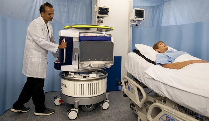
Hyperfine Mobile MRI
For Hyperfine, the approval of its portable MRI system is undoubtedly a major breakthrough; indeed, it represents a milestone product for the medical imaging industry. It is undeniable that over the next decade, the miniaturization of diagnostic medical imaging equipment, early disease detection, and telemedicine will be the three major trends driving improvements in the medical imaging sector.
The trend toward miniaturization and portability is dismantling traditional behemoths, enabling imaging equipment such as CT and MRI to break free from the confines of radiology and imaging departments and become intelligent point-of-care diagnostic devices. While miniaturized equipment cannot yet replace conventional large-scale systems, it possesses unique value and broad application potential. During the global COVID-19 pandemic, mobile imaging devices played a significant role, helping physicians perform necessary examinations more safely and efficiently during urgent public health emergencies.
Can Miniaturized Medical Imaging Devices Become the Next Growth Engine for the Medical Imaging Market? As Multi-Ton Large-Scale Imaging Equipment Becomes More Compact, What Technological Innovations and Iterations Are Driving This Transformation? With These Questions in Mind, VCBeat (WeChat ID: vcbeat) has reviewed the development of miniaturization in global medical imaging equipment.
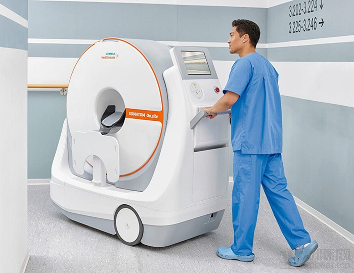
Siemens Mobile CT
For a long time, innovation in global medical imaging has been dominated by large medical imaging companies. However, over the past decade, the system architectures of most medical imaging devices—whether CT, MRI, or ultrasound—have become relatively mature and stable, with most products undergoing little significant change.
Taking MRI as an example, although ultra-high-field MRI systems can achieve magnetic field strengths of 10.5T (compared to the 1.5T or 3T typically found in general hospitals), their weight can be equivalent to that of three Boeing 737 aircraft. However, from another perspective, previous innovations and advancements in MRI have failed to address three major issues, which have also remained unresolved in the medical imaging industry.
First, the high cost means that only large hospitals can afford to use large-scale imaging equipment. The price of an MRI machine ranges from several million to tens of millions, and this substantial cost limits the global accessibility of MRI. Jonathan Rothberg, founder of Hyperfine, stated that 80% of the world’s population lacks access to MRI, with average wait times reaching up to 15 hours. He believes that the price of medical imaging equipment should continue to decrease. Previously, he founded Butterfly Network, which launched a handheld ultrasound device that combines ultrasound imaging with semiconductor chips. By disrupting the ultrasound field, the company raised over $350 million in public funding. Since its IPO in 2018, the Butterfly iQ device has been adopted by thousands of doctors, nurses, and other healthcare practitioners in the United States.
In China, resources for high-end imaging examinations are even scarcer. According to statistics from the Chinese Association of Medical Equipment, as of 2018, there were approximately 22,000 CT scanners and 9,255 MRI scanners in operation nationwide. Each machine served only about 50 patients per day in terms of workload and scan volume, and the waiting time for outpatient MRI appointments sometimes extended up to one month. A set of data presented by Professor Guo Qiyong from Shengjing Hospital Affiliated to China Medical University in his report “Current Status and Future of Radiologists in China” at the 6th Oriental Congress of Radiology in 2016 indicated that Chongqing had the lowest number of CT scanners per million population, at 6.63 units, while Tibet had the lowest number of MRI scanners per million population, at only 0.93 units. By 2017, the number of MRI scanners per million population in the United States and Germany was 37.56 and 34.49, respectively, with most other major developed countries also exceeding 10 units per million population. In contrast, China had only 6.2 MRI scanners per million population.
The second issue is the complex operational workflow. Large-scale MRI and CT equipment require specialized operators, necessitating collaboration among technologists, nurses, and radiologists during procedures. Radiologists must be present during scanning and are also responsible for issuing diagnostic reports.
Finally, there are stringent requirements for patients and the scanning environment. During an MRI scan, patients must remain still and hold their breath. They need to stay alone in a dark and noisy environment for nearly half an hour, which poses challenges for pediatric and elderly patients. As a large, precision instrument, MRI systems have specific requirements for ambient temperature and humidity, thereby imposing high standards on healthcare facilities.
Large MRI and CT scanners are immobile, requiring patients to be transported to radiology or imaging departments for scanning. Transporting hospitalized patients increases the risk of complications; statistics show that over 71% of ICU patients experience adverse events during transport to the CT suite. (Source: Anticipating Unique Disruptions: Key Findings from PwC’s 21st Annual CEO Survey – Healthcare Industry.)
Clearly, the key to better addressing the aforementioned issues and making equipment such as CT and MRI scanners accessible to a broader population lies not in addition, but in subtraction—namely, the miniaturization of large-scale imaging devices.
Over the years, major medical device manufacturers have made limited attempts to miniaturize large-scale medical imaging equipment. In addition to considerations of return on investment, their development has been constrained by the significant technical challenges involved. It remains difficult to overcome the key obstacles inherent in miniaturizing large MRI and CT systems.
Hyperfine has disclosed limited technical details regarding its mobile MRI system; however, the following three factors represent the core reasons enabling the miniaturization of mobile MRI.
Generally speaking, the largest and most critical component of a magnetic resonance imaging (MRI) system is the magnet. The magnet is used to generate a magnetic field, and the unit of magnetic field strength is the tesla, which is defined as the unit of measurement for describing the strength of magnets used in MRI. Most MRI systems have a magnetic field strength of 1.5 T, while more advanced models can achieve 3.0 T, with correspondingly large magnet sizes. The key reason Hyperfine has been able to miniaturize MRI equipment is that the magnet used in its portable device has a field strength of only 0.064 T. In contrast, the neonatal MRI system developed by Aspect Imaging, an Israeli company, operates at 1.0 T.
Magnets are used to generate magnetic fields and primarily fall into three categories: permanent magnets, electromagnets [with magnetic flux density reaching up to 24,000 Gs (2.4 T)], and superconducting magnets [with magnetic flux density reaching up to 190,000 Gs (19 T)]. Magnetic flux density is closely correlated with imaging quality. Both superconducting and electromagnetic systems require cooling mechanisms, which is one of the reasons why large-scale MRI equipment is bulky. In contrast, mobile MRI systems utilize permanent magnets, eliminating the need for cooling systems and thereby reducing the overall size of the equipment.
Electromagnets and superconducting magnets generate powerful magnetic fields that attract surrounding metallic objects, necessitating that MRI procedures be conducted in enclosed rooms. In contrast, miniaturized MRI systems utilize magnets with lower magnetic flux density, eliminating the need for enclosed spaces during startup and operation, thereby reducing the spatial and environmental requirements for MRI equipment.
Overall, Hyperfine uses permanent magnets that do not require specialized power or cooling systems, enabling image generation using low-power radio waves and magnetic fields.
In the field of miniaturized CT scanners, numerous companies have successfully launched products, including NeuroLogica (a subsidiary of Samsung) and Siemens, which unveiled its mobile CT system, SOMATOM On.site, at the 2019 RSNA Annual Meeting. The SOMATOM On.site is a head CT scanner designed for free mobility across various departments and operating rooms.
In the design of mobile CT systems, addressing radiation exposure is a critical challenge. Siemens’ mobile CT employs a telescoping gantry design, where the X-ray source and gantry move away from the patient during scanning to reduce radiation scatter, while the base of the mobile scanner remains stationary. Since only the gantry moves during image acquisition, this design improves workflow and reduces motion-induced image artifacts. Additionally, the SOMATOM On.site is equipped with protective curtains that can be attached to the front and rear of the gantry opening, further reducing radiation exposure to staff and nearby patients. In contrast, the BodyTom, a whole-body 32-slice CT scanner launched by NeuroLogica in 2011, incorporates built-in internal lead shielding of 0.75 mm thickness.
VCBeat interviewed Dr. Zhang Minglong, Executive Partner at Chuangrui Capital, on the trend toward miniaturization and portability of CT and MRI systems. Dr. Zhang previously served as a Principal Researcher and Project Manager at GE Global Research Center, bringing 12 years of extensive experience in R&D and market development across advanced medical devices, new energy, new materials, and advanced manufacturing. His expertise spans core technologies and products in nearly all major medical imaging modalities, including CT, PET, SPECT, DR, MRI, and ultrasound.
He stated, “For hospitals, MRI and CT systems must meet the needs of a broader patient population, particularly obese patients and those requiring full-body scanning, which necessitates larger bore sizes and thus limits the miniaturization of MRI and CT devices. Furthermore, CT operates on the principle of scanning the human body with X-ray beams; its core hardware includes opposing X-ray tubes and detectors along with a rotating mechanism, which also constrains the potential for CT device miniaturization. Essentially, DR (Digital Radiography) equipment represents a miniaturized form of CT technology. Without rotating components, DR systems can be made compact and mobile, feature lower radiation doses, and require simpler post-exposure shielding. Although they cannot perform tomographic imaging, they still provide basic diagnostic information. The principle of MRI relies on using magnetic fields to excite hydrogen protons in the human body, inducing magnetic resonance. Image quality is directly correlated with magnetic field strength; achieving high-quality images requires stronger magnetic fields. Superconductivity is the only technology capable of generating fields above 1.5 Tesla, and superconducting magnets necessitate bulky cooling systems.”
Another core technology driving the miniaturization of medical imaging equipment is the integration of cross-disciplinary technologies, a trend that is gaining momentum across the entire medical device sector. Technologies such as biosensors, micro-electromechanical systems (MEMS), solid-state batteries, and artificial intelligence are transforming the medical device landscape. Among these, AI-driven digitalization has played a pivotal role in enabling the portability of medical imaging devices. Since miniaturization inevitably leads to some degree of image quality degradation, artificial intelligence and deep learning technologies help achieve superior diagnostic accuracy despite limited imaging quality.
Hyperfine is currently developing 3D rendering capabilities for MRI, which can generate standard clinical contrast images as well as richly detailed 3D renderings, enabling physicians to transition from interpreting 2D MRI scans to directly diagnosing using intuitive 3D images. Additionally, Hyperfine is developing software that leverages deep learning algorithms to continuously improve with each use, reconstructing images and aiding in diagnosis; however, this AI-powered product has not yet received FDA approval.
In the application of mobile CT, Siemens’ mobile CT leverages a digital system to simplify physician examinations. Radiologists can perform scans via myExam Companion, the intelligent user interface of SOMATOM On.site, which optimizes scanning parameters based on the patient’s specific clinical questions, ensuring consistent imaging results regardless of the physician’s level of experience. Once the examination is completed, the patient can be slid back from the headboard position onto the hospital bed, and the scan images are automatically transmitted to the PACS within minutes.
Samsung’s NeuroLogica has partnered with the Israeli artificial intelligence company MedyMatch to leverage AI for assisted diagnosis, enabling physicians to rapidly determine whether a stroke patient is suffering from intracerebral hemorrhage or thrombosis.
Thanks to innovations in visual sensors and AI algorithms, modern medical imaging is undergoing a paradigm shift, moving from large, meticulously calibrated machines to flexible, self-correcting multi-sensor devices.
Although mobile medical imaging equipment integrates numerous cutting-edge technologies, the image quality of mobile MRI systems still cannot compare with that of large-scale imaging systems. While mobile imaging devices cannot replace large-scale equipment, they can complement the limitations of existing MRI and CT scanners, thereby expanding the application scenarios for imaging equipment. In summary, mobile MRI and CT systems can play a vital role in settings such as emergency departments, intensive care units (ICUs), and point-of-care diagnostics, enabling medical imaging to move beyond traditional radiology departments to wherever patients are located.
Emergency Applications:In emergency settings in the United States, mobile CT scanners are primarily deployed within Mobile Stroke Units (MSUs) to facilitate rapid diagnosis and treatment of stroke patients. An MSU is a specialized ambulance equipped with mobile CT and other essential diagnostic instruments, enabling more stroke patients to receive timely and effective intervention within the critical 60-minute "golden hour." Within an MSU, physicians can utilize mobile CT for imaging-based diagnosis to determine whether thrombectomy is indicated, thereby saving precious time in the race against the clock during stroke care.
ICU Applications: In the United States, mobile CT scanners are also utilized for bedside diagnostics and in intensive care units (ICUs), reducing patient transport and thereby lowering the risk of adverse events. In ICUs as well as neurosurgical and internal medicine departments, patients are often difficult to move and require meticulous care. Traditional fixed imaging equipment necessitates patient transport, which can pose significant risks to patients. In ICUs and neurosurgery, CT scans are frequently required for routine follow-up. However, because MRI and CT scanners are sophisticated instruments with stringent stability requirements, many MRI and CT suites are located on the first floor or in the basement of hospitals, posing considerable challenges for patient transport. Data indicate that 71% of ICU patients experience adverse events during transport to the CT scanning room.
Therefore, the ICU and neurosurgery departments represent major application scenarios for mobile MRI and CT systems. In clinical neurosurgery, some physicians have expressed a preference for using MRI equipment over CT for bedside diagnostics. This is because MRI is more sensitive to brain injury, enabling earlier and more accurate detection of ischemia. It is foreseeable that mobile MRI will capture a certain share of the market for bedside diagnostics in neurosurgery.
Taking the collaboration between Hyperfine and Yale New Haven Hospital as an example, the hospital uses mobile MRI to scan patients in the neuro-intensive care unit. Physicians can utilize mobile MRI to evaluate patients with ischemic stroke, tumors, and hydrocephalus, thereby providing clinicians with more timely and comprehensive data to inform diagnosis and treatment decisions.
Operating Room:Mobile CT units installed in operating rooms enable preoperative, intraoperative, and postoperative imaging, providing surgeons with more accurate pathological data. Furthermore, surgical patients no longer need to be transported to the Radiology Department on the first floor of the outpatient building for scans, thereby eliminating transportation risks and improving surgical quality. It is reported that the Chinese People's Liberation Army General Hospital (Beijing 301 Hospital) has already deployed two of NeuroLogica’s latest mobile spiral whole-body CT systems, the BodyTom, and one mobile spiral head CT system, the CereTom.
Pediatrics: In addition to its applications in neurology, another major use case for compact MRI systems is in neonatology. In pediatric care, newborns who experience prematurity, asphyxia, or hypoxia at birth are recommended to undergo magnetic resonance imaging (MRI) to assess cranial structures. Furthermore, multiple MRI scans are often required during the course of treatment. Similar to ICU patients, neonates face an increased risk of adverse events during transport. Aspect Imaging, a high-performance MRI manufacturer from Israel, has developed an MRI device specifically designed for newborns. Featuring lower noise levels and a more compact footprint, this system is better suited for neonatal care.
Primary Healthcare Institutions:From a broader perspective, mobile MRI systems have the potential to expand into the extensive primary care market. Some may question whether mobile MRI units are functionally inadequate or unable to meet the needs of primary care settings. However, in the past decade, the scope of clinical MRI examinations has remained largely unchanged. Brain and spinal studies account for more than 50% of all examinations, while breast, cardiac, and interventional studies constitute less than 5%. Among all clinical MR examinations, functional MRI (fMRI) and other complex procedures together make up less than 1% (data sourced from Magnetic Resonance). Mobile MRI systems are currently capable of performing brain examinations. Although they cannot fully replace large-scale MRI imaging equipment, they help primary care hospitals handle the majority of MRI examinations. For hospitals that previously lacked MRI equipment, mobile MRI represents a viable supplementary option.
However, in addition to developing portable devices to expand the application scenarios of imaging equipment, CT scanners are also being deployed in emergency settings and primary healthcare facilities through mobile solutions such as vehicle-mounted CT and modular container-based CT. During the fight against the COVID-19 pandemic, domestic CT manufacturers including Mingfeng Medical, United Imaging, and Neusoft introduced vehicle-mounted CT systems to alleviate the shortage of imaging equipment. Nevertheless, vehicle-mounted and modular container-based CT systems involve minimal changes to the CT technology itself and still require shielded rooms. This article focuses on products that truly mobilize the CT scanner itself.
Although mobile CT and MRI systems offer significant potential for diverse application scenarios, they are unlikely to become mainstream products akin to large-scale systems at the current stage.
Zhang Minglong stated: “The miniaturization and mobility of MRI and CT equipment aim to enhance operational flexibility and convenience to meet the needs of specific application scenarios, such as bedside monitoring, intensive care units (ICUs), emergency rescue sites, and stroke care. While there is a certain level of demand, it remains fragmented, making it difficult for hospitals to achieve significant economic benefits. Additionally, physician adoption poses another challenge. In the future, driven by policy support, consumption upgrades, and artificial intelligence, these devices will hold supplementary value and capture a niche market, but they are unlikely to become mainstream.”
The primary market for miniaturized CT scanners is likely the small-animal imaging sector, which does not require large bore diameters. Digital radiography (DR) can fulfill the preliminary functional and diagnostic roles of CT; therefore, miniaturizing CT scanners for specific human body regions offers limited clinical significance. Mobile CT units address special on-site needs, such as pandemic response (e.g., during the COVID-19 outbreak) and health screenings, where vehicle-mounted CT systems can be deployed; however, this mobility should not be conflated with miniaturization. From a cost-effectiveness perspective, mobile CT serves merely as a supplement to the existing healthcare system.
“The miniaturization and mobility of MRI can be achieved through the use of permanent magnets. Since permanent magnet-based MRI systems do not require cooling systems and operate at lower magnetic field strengths, they can be made compact and portable. However, this comes at the expense of image quality to meet specific application scenarios such as bedside imaging, emergency rescue settings, and stroke care. These systems are generally limited to localized examinations, such as of the head or extremities, or open-bore imaging for small animals. While there is some value and market demand for such devices, it will not be substantial, given the suboptimal image quality; they serve primarily as tools for emergency diagnosis.”
A significant portion of the market potential for mobile MRI systems lies in the point-of-care segment. However, clinicians in emergency departments and intensive care units (ICUs) require a certain learning curve to become proficient in interpreting diagnostic MRI images. Furthermore, point-of-care diagnostics are dispersed across various clinical departments, making market education challenging and thereby posing considerable difficulties for market promotion.
Therefore, we should take a measured view: while mobile MRI has achieved significant breakthroughs, it will not overnight transform current clinical practices and habits in the conservative healthcare market. The market transformation driven by mobile devices will be gradual, and the COVID-19 pandemic may accelerate this process.
In February this year, China ordered 10 mobile CT units from NeuroLogica, a subsidiary of Samsung, to alleviate the strain on medical resources amid the COVID-19 pandemic.
Improving the accuracy of medical imaging remains a key driver for the future development of the medical imaging industry. However, the trends toward miniaturization, portability, and digitalization in medical imaging cannot be overlooked. Miniaturized devices not only enhance the efficiency of image-based diagnosis but also enable hospitals to expand their billable testing services. In the future, as miniaturized and mobile equipment becomes more cost-competitive, the adoption of mobile medical imaging devices will accelerate further.