Tencent Tianyan Lab Achieves Breakthrough with 14 Papers Accepted at MICCAI 2020, Advancing AI in Medical Imaging

Tencent
Internet Comprehensive Service Provider
Recently, the acceptance results for papers at the 23rd International Conference on Medical Image Computing and Computer-Assisted Intervention (MICCAI 2020) were announced. Tencent’s Tianyan Laboratory, which focuses on research in medical artificial intelligence and big data technologies, had a total of 14 papers accepted, a significant increase from the four papers accepted last year. The research areas cover application scenarios such as classification, segmentation, detection, and domain adaptation in medical imaging, achieving comprehensive breakthroughs in AI technology for medical imaging.
In recent years, AI technology has become deeply integrated with the healthcare industry, with its most extensive application found in medical imaging. Among the papers accepted to MICCAI 2020, Tencent’s Tianyan Laboratory, as one of the algorithmic technology providers behind Tencent Miying, conducted innovative research on various machine learning methods tailored to diverse clinical scenarios in medical imaging. This work holds promise for accelerating the practical implementation of AI in the healthcare sector.
Dr. Zheng Yefeng, Head of Tencent’s Tianyan Laboratory, has been engaged in intelligent medical image analysis for many years. His invented projection space learning method received the U.S. Thomas Edison Patent Award in 2011, and his related research findings were compiled and published in 2014 as the book *Projection Space Learning for Medical Image Processing: Rapid Detection and Segmentation of Organs*. He is also a Fellow of the American Institute for Medical and Biological Engineering (AIMBE) and an Associate Editor of the IEEE Transactions on Medical Imaging (TMI, IF=7.8).
In the learning and training of AI-based medical imaging, facing the industry-wide challenges of insufficient annotation resources and inconsistent quality of medical imaging data, Tencent Tianyan Laboratory has sought breakthroughs through technological innovations in various machine learning methods to maximize the utilization of annotated data and unlock the potential of AI in the field of medical imaging.
Compared with traditional supervised learning, weakly supervised learning employs limited, noisy, or inaccurately labeled data for model parameter training, representing a common approach in AI-based medical image analysis. Among the papers accepted this year, Tianyan Laboratory proposed a novel weakly supervised framework comprising two components: a macro-network and a micro-network. The macro-network is responsible for learning positional and regional information from a large volume of weakly labeled images, while the micro-network focuses on learning fine-grained structural information from a small number of fully labeled images. Building upon this framework, the researchers implemented an uncertainty-based macro-micro data flow, leveraging parameter sliding average methods and uncertainty-guided KL-loss to facilitate knowledge exchange between the two models. Extensive experimental results demonstrate that this method outperforms traditional single semi-supervised and weakly supervised approaches, offering new possibilities for more efficient utilization of segmentation annotations.
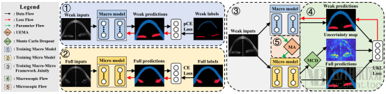
Figure Caption: Macro-Micro Weakly Supervised Learning for Retinal OCT Tissue Segmentation
Addressing the susceptibility of neural networks to noise interference during medical image acquisition, Tencent’s Tianyan Laboratory has proposed an anti-interference endogenous neuron learning method. This approach generates correct response maps using accurate labels and then randomly generates incorrect labels to produce erroneous response maps as interference. During neural network training, the discrepancy between the two response maps is maximized, enabling the network to learn from interference information and thereby enhance its anti-interference capability. Unlike previous methods, the proposed approach generates interference in the feature space rather than at the image level. Since the interference generated in the feature space is determined entirely by the network’s internal mechanisms, the network achieves stronger anti-interference performance and robustness. In the current context of scarce high-quality annotated data resources, this research offers new explorations for enhancing the value of noisy data and expanding the scope of usable data for machine learning.

Figure Caption: Interference-Resistant Endogenous Neuronal Learning Aids Medical Image Classification
High annotation requirements and difficulties in data collection for medical imaging data have made the utility and potential of unlabeled data a new direction for AI research in medical imaging.
In the task of automated glaucoma diagnosis based on deep learning, the “Learning-to-Teach Knowledge Transfer (L2T-KT)” training strategy and “Quiz Pool” proposed by Tianyan Laboratory leverage undiagnosed fundus images to upgrade the teacher network, representing an attempt to apply unlabeled data in AI medical imaging. This approach enables the teacher network to encode information from undiagnosed fundus images into a latent feature space, allowing the student network to be trained on these images by learning from the teacher. Experimental results on both proprietary datasets and the LAG dataset demonstrate that this method significantly improves the performance of glaucoma diagnosis by utilizing undiagnosed fundus images.
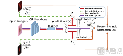
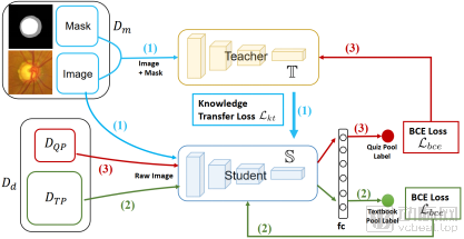
Figure Caption: Glaucoma classification learning model trained on undiagnosed data based on the teacher–student network framework
In the era of deep learning, pre-trained models have played a crucial role in medical image analysis. However, significant domain shifts exist between natural images and medical images, leaving substantial room for improvement. Research demonstrates that the Comparing to Learn (C2L) model pre-training framework, proposed by Tianyan Laboratory, leverages 700,000 completely unlabeled radiological images. By comparing differences in image features, C2L achieves performance superior to models pre-trained on ImageNet with supervised learning, as well as other current state-of-the-art self-supervised models.
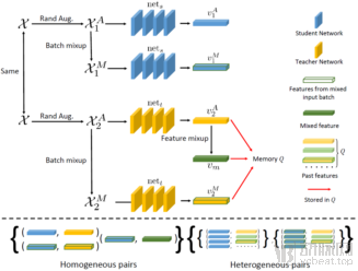
Figure Caption: Contrastive Learning-Based Pretraining of Radiological Image Representations Surpassing ImageNet Pretraining
In addition to leveraging as much data as possible for AI learning, it is also crucial to further enhance the effectiveness of AI model training based on existing data annotation and algorithms.
Medical images are typically annotated by multiple experts or physicians, and the final gold-standard labels are determined by averaging the multi-rater annotations or using majority voting. When these annotation results are used for AI model training, usually only the final gold-standard labels are utilized, while the value of the original raw annotations beyond the gold standard is often overlooked. In their study “Development of a Hard-Example-Aware Glaucoma Classification Model Based on Multi-Annotation Consistency,” Tianyan Laboratory proposed a new deep learning-based model framework that leverages raw multi-expert annotations to enhance glaucoma classification performance. Furthermore, it predicts whether an image represents a simple or difficult case based on the consistency or inconsistency of annotations provided by different experts for each image. This research is also expected to improve the efficiency and accuracy of clinicians’ diagnostic analyses, alerting them to pay special attention to difficult cases.
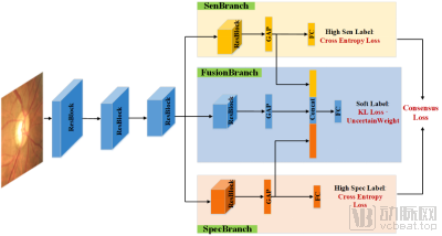
Figure Caption: Hard Example-Aware Glaucoma Classification Model Based on Multi-Annotation Consistency
In the task of target region segmentation for medical image analysis, manual annotations are typically used as the ground truth for model learning. Although most regions of the segmentation target are relatively easy to label, the boundary areas between different tissues are often difficult to handle due to factors such as ill-defined boundaries and partial volume effects. Hard labels in these regions may carry considerable uncertainty, and existing edge detection algorithms cannot accurately locate and align with the true edges of objects. This uncertainty further compromises the performance of the trained models.
In the accepted paper “Superpixel-Guided Label Softening for Medical Image Segmentation,” Tianyan Laboratory proposed a superpixel-based label softening technique. This method softens labels within a region based on the distance from voxels to annotation boundaries within each superpixel, and trains the segmentation model using both the softened labels and the original hard labels. In another paper, “Logic Refinement Network: An Edge Detector for Extracting Fine Edges,” researchers designed a logic refinement network and a logic valve operation function (developed based on the logical relationship between segmentation maps and edge maps). This approach applies the object detection results (i.e., segmentation maps) and the coarse edge detection maps from the initial stage to the original image via the logic valve operation function, then feeds them into the edge refinement network. The network progressively reinforces object boundary locations and refines the edge map, ultimately outputting an accurate and fine-grained edge map. Experimental results demonstrate that these methods effectively enhance the performance of AI networks in edge detection for 2D and 3D medical images, building upon existing hard labels and edge detection algorithms.
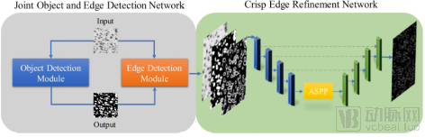
Figure Caption: Logic Refinement Network: Edge Detector for Extracting Fine Edges
As the technology provider behind “Tencent Miying,” Tianyan Laboratory’s research and innovation continuously expand the boundaries of AI medical technology, while also progressively translating research achievements into clinical research and practical applications.
During the COVID-19 outbreak this year, Tencent Tianyan Laboratory leveraged AI and big data technologies to deliver outstanding performance in technology-driven epidemic response. Zheng Yefeng, Director of Tencent Tianyan Laboratory, stated that among the 15 tools launched in the epidemic response section of the Tencent Health mini-program, Tianyan Laboratory contributed to five, including Q&A on epidemic knowledge, identification of patients in the same residential community, self-check for fever, locator for fever clinics, and guidelines for mask usage. In the epidemic Q&A service, the laboratory employed its self-developed LTD-BERT model to identify user intent, increasing inference speed by 40-fold and providing users with precise epidemic-related information.
In the development of CT-assisted diagnostic products for COVID-19, Tianyan Laboratory addressed challenges such as insufficient data volume and limited annotation resources by employing a “Magic Cube” self-supervised learning approach to train its models. The models were fine-tuned on small datasets to classify pneumonia versus non-pneumonia cases, as well as viral pneumonia versus non-viral pneumonia cases. During the pandemic, Tencent Miying’s AI-assisted diagnostic solution for COVID-19 was deployed at Zhongnan Hospital of Wuhan University. Leveraging robust technical support, the system completed AI-based pattern recognition within as little as 2 seconds after a patient’s CT scan, providing physicians with auxiliary diagnostic references within one minute. Over a two-month period, it assisted multiple hospitals in Hubei Province in performing lung CT diagnostics for more than 24,000 patients.
Tencent Miying’s application in fundus disease screening holds particularly broad social significance, as it helps address the shortage of ophthalmologists at the primary care level and enables the widespread adoption of low-cost fundus examinations in communities and grassroots healthcare settings. Currently, the system has been validated in collaboration with more than 30 hospitals across China and is being piloted within primary healthcare systems in provinces such as Guangdong, Guangxi, and Shandong. It is expected that an increasing number of people will benefit from this initiative.