AI-Powered Head and Neck CTA Reconstruction System Developed by Xuanwu Hospital and Shukun Technology Published in Nature Communications

SHUKUN
Provider of Intelligent Products and Innovative Solutions
Recently, the Nature sub-journal Nature Communications published online a research paper titled “Rapid vessel segmentation and reconstruction of head and neck CTA using 3D convolutional neural network” (IF=12.121). The paper was jointly released by Professor Lu Jie’s team from Xuanwu Hospital of Capital Medical University and SHUKUN (Beijing) Network Technology Co., Ltd. Dr. Fu Fan is the first author, and Professor Lu Jie is the corresponding author.
This study is the first to employ a 3D convolutional neural network to design a post-processing system for head and neck CTA vessel segmentation, capable of automatically removing bone structures and completing automated vascular reconstruction. Through this approach, the system has, to some extent, reshaped the clinical workflow for head and neck CTA.
Generally, head and neck CTA examinations require patients to undergo two CT scans; however, with the assistance of this model, only a single contrast-enhanced scan is needed to obtain images suitable for reconstruction. Furthermore, over the two-year study period, the precision and accuracy of the AI-based reconstruction continuously improved with ongoing training, gradually reaching a level comparable to that of senior physicians.
As a global disease characterized by high incidence, high mortality, high recurrence, and substantial medical burden, cerebrovascular diseases such as stroke affect tens of millions of patients. The innovative breakthroughs in the clinical pathway for head and neck CTA, along with their underlying clinical value, constitute one of the key reasons why this research was published in a Nature subsidiary journal.
Furthermore, the research process of this paper also reflects the continuous growth and improvement of artificial intelligence—developing an effective model is not achieved overnight; it is a gradual, step-by-step process.
The latest Global Burden of Disease Study (GBD) indicates that China’s overall lifetime risk of stroke is 39.9%, ranking first globally. This means that approximately two out of every five Chinese individuals will develop stroke during their lifetime. Furthermore, stroke is the leading cause of years of life lost due to disease in China. According to data from the “2019 Statistical Bulletin on China’s Health Development,” cerebrovascular diseases accounted for more than 20% of all deaths among Chinese residents in 2018, implying that at least one out of every five deaths was attributable to stroke.
Globally, according to data published in *The Lancet Neurology* on March 11, 2019, the number of stroke cases reached 80.1 million in 2016, making it the second leading cause of death worldwide.
In the diagnosis and treatment of ischemic stroke and various cerebrovascular diseases, head and neck CT angiography (CTA) is a routine examination method. However, the growing volume of examinations coupled with a shortage of CTA-interpreting physicians has left hospitals struggling to meet patient demand. The workload within departments is gradually increasing, potentially resulting in prolonged wait times for patients to schedule CTA exams and receive their reports.
Deep learning algorithms based on convolutional neural networks may resolve this contradiction. Since the wave of artificial intelligence swept through the medical field, many healthcare technology companies, hospitals, and scholars have attempted to reshape the CTA examination process using AI. Improving the efficiency of CTA examinations and enhancing diagnostic accuracy could benefit hundreds of millions of patients—this is one of the key reasons why Professor Lu Jie’s team chose head and neck CTA as their research focus, reflecting the global trend of healthcare development toward social good.
After years of development, the capability of artificial intelligence (AI) in coronary computed tomography angiography (CTA) has been validated in clinical practice. Many cardiology departments at Grade A tertiary hospitals have already implemented “AI+CTA” products developed by AI companies such as SHUKUN. However, compared with coronary CTA, the reconstruction process for head and neck CTA is more complex. This increased difficulty stems from the intricate vascular anatomy of the head and neck region and the interference caused by bone visualization in CT images.
“Since the skull cannot be excluded during head and neck CT scans, and due to its high density, it appears as bright areas on CT images similar to contrast agents, with very close Hounsfield unit values. Therefore, physicians must employ specialized methods to differentiate blood vessels from the skull,” explained Guo Ning, Head of the Clinical Research Institute at SHUKUN.
Specifically, to eliminate the impact of skull visualization on reconstruction, physicians often require patients to undergo two CT scans: the first without contrast agent injection and the second with contrast agent injection. In the first CT scan, only the high-density skull structures are visualized, whereas the second scan captures both the skull and blood vessels. After completing both scans, image subtraction is performed on the results; by subtracting the skull and other structures that appear hyperdense in both images, the remaining data constitute the vascular imagery required for reconstruction.
In practice, such procedures often encounter numerous challenges. First, this approach demands a high level of patient cooperation; the patient’s position must be precisely matched between the two scans, and any movement can compromise the quality of the subtraction imaging. Second, undergoing two CT examinations inevitably increases the patient’s radiation exposure. Although the dose remains within safe limits, it may still raise patient concerns.
Following the integration of AI, the workflow for head and neck CTA has undergone significant changes. This approach eliminates one CT scan, thereby enhancing the patient experience, while also substantially reducing the time required for image reconstruction.
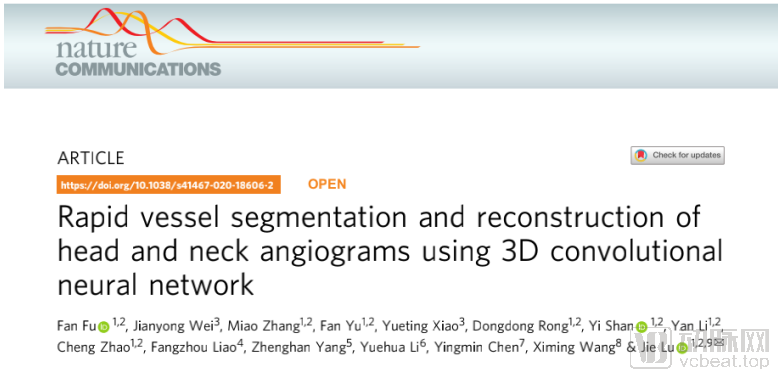
Returning to the paper, the entire experiment can be broadly divided into two parts: model training and model validation. The dataset of 18,259 head and neck CTA cases was collected between June 2017 and November 2018, provided collaboratively by five top-tier tertiary hospitals in China. Based on a calculation of 600 images per case and 10 vascular regions per image, the AI delineation and segmentation of over 100 million vascular regions were performed throughout the experimental process.
To ensure the validity of the sample data, researchers manually reviewed and excluded 507 samples with poor image quality, leaving 9,370 male and 8,889 female participants. All participants were aged 63 ± 12 years.
 Study Design
After completing sample construction, the researchers used the CerebralDoc AI model developed by SHUKUN to perform post-processing reconstruction of the images.
For general deep neural networks, achieving high-standard segmentation in medical images with extremely high precision requirements is quite challenging. For instance, head and neck vessels, particularly intracranial vessels, are so fine that they occupy only dozens or even just a few pixels in an image. Prior to SHUKUN, no superior network capable of large-scale clinical application was available either domestically or internationally.
SHUKUN’s CerebralDoc model fully accounts for the three-dimensional characteristics of CT images and the anatomical features of human tissues and organs. It amplifies various features of the target tissues to be learned (such as blood vessels), enabling comprehensive learning and extraction of the required characteristics during training. SHUKUN’s specially designed training process allows the network to observe blood vessels from both global and local perspectives, thereby continuously enhancing its robustness—a distinctive feature of this approach.
Compared to the previous generations of networks independently developed by SHUKUN, the new neural network has achieved breakthroughs in tracking small intracranial vessels and excluding bone structures.
Guo Ning, head of the SHUKUN Technology Research Institute, told VCBeat, “This experiment has been underway for more than two years. When the model was first established, it was like a newborn child. With the rapid growth of the network, the accuracy and efficiency of image reconstruction gradually improved. By the time the paper was published, the AI’s reconstruction accuracy had approached 100%.”
At the time of publication, the AI’s algorithm achieved performance metrics exceeding 90% for the Dice correlation coefficient, vessel-weighted score, and recall rate.
Specifically, the AI achieved a reconstruction accuracy of 93.1% on an independent test set. In comparison with 152 manually reconstructed cases, the AI reconstruction pass rate reached 92.1%. Furthermore, the vascular boundaries in the AI-reconstructed VR images were smoother than those from manual reconstruction, and the bone removal effect in the Maximum Intensity Projection (MIP) images was superior.

a. Occlusion of the right middle cerebral artery without establishment of collateral circulation; b. Basilar artery aneurysm with thrombosis and calcification, observable in the MIP reconstructed by CerebralDoc; c. Post-atlanto-occipital surgery, metal artifacts in AI images are better suppressed; d. Severe stenosis at the bifurcation of the right common carotid artery and left vertebral artery directly caused by the aorta
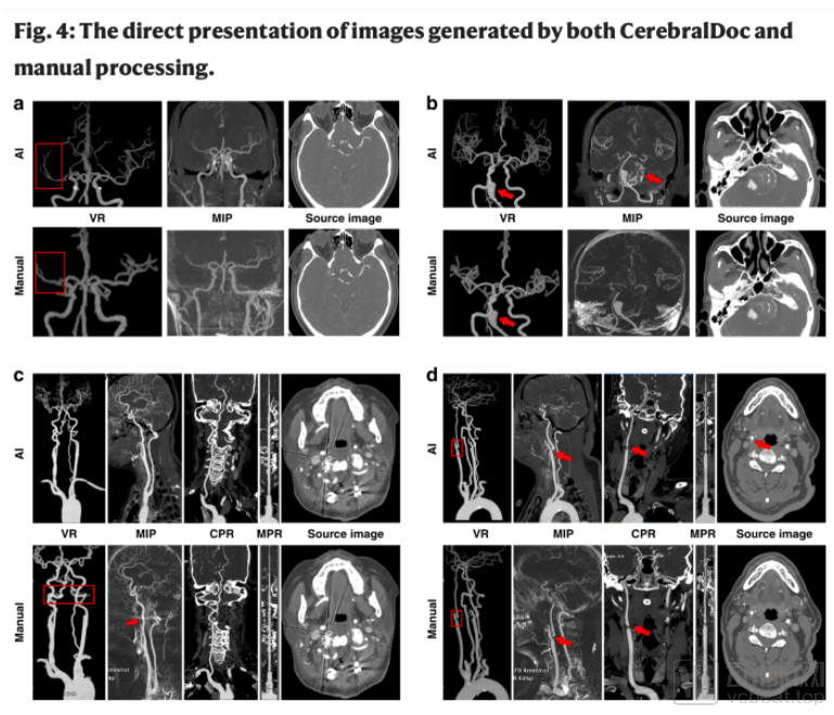
Comparison of Image Quality Between AI Reconstruction and Manual Reconstruction. The first row shows that the AI-reconstructed images have smoother vessel walls and display more distal branches; the second row shows that manual bone removal is significantly affected by scanning artifacts.
In terms of efficiency improvement, AI also demonstrated excellent performance. After the system was implemented at Xuanwu Hospital, the average post-processing time for imaging decreased from 14.22 ± 3.64 minutes to 4.94 ± 0.36 minutes, reducing the time to one-third of the original duration. Meanwhile, the number of clicks required by technicians dropped rapidly due to AI integration, decreasing from 115.87 ± 25.9 to just 4 clicks.
Furthermore, after five months of use, the number of CTA post-processing technicians at Xuanwu Hospital has been reduced from three to one. Guo Ning stated, “The change in technician staffing reflects the growing capabilities of AI. At the beginning of the trial, AI-generated results still required physicians to make corrections and confirmations. However, as the model has matured, physicians have been able to delegate most of the workload to AI, allowing them to focus on more meaningful analysis and research. At this stage, AI technology has become deeply integrated into physicians’ workflows.”
For a long time, we have been unable to quantitatively measure the value that AI brings to physicians, and the research methodology presented in this paper undoubtedly provides a rational pathway.
The experiments have effectively validated the superiority of AI in post-processing head and neck CTA images, and the research team continues to seek further improvements.
Guo Ning told VCBeat, “This experiment excluded cases with artifacts or congenital vascular anomalies, which will be the key focus of our next phase of research.”
This principle applies equally to the broader field of artificial intelligence. After years of development, AI requires continuous in-depth exploration by teams of physicians and scientists to expand its scope and depth of application. Moving forward, we need more such outcomes from industry-academia collaborations to empirically demonstrate the superior efficacy of AI.