UCSF Researchers Leverage NVIDIA GPUs to Enhance Cryo-EM Image Clarity for Accelerated Drug Discovery

NVIDIA
Artificial Intelligence Computing Service Provider
When photographers take photos that require long exposures, they maximize the amount of light received by the camera sensor. This technique helps capture scenes such as the night sky,However, as shown in the figure below, this results in a blurred appearance of the final image.。
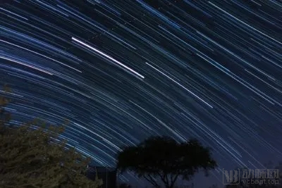
In fact, this technique is not significantly different from cryo-electron microscopy (cryo-EM), which scientists use to study the microscopic molecular structures of specimens vitrified through cryopreservation. While motion blur in photography can yield aesthetically pleasing images, it constitutes an undesirable artifact in structural biology.
Protein samples for cryo-EM are frozen at -196°C to preserve their biological structures, which would otherwise be destroyed by the high-energy electron beam of the microscope. However, even when frozen, the samples are subject to perturbation by the intense electron dose, causing motion that blurs images acquired during long exposures.
To address this issue, researchers at the University of California, San Francisco, used specialized cameras to capture video footage of biomolecules, which remain nearly stationary in each frame. Cross-frame motion correction is a computationally intensive task, but it can be completed in just seconds on NVIDIA GPUs.
Shawn Zheng, a scientific software developer at the University of California, San Francisco and the Howard Hughes Medical Institute, stated, “Without motion correction, we would lose high-resolution images of the 3D structures of molecules. Understanding molecular structure is crucial for comprehending their function.”
Shawn and his colleagues run MotionCor2, the world’s most widely used motion correction application, on NVIDIA GPUs to align each molecule in the video frame by frame, thereby generating clear images that researchers can convert into 3D models.
These 3D models are essential for scientists to understand the complex chains of interactions occurring within individual proteins, thereby accelerating drug and vaccine discovery, such as for the spike protein on the COVID-19 virus.
As a leader in cryo-electron microscopy research, the University of California, San Francisco has consistently been at the forefront of enhancing microscope image resolution. This technology enables atomic-level visualization of proteins, an achievement deemed impossible a decade ago.
However, the entire workflow enabled by this technology is lengthy and involves freezing samples. It requires the use of cryo-electron microscopy (cryo-EM) instruments, which cost millions of dollars, to capture images of the samples. After motion correction, detailed 3D models of the molecules are reconstructed. To ensure the smooth operation of the workflow, it is critical that the motion correction process be sufficiently rapid to keep pace with the rate at which new data are being collected.
Shawn stated, “Cryo-EM is an extremely expensive instrument, and no one wants it to sit idle. However, if our machine’s data storage becomes clogged with a backlog of videos, there will be no space left to store additional footage. This would result in significant underutilization of this costly instrument and delay research on other instruments.”
To achieve rapid motion correction, the Advanced Electron Microscopy Center at the University of California, San Francisco, has equipped each workstation with eight NVIDIA GPUs for every microscope. These workstations are essential to maintain the speed of cryo-EM data collection, ensuring that each microscope can acquire four video segments per minute.
GPU settings enable the simultaneous execution of eight tasks for the iterative motion correction of videos comprising up to 400 frames (nearly 100 million pixels per frame).
To accelerate the development of new applications, Shawn, who has a decade of experience in NVIDIA GPU research, adopted a workstation equipped with two NVIDIA Tensor Core GPUs. This system can analyze 70 GB of microscopy video footage within one minute.
Shawn and his colleagues also use GPUs to run alignment software for cryogenic electron tomography, or cryo-ET. This technique is better suited for studying slightly heterogeneous specimens, such as macromolecules and cells. As the sample is tilted at different angles, a series of collected images can be aligned and reconstructed into detailed 3D models.
He stated that NVIDIA GPUs can fully automate the reconstruction process, with a single GPU requiring only half an hour.
In a recent issue of Science, Shawn collaborated with lead researchers from the Leiden University Medical Center in the Netherlands to use cryo-electron tomography (cryo-ET) to study the molecular pores involved in SARS-CoV-2 replication within cells. A better understanding of this pore structure could help scientists develop drugs targeting it, thereby blocking viral replication in patients.
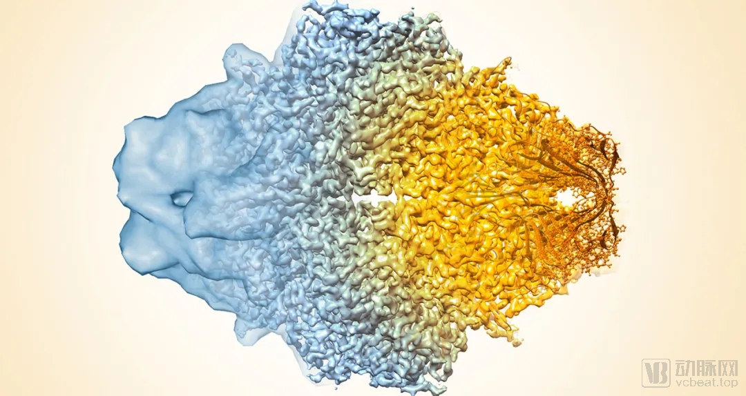
*This image displays the cryo-EM density map of β-galactosidase, illustrating the progressive improvement in cryo-EM structural quality from low to high resolution. Image courtesy of Veronica Falconieri and Sriram Subramaniam, released into the public domain by the National Cancer Institute (NCI).