Gaocheng Capital-Backed Nanovision Unveils World's First Stationary CT at CMEF: A Breakthrough Solution to Overcome the Bottlenecks of Conventional CT Technology?

Nanovision
X-ray Detector and Static CT Product R&D Provider
In 1965, Intel CEO Gordon Moore observed that the number of transistors on an integrated circuit would double approximately every 18 months.
Fifty years later, while Moore’s Law retains its magic, it may ultimately fade away due to the existence of physical limits.
Spiral CT is facing the same issue. The world’s first spiral CT acquisition system, SOMATOM Plus, was introduced in 1988. This CT scanner replaced the previous translate-rotate method, reducing scan time from tens of seconds to just 0.25 seconds.
However, over the next 30 years, spiral CT underwent only two major changes while its fundamental principles remained unchanged. The first was the transition from single-slice to multi-slice data acquisition, accompanied by a continuous increase in detector width. The second was the shift from single-tube to dual-tube systems, which enabled advanced spectral imaging technologies.
Throughout the entire process, the physical factors limiting the development of spiral CT have remained unchanged—The rotational speed of the X-ray tube-detector assembly and the detector width are two major hurdles that are difficult to overcome.
For physicians, the more abundant the information, the more precise the diagnosis. Therefore, since the inception of spiral CT, CT manufacturers have been engaged in a race to increase the rotation speed of the X-ray tube-detector system and widen the detector coverage. However, the competition over rotation speed had largely reached its end by around 2010.
Components such as the X-ray tube, detector, and high-voltage generator can weigh up to 500 kilograms, generating substantial centrifugal force during high-speed rotation around the human body. Generally, the maximum centrifugal force that a CT structure can withstand is 75 G, corresponding to a maximum rotational speed of 0.25 seconds per revolution, at which point the temporal resolution approaches its limit.
On the other hand, the unit pixel size of the detector and the amount of effective information obtained per unit pixel determine the spatial resolution of each CT image acquisition. The more pixels the detector captures in each projection, the more data are available for computerized image reconstruction, resulting in clearer images. Currently, Toshiba’s 320-slice, 640-row CT scanner has compressed the detector width to its physical limit, making further reduction in detector size virtually impossible.
From the perspective of exploring the unknown, spiral CT appears to have reached the limits of its capabilities. If manufacturing processes cannot be optimized, abandoning the “spiral” design may be the only solution.
The industry’s pursuit of more advanced CT systems has never ceased. Prior to the advent of multi-slice spiral CT, Dr. Douglas Boyd in the United States developed a system known as “electron beam CT” in 1983, which was subsequently commercialized by Imatron Corporation. Later, GE acquired Imatron for $1.5 billion, further advancing the research and development of electron beam CT. In the history of CT evolution, electron beam CT is classified as fifth-generation CT.
The X-rays in electron beam CT are generated when a continuously emitted electron beam is deflected by a magnetic field and subsequently strikes a ring-shaped tungsten target.Its distinguishing feature is the ring-shaped arrangement of detectors, which can continuously receive X-rays emitted from the opposite side. With this configuration, the CT scanner achieves an extremely high scanning speed of 50–100 ms, offering unique advantages for imaging the heart and major blood vessels, as it is virtually unaffected by cardiac motion or vascular pulsation.
However, due to its high manufacturing and patient examination costs, as well as excessive maintenance expenses, purchasing electron beam CT (EBCT) is far less cost-effective for hospitals compared to spiral CT. Especially when conventional CT scanners with sub-second scan times and multi-slice spiral capabilities can also obtain high-quality cardiac images, the advantages of EBCT have completely disappeared. Under this declining trend, GE had no choice but to shut down its EBCT product line.
"In the long run, the principles of electron beam CT mean it will not encounter the two current hurdles faced by spiral CT. If its cost can be reduced, it will have unlimited potential."
By 2013, the focus of competition had shifted from the United States to China. Cao Hongguang, Li Yunxiang, and others, with decades of experience in medical imaging, founded in BeijingNanovision, attempting to reinvent a new generation of CT. After years of ups and downs, the founding team, having accumulated extensive experience and lessons learned, gradually charted a clear course and set its goal on developing the sixth-generation CT—static CT.
Nanovision understands that in-house research and development is a crucial step for cost control in achieving the commercialization of static CT, given the high demand for detectors and X-ray tubes. This time, Nanovision finalized the concept of static CT before embarking on the project, starting from the most fundamental core components and progressively completing the R&D of parts such as CT chips, detectors, scintillators, and high-speed narrow-pulse high-voltage generators.
After seven years of dedicated development, the world’s first static CT scanner, developed by Nanovision, was finally unveiled in 2020 and made its debut at the 2020 RSNA Annual Meeting. This static CT system, named “Compound Eye 24” by Nanovision, was publicly displayed for the second time at the currently ongoing 2021 CMEF.
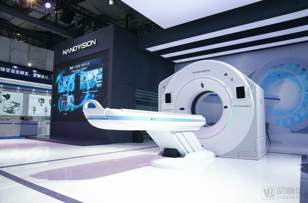
World’s First Static CT “Compound Eye 24” Unveiled at Nanovision Booth During 2021 CMEF
Compared with the structure of spiral CT, the static CT "Compound Eye 24" innovatively adopts a completely different dual-ring structure, including an X-ray source ring and a detector ring.
The X-ray source ring consists of hundreds of independent X-ray sources arranged in a circular configuration. Each X-ray source can be activated independently, supporting simultaneous exposure of up to six sources with sequential switching. Furthermore, the exposure time and energy of each X-ray source can be customized according to specific clinical requirements, meaning that this CT system is not limited to imaging a single anatomical region.
The detector ring is distinguished by its unique, modular photon flux detectors. Unlike the integrating detectors used in conventional spiral CT systems and distinct from photon-counting detectors, this design marks the first international introduction of the photon flux concept into the CT field. Nanovision’s self-developed, highly integrated signal processing module enables hardware-accelerated image acquisition, achieving zero-distortion data transmission.
The dual-ring structure innovatively resolves the limitations of mechanical rotation speed and centrifugal force tolerance in conventional spiral CT scanners by employing stationary imaging. This design enables high-speed rotation of dozens of revolutions per second with ease, while eliminating the trailing artifacts associated with the high-speed rotation of spiral CT systems during scanning. As a result, image capture is significantly clearer, and radiation exposure is substantially reduced. Currently, the detector’s inner layer of the “Compound Eye 24” system is capable of acquiring 288 layers of signal data.
In brief: Compared with top-tier spiral CT, static CT technology offers a 10-fold improvement in temporal resolution and a 64-fold improvement in spatial resolution, while also enabling multi-energy spectral imaging.
According to Nanovision, the “Compound Eye 24” does not represent the limit of static CT technology. In fact, Nanovision’s next-generation static CT system, the “Compound Eye 36,” is currently under development. The new generation will deliver further improvements in temporal resolution, spatial resolution, and the range of elements identifiable via mass spectrometry. Developed entirely in-house, the “Compound Eye 24” can enter the market at a price point comparable to that of high-end CT systems from GPS (GE Healthcare, Philips, and Siemens Healthineers).
A disruptive technological roadmap embodies a strategy of “overtaking on a new track.” Static CT represents a complex system fundamentally different from spiral CT, with none of its required components available for procurement on the open market. The existing international supply chain cannot provide core components essential for static CT, such as chips, detectors, scintillators, compact array X-ray tubes, and narrow-pulse high-voltage generators. Achieving static CT necessitates independently overcoming the technical barriers for each of these core components, thereby realizing this milestone innovation.
“China is the country with the largest number of CT equipment manufacturers globally, yet it has long relied on imports from the United States and Germany for core components such as high-end X-ray tubes and high-end detectors. In other words, domestic enterprises have lacked the capability to independently develop top-tier CT systems,” Cao Hongguang told VCBeat. “To prevent technological chokepoints and achieve self-reliance in upstream core components, Nanovision has consistently pursued this goal and made it the focus of its ongoing efforts.”
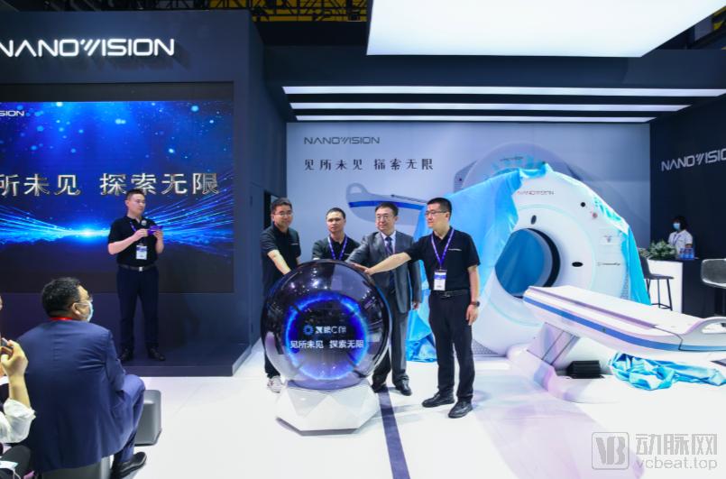
At the launch event of CMEF “Compound Eye 24,” Dr. Cao Hongguang is second from the right.
Therefore, since its establishment, Nanovision has embarked on a series of research and development initiatives for core components of static CT scanners.
First, X-ray segmentation is performed.On spiral CT scanners, scintillators typically provide a complete beam of laser light, which is sliced into individual strips by a grid to achieve pixelation. Since the pixel size of static CT is approximately 1/64 that of spiral CT, this means that to image using the same process, the grid must be further refined.
Regarding this issue, Nanovision has revealed a new material growth technology. Through this technology, Nanovision can cultivate materials with finer resolution than the pixels of static CT, thereby achieving precise segmentation of X-rays.
Next is the core component, the detector.Internationally, it is widely acknowledged that the concept of static CT must be supported by photon-counting detectors. However, Nanovision believes that photon-counting detectors capable of detecting individual photons fail to meet the requirements of static CT. To address this, with the support of the Ministry of Science and Technology, Nanovision has developed the world’s first photon-flux detector, which is key to building static CT systems.
The X-ray tube is independently developed.The core, involving the cost of static CT and its scalability. Since static CT replaces the rotation of spiral CT with sequential exposure switching of a ray source array, there is no need to consider centrifugal force issues. Therefore, the X-ray tubes of the CT are arranged around the human body, with up to 24 tubes in one circle.
Since the number of X-ray tubes is positively correlated with both equipment cost and imaging quality, Nanovision must independently develop technologies to reduce tube costs and miniaturize them as much as possible, thereby enabling the integration of a greater number of tubes to achieve commercialization of static CT.
Miniaturization is no easy feat. For Siemens’ dual-source CT, the internal space becomes fully occupied after installing two X-ray tubes and two detectors. In contrast, Nanovision can integrate 24 sets of X-ray tubes and high-voltage components, demonstrating its design advantages and robust technical capabilities.
The final core component is the narrow-pulse high-voltage power supply.. The X-ray tube in a spiral CT emits continuous X-rays, whereas a static CT does not rotate; instead, each X-ray tube must sequentially emit instantaneous pulsed X-rays. This requires narrow-pulse rapid switching technology to precisely control the flash duration and maximize the flash frequency as much as possible.
The term "switch" may seem simple, but it requires a 140 kV high-voltage power supply to support X-ray emission and demands rapid switching on the microsecond scale. The successful development of this technology has also established an extremely high technical barrier for Nanovision.
With these steps completed, the core technologies of the static CT system have been fundamentally established. However, in Cao Hongguang’s view, the “spirit of craftsmanship” should not lead to neglecting minor details simply because the core components have been manufactured to perfection. “Every core component you see is manufactured in-house, and our supply chain is based entirely within China. This is Nanovision’s steadfast commitment.”
At the CMEF, following the static CT’s debut in China, crowds of attendees flocked around the exhibition booth. Yet two critical questions remained for Nanovision to address: How to achieve mass production? And how to drive future development?
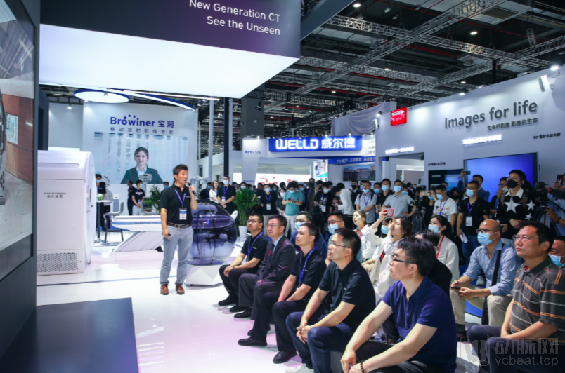
CMEF Nanovision Booth
In March 2020, Nanovision officially commenced construction of its “Static CT R&D and Production Base” in Chengdu. The project aims to establish the world’s first independently developed production line for static CT scanners, addressing mass-production challenges. Upon completion, Nanovision plans to achieve an annual output of 1,000 units, thereby responding to market concerns regarding mass production capabilities.
As for the company’s future development, Cao Hongguang believes that the story behind the name “Nanovision” is its best answer.
“Based on the principles of optical imaging, we can visualize cells at the micrometer scale. For instance, under a microscope, a single blood cell measures approximately 7 micrometers, while a skin somatic cell is around 12 micrometers. In contrast, CT scanning relies on radiation imaging using X-rays, with wavelengths ranging from 0.1 to 10 nanometers. Theoretically, such short wavelengths enable the identification of finer structures within the human body. Therefore, the current ‘Compound Eye 24’ system represents only the beginning; our ultimate vision is nanometer-scale imaging. This is Nanovision.”
As a cutting-edge innovation, static CT is currently in the pre-commercialization stage. With support from national policies, ultra-high-end CT systems are entering a period of market growth. Backed by a global market worth hundreds of billions, Nanovision, as part of the Hillhouse Healthcare portfolio, holds promising prospects for the future.