From Single-Disease AI to Digital Human: How Far Is Shukun Technology from the Next Stage of Medical AI?

SHUKUN
Provider of Intelligent Products and Innovative Solutions
As the largest medical device exhibition in China, the China Medical Equipment Fair (CMEF) Spring Session annually showcases advanced medical devices and equipment from across China and around the world. However, since AI-assisted diagnostic software for healthcare has been classified as a medical device, an increasing number of medical imaging AI companies have begun presenting their artificial intelligence solutions at the event. Non-hardware-focused enterprises are gradually establishing themselves as independent and competitive players in the healthcare market.
From a developmental perspective, the rise of artificial intelligence in medicine appears to be inevitable. Behind this trend, the continuous upgrading and iteration of medical imaging equipment provide an inexhaustible impetus for the advancement of AI.
On one hand, there is a growing expectation to extract more valid information from a single image to assist physicians in making precise diagnoses. For instance, traditional CT imaging could only reveal larger lesions, with its limited resolution restricting further diagnostic interpretation. In contrast, current CT technology can clearly depict every detail of the patient’s scanned anatomical region.
This means that high-resolution imaging can detect smaller lesions, but it also increases the workload for doctors—they need to make more judgments on whether suspicious lesions are benign or malignant. Therefore, AI-based assisted diagnosis has become increasingly necessary due to the upgrade of imaging equipment.
On the other hand, as the precision of imaging equipment continues to improve, the decision-making weight of imaging evidence has increased significantly, leading to a growing demand for medical imaging among patients. In response to this trend, physicians are seeking more efficient, precise, and rapid methods for the interpretation, reconstruction, and analysis of acquired images, thereby addressing the widening gap between the supply and demand for imaging services.
The CMEF also welcomed numerous AI medical imaging companies. During the event, VCBeat interviewed SHUKUN, a leading enterprise in the field of AI-powered imaging. As one of the earliest companies to obtain the Class III Medical Device Registration Certificate for auxiliary diagnosis from the National Medical Products Administration (NMPA), SHUKUN’s growth trajectory offers valuable insights into the development of artificial intelligence in healthcare. The strategic path showcased by SHUKUN at this year’s CMEF may provide meaningful inspiration for the industry.
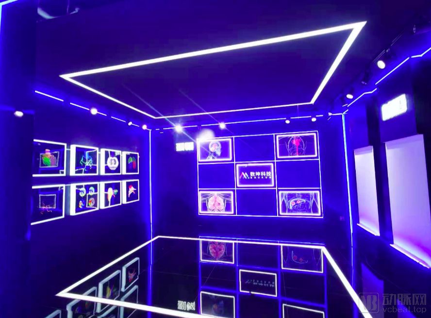
CMEF SHUKUN Exhibition Area
SHUKUN entered the medical AI imaging sector in June 2017, somewhat belatedly. Although no company had yet achieved a proven business model in the pulmonary nodule segment at that time, the large number of participants had already turned it into a red ocean. Therefore, SHUKUN adopted a differentiated R&D strategy, starting with the less-explored cardiac field to pioneer the intelligent analysis of coronary CTA.
Performing coronary CTA is no simple task. First, it is essential to engage senior experts in cardiology who can assist algorithm researchers in dissecting cardiac anatomy, clarifying regional segmentation and vascular delineation. Second, algorithm researchers cannot simply transfer existing computer vision algorithms as they might for pulmonary nodules; instead, they must iteratively develop deep learning-based model reconstruction methods through exploratory research. Finally, and most importantly, unlike conditions such as pulmonary nodules or fundus diseases that benefit from numerous large, publicly available datasets, coronary CTA data are limited, fragmented, and unstructured. This necessitates collaborative efforts between enterprises and physicians to build standardized databases to fuel AI development.
Over the course of two years of exploration, SHUKUN has systematically addressed the three aforementioned challenges. Its coronary CTA solution leverages artificial intelligence to automate processes traditionally performed by physicians, including 3D reconstruction, interpretation, assessment, and report review of coronary CTA images, thereby reducing the image post-processing time from 30–40 minutes to just a few minutes.
Compared with coronary CTA, the reconstruction process of head and neck CTA is more complex, with the increased difficulty stemming from the intricate vascular anatomy of the head and neck in CT images and interference caused by bone visualization.
“Since the skull cannot be excluded during head and neck CT scanning, and given its high density, it appears as bright areas on CT images similar to those produced by contrast agents, with very close Hounsfield unit values. Therefore, physicians must employ specialized methods to differentiate blood vessels from the skull,” explained Guo Ning, Head of the Clinical Research Institute at SHUKUN Technology.
Specifically, to eliminate the impact of skull imaging on reconstruction, physicians often require patients to undergo two CT scans: the first without contrast agent injection and the second with contrast agent injection. In the first CT scan, only the high-density skull structures are visualized, whereas the second scan captures both the skull and blood vessels. After performing both scans, image subtraction is applied to the results; by subtracting the skull and other structures that appear hyperdense in both images, the remaining data constitute the vascular images required for reconstruction.
In practice, such procedures often encounter numerous challenges. First, this approach demands a high degree of patient cooperation; the patient’s position must be precisely aligned between the two scans, and any movement can compromise the quality of the subtraction images. Second, undergoing two CT examinations inevitably increases the patient’s radiation exposure. Although the dose remains within safe limits, it may still raise concerns among patients.
With the integration of AI, the workflow for head and neck CTA has undergone significant changes. This approach eliminates one CT scan, thereby enhancing the patient experience, while also substantially reducing the time required for image reconstruction.
In September, SHUKUN collaborated with Professor Lu Jie’s team at Xuanwu Hospital of Capital Medical University to conduct research on the application value of artificial intelligence in head and neck CTA vessel reconstruction. The research findings, titled “Rapid vessel segmentation and reconstruction of head and neck CTA using 3D convolutional neural network,” were published online in Nature Communications, a Nature portfolio journal.
This is the first large-scale study in China focused on the segmentation and extraction of head and neck vessels. The results not only demonstrate the advantages of convolutional neural networks in post-processing medical images, but also reflect the clinical application value and potential of AI.
。
In summary, the success of coronary CTA and head-and-neck CTA can be attributed to SHUKUN’s effective execution of the following three key strategies.
First, efficiency improvement. For a single disease type with positive revenue, it previously took three days to issue CTA examination results. With AI intervention, patients can obtain results in just half a day, which means that the hospitalization process for each patient will be shortened by two days. This allows hospitals to admit more patients within the same time frame.
Second, quality improvement. The high-precision reconstruction capabilities of coronary CTA and head-and-neck CTA provide physicians with richer clinical decision-making information, indirectly enhancing the quality of medical decisions, reducing patient readmission rates, and thereby lowering the per-disease treatment costs for hospitals.
Third, operational improvement. The implementation of DRG has imposed performance assessment pressures on hospitals; the reduction in treatment costs enabled by the two AI solutions can help hospitals better meet their performance targets.
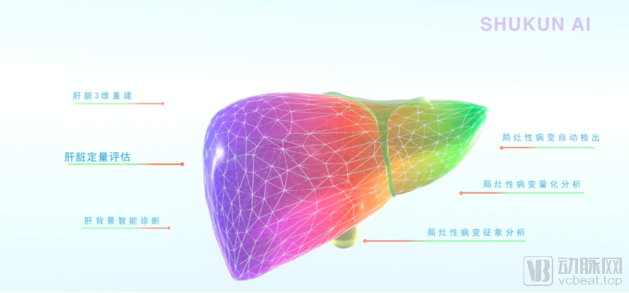
After establishing market recognition through CTA, SHUKUN further expanded its cardiovascular and cerebrovascular product line, successively developing AI products such as head and neck CTA and an intelligent auxiliary diagnosis system for stroke. At this CMEF, SHUKUN once again launched new products, introducing the industry’s first AI-powered solution for liver MR.
Liver disease is a common condition in China, characterized by its insidious onset and rapid progression, posing a significant threat to socioeconomic stability and public health.
Among the myriad of liver diseases, liver cancer is particularly challenging.
According to data from the "2019 Annual Report of Cancer Registration in China," approximately 3.929 million new cases of malignant tumors were reported nationwide in 2015 (data from the National Central Cancer Registry are generally lagged by three years). Among these, there were approximately 365,000 new cases of liver cancer in China, accounting for 50% of the global new cases. Furthermore, liver cancer is the second leading cause of cancer-related deaths in China. Among individuals under the age of 60, it is the most common malignancy and has the highest mortality rate.
Early diagnosis and treatment, prognostic assessment, and personalized therapy for this patient population have remained a focal point of clinical research. Despite the rapid advancement of artificial intelligence technologies, systematic studies in the field of liver tumor diagnosis and treatment are still lacking.
In pathological conditions, patients can maintain their body’s physiological needs even with only 30% of normal hepatocytes remaining. Early-stage liver diseases, such as hepatitis and cirrhosis, do not present obvious symptoms in patients. Consequently, it is difficult for patients to recognize the onset of the disease based on their own sensations; noticeable symptoms typically emerge only when the liver disease has progressed to the middle or late stages. By this time, patients have already missed the optimal window for treatment.
Due to its high soft-tissue resolution, multiplanar imaging capabilities, multiparametric nature, and rich information content, magnetic resonance (MR) imaging has become a commonly used method for hepatic imaging. In recent years, with advancements in MR software and hardware, particularly the development and application of rapid scanning sequences and liver-specific contrast agents, the speed of liver MR examinations has significantly increased, and image quality has continually improved, leading to a marked enhancement in the sensitivity and specificity of liver MRI diagnosis.
“The benefits of liver MRI are obvious, but the drawback lies in the scarcity of radiologists who are proficient in operating MRI and continuously innovating. Due to the inherent complexity of the liver, a single organ can present with numerous types of lesions, and MRI involves many techniques that require learning and interpretation; therefore, the learning and professional development process for radiologists is extremely lengthy.” Professor Yang Zhenghan, Director of the Department of Radiology at Beijing Friendship Hospital, remarked when discussing the shortcomings of liver MRI.
According to Professor Yang Zhenghan, there is a significant disparity in imaging interpretation skills among hospitals of different tiers, and even among physicians within the same tertiary Grade A hospital, particularly across different age groups. Ultimately, this stems from the inherent complexity of MR technology and the considerable challenges in training experienced physicians, resulting in slow professional development and preventing MR technology from realizing its full potential.
“Since high-level experts can make effective diagnoses using MR, this indicates that these diseases still follow discernible patterns,” stated Professor Yang Zhenghan. “We can leverage the image recognition capabilities of computers to establish an effective model, thereby transferring the knowledge and experience of highly skilled and experienced physicians to machine learning systems, enabling artificial intelligence to automatically identify liver lesions.”
Faced with the talent shortage in MR, Yang Zhenghan turned to SHUKUN. Previously, Beijing Friendship Hospital and SHUKUN had already engaged in in-depth collaboration in the field of AI for coronary CTA. In Yang Zhenghan’s view, SHUKUN was the only suitable partner for developing an AI-assisted diagnostic system for liver MR.
Upon recognizing the demand, SHUKUN swiftly engaged in the collaborative development of MR solutions. Compared to coronary CTA, the liver presents a far greater level of complexity, with numerous types of diffuse lesions and over a hundred varieties of focal lesions. Nevertheless, leveraging its extensive experience in AI development, SHUKUN successfully developed an AI-powered liver MR product within a few months, achieving considerable success in clinical applications.
“SHUKUN previously developed an intelligent AI-assisted diagnostic system for coronary CTA, achieving excellent performance in vascular segmentation. Since hepatic page-based segmental analysis is based on the vasculature, we can perform 3D reconstruction after extracting the blood vessels, thereby establishing standardized models for subsequent procedures such as liver transplantation and hepatobiliary surgery. Once the vascular structures are delineated, the non-vascular regions correspond to lesions. We tested our trained model, with mean Dice scores exceeding 0.9 and the highest reaching above 0.96, which is sufficient to address general clinical applications at present,” said Professor Yang Zhenghan.
A Review of the History of Artificial Intelligence in Medical Imaging. With continuous advancements across AI generations, the application scenarios of intelligent medical imaging have expanded from radiology departments to multiple clinical specialties.
From pulmonary nodules to cardio-cerebral CTA, the application scenarios of imaging AI remain confined to detection, reconstruction, printing, and reporting within radiology departments. In contrast, one-stop solutions for stroke and liver MRI extend beyond radiology applications, transforming imaging AI into a tool that enables clinical departments to shift from image-dependent decision-making to image-guided treatment planning.
Throughout this generational iteration, the scale of image processing by the AI engine has evolved from 200+ single sequences, to 250–700 single sequences, then to 1,000–2,000+ multi-sequences, and finally to 2,000–3,000+ multi-parameter, multi-sequence datasets.
However, whether in head and neck CT or liver MRI, companies such as SHUKUN have made significant breakthroughs in both the depth of AI technology and the breadth of application scenarios, keeping pace with advancements in imaging technology, and have successfully applied these innovations in clinical practice. Nevertheless, the entire AI industry remains in a “point-specific breakthrough” mode, failing to form hospital-level solutions.
So, where will the next phase of AI in medical imaging be?
Different companies hold varying perspectives on this issue, but from SHUKUN’s standpoint, the “Digital Human” represents the future of imaging AI.
“Digital Human” is not merely about digitizing human body data. In SHUKUN’s view, next-generation artificial intelligence should break through the limitations of individual departments, starting from patients’ physiological functions and adopting a holistic perspective to detect potential diseases.
Specifically, SHUKUN integrates its digital products for the “Digital Heart,” “Digital Brain,” “Digital Chest,” “Digital Musculoskeletal System,” and “Digital Abdomen” to establish the foundation of its “Digital Human” platform. Currently, the Digital Human platform is powered by multi-modal imaging technologies, including non-contrast cardiac CT combined with CT angiography, non-contrast chest CT, non-contrast head and neck CT combined with CT angiography, cerebral CT perfusion imaging, and non-contrast plus contrast-enhanced liver MRI. It encompasses intelligent imaging solutions for multiple anatomical regions, such as the cardiovascular system, cerebrovascular system, lungs, liver, and musculoskeletal system. These capabilities are applicable to a wide range of clinical scenarios, including coronary artery disease, cerebral infarction, tumors, trauma, chest pain centers, stroke centers, and cardiology specialties, thereby forming a comprehensive hospital-level intelligent imaging solution.
Through integration, this solution combines morphological and functional disease detection to generate disease risk predictions, and leverages structured anatomical, physiological, and pathological databases to establish an imaging platform-supported multidisciplinary diagnosis and treatment model.
Looking back on SHUKUN’s development journey, the key to its success lies in uncovering genuine yet often overlooked clinical needs. These needs are not merely the individual desires of hospitals, patients, or physicians, but rather shared priorities among all stakeholders. This alignment has enabled SHUKUN’s products to effectively address two critical objectives: enhancing efficiency and achieving commercial viability.
Next is the continuous maturation of technology. When performing coronary CTA, a single case requires processing approximately 200 images; subsequent head and neck CT scans can reach nearly 1,000 images. Today, liver MR imaging requires SHUKUN to process thousands of images simultaneously. Therefore, for medical imaging AI, although we often discuss breakthroughs in commercialization, technology itself remains the core competitiveness of every enterprise.
In the long run, SHUKUN’s Digital Human is essentially reshaping clinical workflows. This is a challenging endeavor that requires concerted efforts from healthcare institutions, medical device manufacturers, and artificial intelligence companies alike, to gradually foster acceptance of digital diagnostic and treatment processes among physicians and patients. While this transformation takes time, it will truly unlock the value of medical data.