Tuge Medical Launches China's First Native Dual-4K Fluorescence Endoscopy Camera System — A Milestone in Hard-Tech Innovation
Recently, following its original 4K endoscopic camera system, Tuge Medical has once again pioneered innovation by launching the first-of-its-kindNative Dual 4K FluorescenceEndoscopic camera system approved for market launch, with simultaneous CE and FDA certifications. Independently developed, achieving breakthroughs and mastering2μm Ultra-High-Precision 3D Registration Technology, reaching an advanced global level,It has filled the long-standing gap in core technologies for high-end medical imaging equipment, further accelerating the pace of domestic substitution for high-end medical endoscopes.Letters of intent are pouring in.
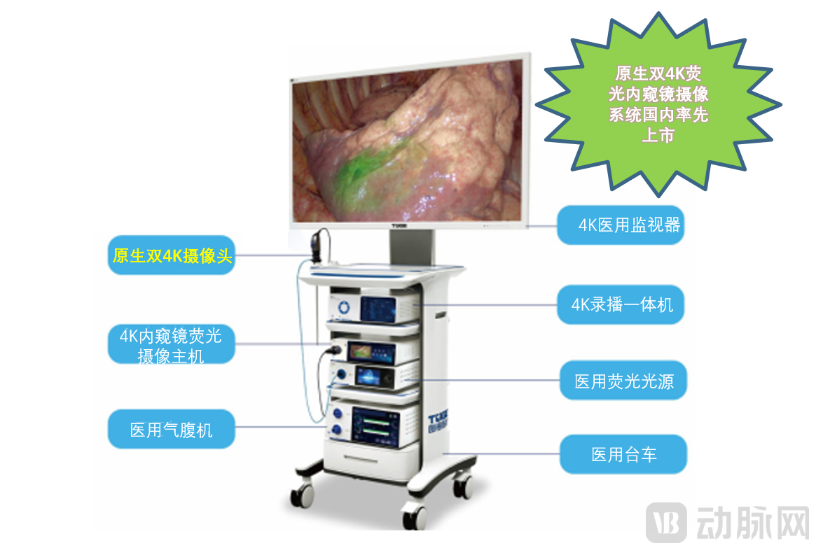
The Tuge Medical Native Dual 4K Fluorescence Endoscopic Camera System FeaturesFive Key Technical Highlights: Clarity, Precision, Speed, Stability, and IntelligenceFor example, in terms of “speed,” Tuge Medical’s integrated system has a latency of only slightly over 40 milliseconds, whereas a certain foreign brand’s system exhibits a latency of approximately 110 milliseconds. These technological highlights are designed to better serve clinical practice, providing physicians with “keen insight.”
This system’s dual 4K imaging pathways ensure true 4K imaging, providing not only excellent “visual acuity” but also enablingFull Autoclaving of the CameraAs one of the few manufacturers worldwide capable of meeting this requirement, Tuge Medical strives for perfection by fully considering doctor-patient relationships and hospital infection management, thereby ensuring the safety of patients seeking medical care and reducing the risk of cross-infection among them.
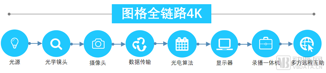
Furthermore, this system features a variety of advanced capabilities:Dual-Endoscope Integration and Multi-Screen Asynchronous DisplayFacilitates multi-device image fusion for observation; flexible switching among white light, fluorescence, fusion, intensity, and hybrid modes; features three types of multi-channel 4K transmission interfaces, multi-channel 4K@60Hz photo and video capture, real-time 4K streaming output, superconducting fiber-optic transmission, and TurbOR4K multi-party remote interaction capabilities, benefiting hospitalsSurgical Demonstration and Remote Consultationof significant value.
4K+Fluorescence imaging technology involves injecting indocyanine green (ICG) into target blood vessels or tissues. Leveraging the property of ICG to emit fluorescence at different wavelengths after absorbing near-infrared light, image sensors capture the fluorescent signals and transmit them to a camera control unit for processing. This enables real-time visualization on a monitor, facilitating intraoperative targeted marking of specific tissues or tracking of blood flow. ThroughIntraoperative MarkingandFluorescence NavigationFunctionality: Real-time, precise localization of organs, tumors, nodules, and lymphatic system visualization, assisting surgeons in performing accurate resections during surgery. It is widely applied in various medical specialties, including hepatobiliary surgery, thoracic surgery, gastrointestinal surgery, urology, and gynecology.
The 4K fluorescence endoscopic camera system approved for market launch adoptsNative Dual 4K CMOS Technology, the inputWhite LightandNear-Infrared Light (NIR)Perform spectral imaging, utilizingSelf-developed optoelectronic registration algorithmand2μm Ultra-High-Precision 3D Registration Technology, integrating true 4K white light with fluorescence, offering advantages such as high clarity, large data volume, rich details, and high color fidelity, thereby completely resolving the inherent limitation of single-CMOS sensors in simultaneously achieving high-resolution, clear imaging for both white light and fluorescence, such as “Dim image quality and weak fluorescence effect” and other core issues, it has also overcome the “limitations of 4 CMOS due to the adoption of full-HD imaging sensors”Insufficient resolution,Poor Consistency” and other bottlenecks.
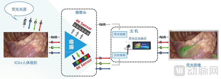
Schematic Diagram of Fluorescence Imaging System
As illustrated in the schematic diagram of the fluorescence imaging system, such systems generally acquire both white-light and fluorescence images simultaneously. These signals undergo separate photoelectric conversion followed by corresponding image processing. The fluorescence imaging system primarily consists of a fluorescence light source, camera, main processing unit, and display. A fluorescence light source of specific wavelengths illuminates human tissue containing contrast agents. The camera captures tissue images through an optical pathway (comprising components such as mirrors and optical adapters) and separates the white-light and fluorescence images via spectral splitting. Subsequently, these images are individually processed, fused, and displayed to provide surgeons with precise intraoperative imaging.
The fluorescence endoscopy camera system is the “crown” of minimally invasive surgical imaging equipment, and the spectral splitting module is the pearl in this crown. The terms “challenging,” “critical,” and “high-value” all aptly describe the spectral splitting module. The Tuge Medical team has risen to these challenges, striving for excellence.
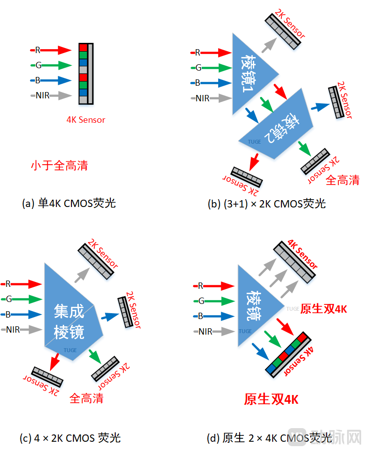
Schematic Diagram of Common Fluorescence Imaging Spectral Techniques
As illustrated in the schematic diagram of common fluorescence imaging spectral splitting principles, various solutions are currently available on the market. Figure (a) depicts imaging using a single 4K CMOS sensor, which eliminates the need for spectral splitting prisms and precise sensor alignment, thereby simplifying implementation. However, this approach fails to accurately separate fluorescence from white light, and the resolution of the R/G/B/NIR channels is limited to 2K (Full HD), resulting in low image clarity and poor image quality. Figures (b) and (c) employ spectral splitting prisms to optically separate the R/G/B/NIR channels of the image, followed by separate imaging using four 2K sensors and subsequent image fusion. While sharing the same fundamental principle, this method avoids issues such as crosstalk caused by poor optical separation in Figure (a). Nevertheless, it remains essentially a Full HD imaging system. Specifically, Figure (b) utilizes a mature 3-CMOS white light optical path with two separate prism assemblies, which presents relatively lower technical difficulty but often leads to poorer consistency. In contrast, Figure (c) adopts a more scientifically rigorous integrated prism design, which is technically more challenging but offers superior consistency; this configuration represents the primary board kit solution promoted by certain overseas vendors.
Figure (d) employs a beam-splitting prism and utilizes two sets of 4K sensors for separate data acquisition. It demands four times the registration accuracy of Figures (b) and (c), with twice the amount of valid data. Although this approach presents greater technical challenges, it delivers superior performance in key metrics such as resolution and consistency, thereby better meeting clinical needs. This configuration is also the choice of a few leading-tier brands.
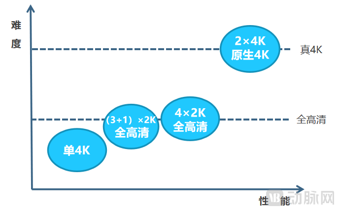
Comparison Chart of Common Fluorescence Imaging Protocols
As shown in the comparison chart of common fluorescence imaging solutions, the native dual 4K imaging solution is highly challenging to implement and is referred to as the “crown jewel,” offering superior performance that meets the demands of precision clinical surgery.
The beam splitter module demands stringent technical specifications across all stages, including product design, R&D, and manufacturing. Our R&D team spent nearly ten months visiting optical manufacturers in Germany, Japan, Italy, and other countries to evaluate comparable products. After rigorous assessment and repeated communications, it was found that most beam splitter modules from these countries offered only full HD resolution and lacked upgradability or expandability. Some vendors’ components exhibited even poorer performance, falling far short of the design requirements for native 4K fluorescence endoscopes. Moreover, there was a prevailing consensus that our proposed specifications were excessively high, with claims that the required precision, resolution, and spectral characteristics were unattainable.
“Can’t find it? We’ll tackle it ourselves, no matter how difficult!” Upholding the corporate mission of “popularizing hard technology to safeguard lives,” the R&D team resolved to conquer this challenge independently.After hundreds of technical seminars and more than 1,000 days and nights of arduous work in the laboratory, this technical bottleneck has finally been overcome, successfully resolving the challenges in white light and near-infrared light imaging.Ultra-High-Definition Micron-Level Image RegistrationofThese technical breakthroughs have reshaped the perception of Tuge Medical among optical manufacturers both in China and abroad, while also giving the Tuge team a profound sense of achievement akin to “holding fast to the green mountains without letting go, where the most breathtaking views lie at the perilous peaks.”
Tuge Medical’s Minimally Invasive Chinese “Chip”
The global “chip shortage” crisis continues to spread. According to Goldman Sachs investment analysis, 169 industries worldwide have been significantly impacted by the chip shortage to varying degrees. Amid this severe “chip shortage,” Tuge Medical has focused on hard-core technologies, shouldering the responsibility of developing China’s own “chips” for minimally invasive procedures, dedicating itself to advancing key technologies in high-end endoscopy, and successfully achieving“Core” — FPGA-Based Real-Time Image Processing System and “Technique” — Image Processing AlgorithmsAchieved a major breakthrough with fully independent intellectual property rights, creating the Chinese “core” for minimally invasive surgery that embodies “rapid chip performance and superior surgical outcomes, integrating chip and technique,” and leading the “core” future of 4K fluorescence imaging technology through intelligent manufacturing in China.
Although many companies directly procure foreign circuit boards to rapidly launch products, and many have advised Tuge to adopt this short, flat, and fast approach—with its low comprehensive cost and risk, enabling the quick release of a “good enough” product—Tuge has not taken shortcuts. Instead, it has persisted in independent research and development, closely monitored technological frontiers, and continuously invested various resources, determined to create high-quality products. The journey has been arduous, but Tuge has never wavered.
By comprehensively considering key factors affecting visual image quality, such as resolution, color deviation, white balance, signal-to-noise ratio, exposure, distortion, and chromatic aberration, Tuge Medical has boldly ventured into “uncharted territory,” taken the lead in China’s “chip” development, and successfully establishedAutonomous and Controllable End-to-End 4K Product Pipeline, equipping domestically produced endoscopes withChina's "Chip"”, which has thoroughly resolved the “chokehold” problem facing China’s high-end endoscopes. Moreover, this core imaging technology can be extended to other high-end imaging applications, earning encouragement and praise from Academician Wu Guanghui of the Chinese Academy of Engineering!
Dr. Wang Yangang, the founder, once said, “The journey of developing high-end endoscopes has been far more arduous than I initially imagined. At first, we were fearless out of ignorance; later, we found ourselves in a position where turning back was no longer an option. What made it even more challenging was the uncertainty surrounding when, or if, our R&D efforts would ultimately succeed. Nevertheless, we firmly believed that someone needed to explore and innovate in this field. Chinese doctors and patients are in need of advanced technologies. We are neither unintelligent nor lacking in focus. Moreover, the broader environment has become increasingly supportive of breakthroughs in core hard technologies. By remaining committed to delivering high-quality products, we hold fast to the conviction that although the road is long and fraught with obstacles, perseverance will lead us to our destination.”
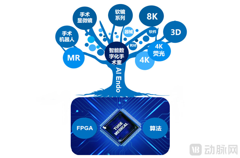
The domestic medical endoscopy industry in China started relatively later than its counterparts in Europe, the United States, and Japan. It has faced numerous challenges, including a shortage of R&D talent, high technical barriers, significant development risks, and scarce interdisciplinary resources. As a result, foreign brands have long monopolized more than 90% of the domestic market share. “Development is the top priority, talent is the primary resource, and innovation is the key driver.” From an initial team of six founders starting from scratch, the company has grown into a team of nearly 100 members today, while also attracting seasoned industry elites to join. This has enabled qualitative leaps in both core technologies and talent development, rapidly enhancing the team’s overall innovation capability. The company possesses reservesOver 70 intellectual property rights, including 39 invention patents and 5 PCT invention patents.Products Coming Soon:4K+3D+Fluorescence Endoscopic Camera System, 8K Medical Imaging System, Flexible Ureteroscope, Flexible Cystoscope, Electronic Choledochoscope and Electronic Bronchoscope, Surgical Demonstration Classroom Software, Medical Imaging Workstation Software, etc.
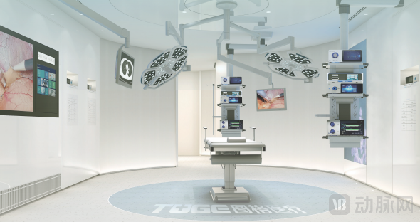
Tuge Medical Innovation Center (Shanghai) Has Established a 4K Endoscopy Demonstration and Experimental Platform
Meanwhile, Tuge Medical has established strategic partnerships with leading domestic and international brands, and has engaged in exchanges and collaboration with numerous research institutes and medical institutions, including the Institute of Automation of the Chinese Academy of Sciences, Southeast University, Fudan University Shanghai Cancer Center, and Zhongnan Hospital of Wuhan University. The company has received guidance and support from many experts, and willContinuously Strengthening Industry-Academia-Research-Medicine-Engineering Collaboration, actively participate in national scientific research projects and local key R&D programs.
Tuge Medical has consistently remained at the forefront of deep-tech innovation, adhering to the philosophy of “entering through narrow gates, treading long paths, and seeking faint glimmers of light.” Dedicated to imaging excellence and the pursuit of knowledge through rigorous investigation, we empower physicians with insightful vision, aspiring to be a leader in medical imaging equipment and a key contributor to the advancement of minimally invasive surgery.