AI-Powered Pathology Breakthrough: Thorough Insights Platform Demonstrates Clinical Impact in Gastric Cancer Diagnosis, Published in Nature Subjournal
Pathological diagnosis is critical to clinical treatment and is regarded as the “gold standard” of medical diagnosis, with pathologists often referred to as “doctors’ doctors.” Regrettably, there is a severe global shortage of pathologists. In China, for instance, only approximately 10,000 pathologists are registered, falling nearly tenfold short of the National Health Commission’s recommended minimum of 100,000. The lengthy training period for pathologists, typically spanning five to ten years, further exacerbates this严峻 reality. This shortage of pathological expertise has given rise to a century-long challenge in pathological diagnosis, urgently necessitating technological innovations to transform the current landscape of pathological diagnosis.
In recent years, traditional pathology departments worldwide have embarked on a digital and intelligent transformation. An increasing number of large hospitals and medical centers have begun digitizing some or all of their physical glass slides, enabling diagnoses to be performed entirely on digital pathology slides. The widespread global adoption of whole-slide imaging has laid a solid foundation for the development and large-scale application of smart pathology systems. It is foreseeable that over the next three to five years, pathological diagnosis worldwide will undergo transformative changes, a process referred to as the “new infrastructure for tumor diagnosis.”
In a study published in Nature Communications in August 2020, we reported an AI-assisted diagnostic system for gastric pathology applied in clinical research at the Chinese PLA General Hospital. The system achieved nearly 100% sensitivity and 80.6% specificity on more than 3,000 real-world test slides from the Chinese PLA General Hospital. Meanwhile, multicenter testing using samples from the Cancer Hospital of the Chinese Academy of Medical Sciences and Peking Union Medical College Hospital demonstrated the stability of this system.
We believe that the sensitivity and efficiency of artificial intelligence (AI) will complement the extensive knowledge of pathologists. In this study, we propose a rigorous multireader-multicase (MRMC) experimental protocol to evaluate the diagnostic performance of 16 pathologists with and without AI assistance. The results demonstrate that AI assistance significantly improves the diagnostic sensitivity of pathologists while substantially reducing slide interpretation time.
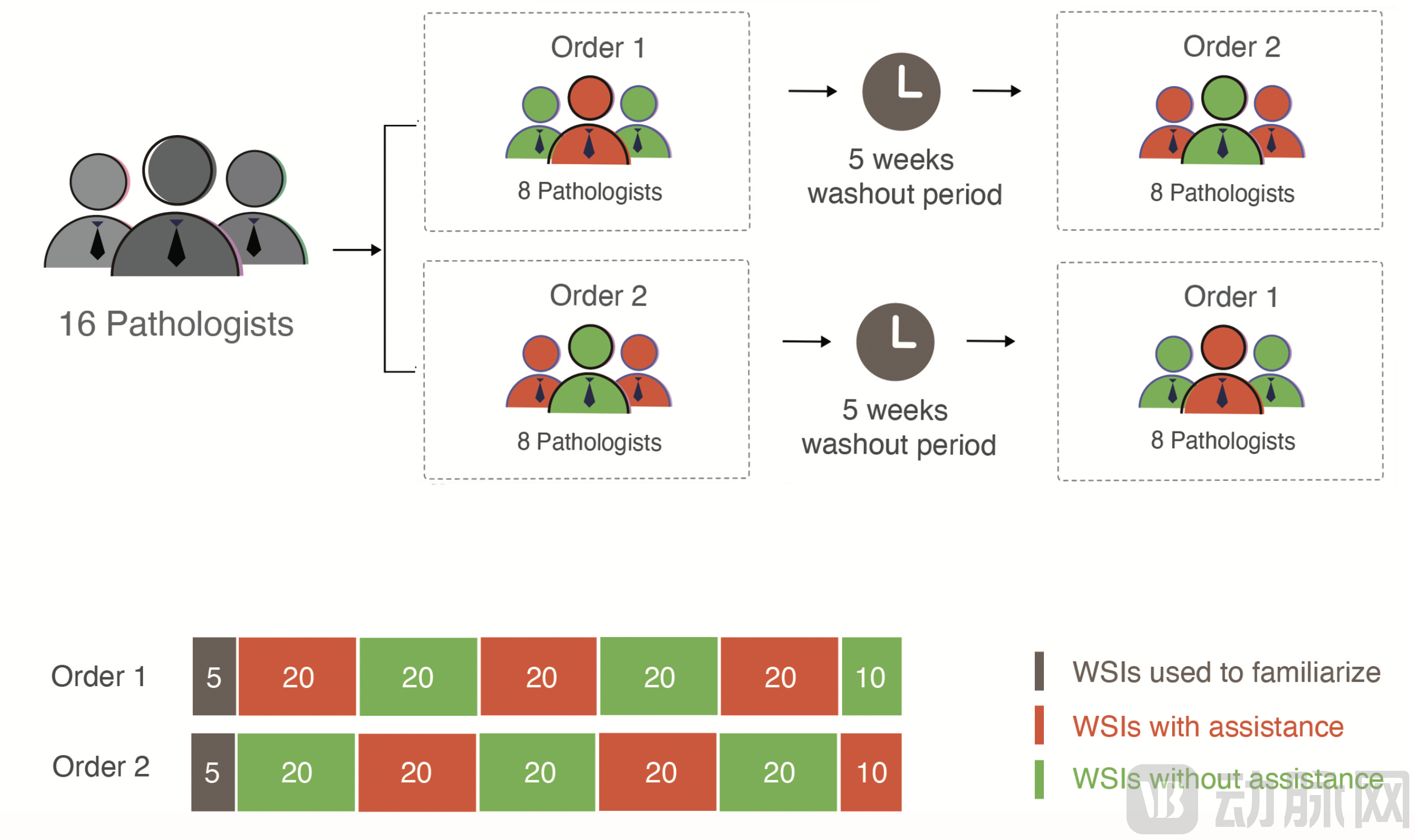
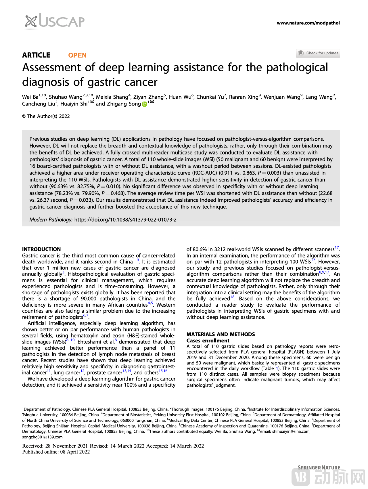
Figure 1. Experimental Design
This study enrolled a total of 110 gastric biopsy pathology specimens, with one slide selected from each case, comprising 50 malignant and 60 benign samples. Sixteen pathologists diagnosed the benign or malignant nature of all samples both with and without artificial intelligence (AI) assistance, categorizing each sample into one of four types: definitely malignant, probably malignant, probably benign, and definitely benign. During the diagnostic process, the assistance mode (with/without AI) was switched every 20 samples, with a five-week washout period in between. For samples with diagnostic assistance, heatmaps highlighting malignant tumor regions could be toggled on or off by pressing the spacebar on the keyboard. To closely mimic clinical diagnostic workflows, there was no time limit for diagnosis; pathologists were allowed to take breaks during the testing process, and such break times were excluded from the total diagnostic duration. This experimental design enabled us to compare the diagnostic results of each pathologist for each sample under conditions with and without AI assistance.
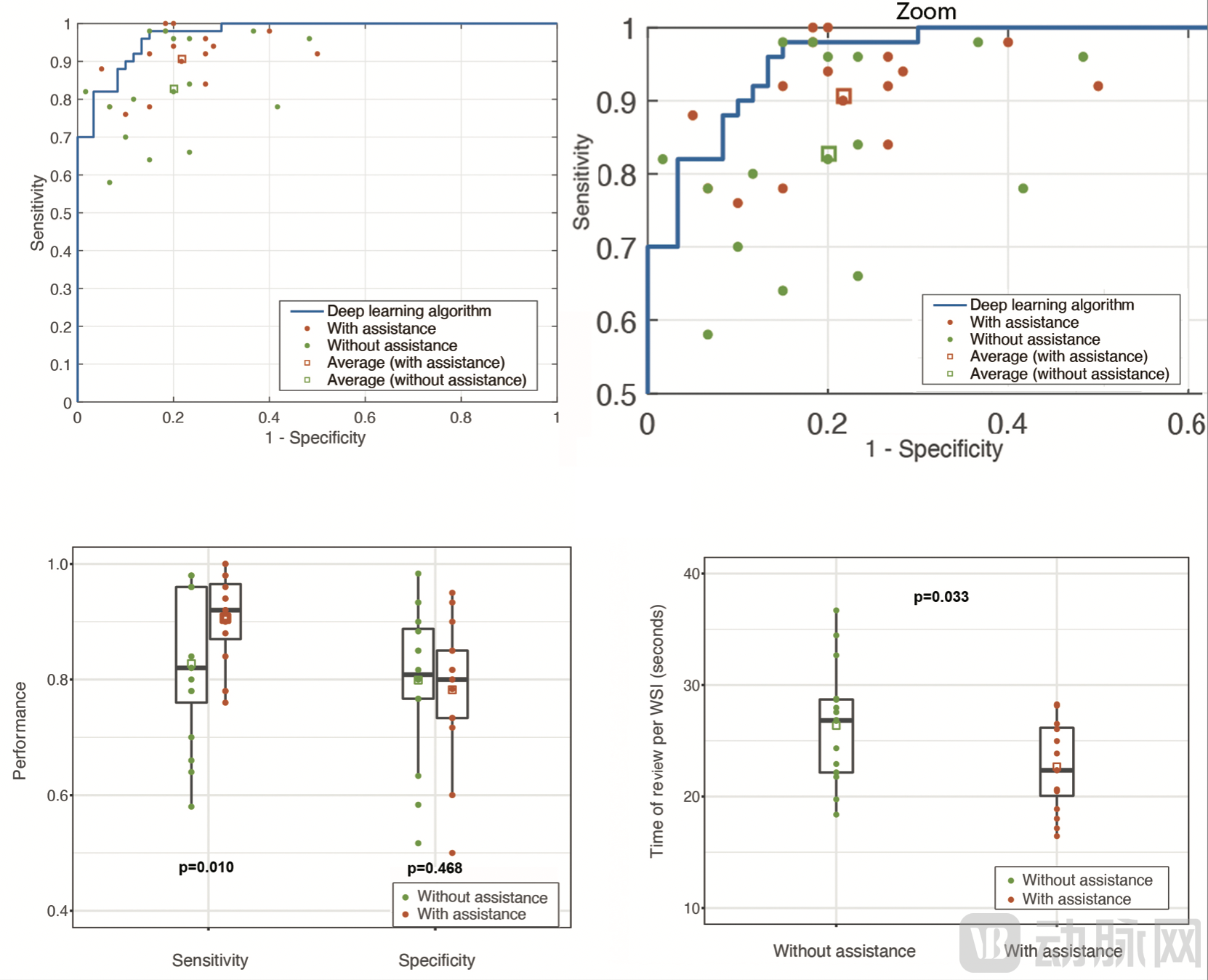 Figure 2. Experimental Results
Data indicate that pathologists assisted by artificial intelligence demonstrate higher diagnostic sensitivity (90.63% vs. 82.75%, P=0.010) and a significantly reduced average diagnosis time per sample (22.68s vs. 26.37s, P=0.033). Notably, AI can substantially improve diagnostic accuracy in cases with small tumor regions or diagnostic challenges.
Figure 3. Pathologists' Evaluation of the AI Platform
Looking ahead to the future of pathology, digital pathology and artificial intelligence will propel traditional pathology into the era of smart pathology. Thorough Imaging has applied cutting-edge AI technologies to the diagnosis of tumors in organs with high incidence rates among the Chinese population. Diagnostic modules for stomach, intestine, lung, prostate, and pan-organ lymph node metastatic carcinomas are already integrated into the Thorough Insights platform, while endometrial and cervical diagnostic modules will be released shortly. In addition to identifying malignant tumors, the AI platform can automatically perform subtyping of malignancies and recognize benign lesions.
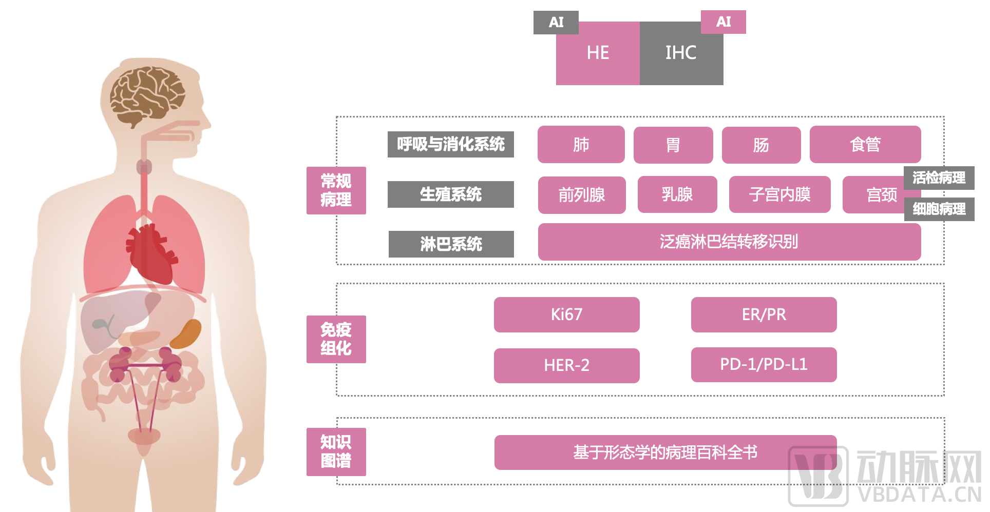
Figure 4. Thorough Insights | In-Depth Analysis
In the near future, by integrating information from pathological superimposition tests, radiology, and clinical manifestations, we will be able to leverage artificial intelligence to provide more personalized diagnosis and treatment services for patients, thereby improving the detection and cure rates for cancer patients.