KR Pharmtech Successfully Hosts NGP-Focused Satellite Symposium on Tumor Microenvironment at 2021 ELab Conference
On September 28, the 2021 ELab Yimao Laboratory Construction and Development Conference and the 5th China Medical Laboratory Summit were successfully held in Nanjing. The “Panorama·Micro-Exploration Thematic Satellite Symposium,” hosted by Kuoran Gene, took place as scheduled. Kuoran joined pathologists, clinical experts, and peers in discussing the clinical applications and development of “multiplex fluorescent immunohistochemistry technology.”
The conference is honored to have Professor Guo Lingchuan, Director of the Department of Pathology at The First Affiliated Hospital of Soochow University, serve as Chair of the satellite symposium, and Professor Wang Hualiang, Dean of Shanghai Institute of Laboratory Medicine and Director of Shanghai Clinical Laboratory Center/Quality Control Center, serve as a Distinguished Guest. Keynote speakers include Director Yang Jun, Head of Molecular Pathology at Nanjing Drum Tower Hospital; Dr. Qian Bangguo, Senior Product Expert at AKOYA; Mr. Chen Yongwu, Medical Director at Kuoran Genomics; and Dr. Pang Zhimin, Technical Expert at IDT.
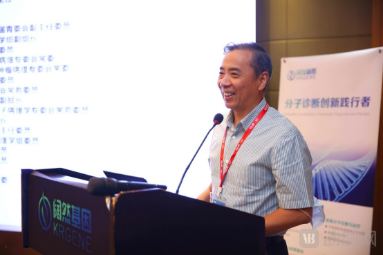
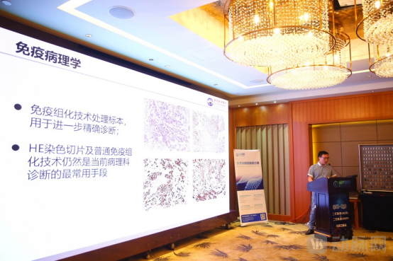
Delivering the opening address as Conference Chair, Dr. Guo Lingchuan, Director of the Department of Pathology at The First Affiliated Hospital of Soochow University, emphasized that immunohistochemistry (IHC) plays a crucial role in the histopathological diagnosis of tumors. He pointed out that only through IHC analysis of tumor tissues can tumors be definitively characterized and graded. Although IHC is widely used in pathological diagnosis, it has certain unavoidable limitations. For instance, IHC can detect only one or two markers per slide (excluding DAPI), making it difficult to fully capture the true characteristics of highly heterogeneous tumors. While multi-marker staining can be achieved through serial sectioning, this approach fails to accurately analyze correlations between proteins. Most importantly, result interpretation relies primarily on manual qualitative and semi-quantitative assessment, introducing a degree of subjectivity. In contrast, Next-Generation Pathology (NGP) technology enables simultaneous detection of multiple markers on a single slide. Coupled with analytical software, NGP allows for absolute quantitative analysis of multiple markers, offering high reproducibility and efficiency. More importantly, it can analyze cellular composition, functional states, and cell-cell interactions. We believe that Next-Generation Pathology (NGP) will significantly facilitate the development and transformation of tumor pathological diagnosis.
Chairman’s Address by Director Guo Lingchuan
Subsequently, President Wang Hualiang delivered the leadership address. President Wang stated that the diagnosis and treatment of cancer represent one of the major global challenges, and research into the tumor microenvironment (TME) has emerged as a cutting-edge field in recent years, offering new hope for cancer diagnosis and therapy. A growing body of evidence indicates that the expression patterns and functions of immune- and tumor-associated molecules within the TME are critical for identifying patient populations most likely to benefit from immunotherapy. A novel multiplex immunohistochemistry/immunofluorescence (mIHC/IF) staining technique enables the acquisition of multichannel information regarding cellular composition and spatial arrangement, thereby facilitating high-dimensional analysis of the tumor microenvironment. Research findings suggest that TME-based assessments may outperform existing biomarkers. Detection of the tumor microenvironment using multiplex immunohistochemistry technology allows for a deeper understanding of tumorigenesis mechanisms and enhances the ability to predict tumor response to treatment.
From the introduction of new technologies to their clinical application, the process involves evaluating test performance, conducting clinical validation, establishing quality standards and protocols, and achieving ongoing consensus among clinical experts. As a pioneer in this field, Kuoran Genomics has undertaken substantial preliminary work; however, the widespread adoption of a diagnostic technology requires collaborative efforts from stakeholders across the entire industry.
We believe that in the future, the integration of traditional molecular testing techniques with novel technologies (NGS + NGP) will bring new perspectives to molecular pathology and clinical molecular research.
Dean Wang Hualiang Delivers Opening Remarks
Next, Dr. Qian Bangguo from AKOYA shared insights on the role of their technology in tumor diagnosis, treatment, and intervention from a technical perspective. Dr. Qian noted that current methods, whether NGS or IHC testing, fail to meet clinical needs for improving tumor treatment response rates. Multiplex fluorescent quantitative pathology analysis technology can detect up to nine colors on a single slide, enabling the acquisition of more information from a single test.
Large-scale biomarker comparative regression analysis indicates that multi-marker technology is superior to other immunotherapy-related biomarkers in predicting drug efficacy.
Dr. Qian then introduced several cutting-edge publications that applied this technology to study the tumor microenvironment, highlighting that mIHC offers significant advantages in acquiring in situ spatial information and characterizing targets, making it particularly suitable for applications in pathology departments.
Remarks by Dr. Qian Bangguo
Director Yang Jun, Head of the Molecular Pathology Group at Nanjing Drum Tower Hospital, delivered a keynote speech titled “The Development History and Trends of Pathological Technology,” reviewing the historical evolution and future trends of pathological techniques. Director Yang first traced the origins and development of pathological technology, highlighting how its advancement has driven progress in the field of pathology, and compared the advantages and disadvantages of modern molecular pathological techniques. He identified three key trends shaping the future of pathology: first, integrated diagnosis, which combines traditional pathological diagnosis with molecular pathology and ultrastructural pathology; second, multiplex immunohistochemistry (mIHC), hailed as the “next-generation pathological technology,” which provides a comprehensive view of the tumor microenvironment; and third, the organic integration of digital pathology with artificial intelligence (AI). These three elements collectively define the future direction of the pathology discipline.
Keynote Address by Director Yang Jun
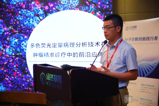
Next, Chen Yongwu, Medical Director of Kuoran Genomics, delivered a presentation titled “Introduction to the Application of Next-Generation Pathology (NGP) in Clinical Oncology Testing and Research.”
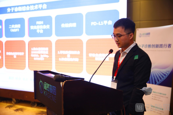
Dr. Chen introduced the comprehensive multiplex fluorescent immunohistochemistry technology platform at the Shanghai laboratory, encompassing pathological diagnosis, multiplex staining and labeling, multispectral imaging systems, and image analysis systems. Six assay products have been developed for predicting response to immune checkpoint inhibitors, with validation results demonstrating high concordance compared to the approved companion diagnostic product for PD-L1 expression.
In addition, Dr. Chen introduced the business model of Kuoran Gene’s NGP technology platform, which plans to collaborate with major hospitals, universities, and pharmaceutical companies to conduct research on the tumor microenvironment (TME) for the discovery of novel molecular biomarkers, and to translate these newly discovered biomarkers into Kuoran’s in vitro diagnostic (IVD) products. Meanwhile, he proposed a three-year plan to promote the establishment of an NGP industry alliance and lead the development of NGP industry standards.
Dr. Chen Yongwu Delivers Keynote Address
mIHCThe technology enables imaging and analysis of 6–8 targets on a single sample, employs flow cytometry-like methods for quantitative assessment of cell phenotype, quantity, and activity, and provides spatial positional relationships among cells. These three technological innovations ensure its unparalleled advantages.
Opal Multiplex Staining TechnologyIt is a multiplex immunofluorescence staining protocol that allows the use of different primary antibodies from the same species to label the same sample. Fluorophores are covalently bound to antigens through HRP enzyme activation on secondary antibodies. Microwave treatment is used to remove unstable binding of primary and secondary antibodies, facilitating subsequent rounds of labeling. This method offers ultra-high detection sensitivity, strong fluorescence signals, and resistance to photobleaching.
The Vectra multispectral imaging analysis system employs high-precision spectral unmixing technology to separate all fluorescent signals within a sample, thereby eliminating fluorophore crosstalk and tissue autofluorescence interference. This enables the acquisition of high signal-to-noise ratio images (with background autofluorescence subtracted) and facilitates accurate batch quantitative analysis.
InFormTM Image Analysis SoftwareIntegrating intelligent tissue recognition algorithms with multiplex analysis, this approach intelligently transforms researchers’ interpretative expertise into objective algorithms, thereby enabling quantitative statistical analysis of specific regions. It yields information on various target categories, components, and expression levels of tissue cells in situ, as well as spatial localization, qualitative, and quantitative data on each target and their interactions. Comprehensive, systematic, intuitive, and scientific analysis of these data holds significant importance for elucidating the mechanisms underlying disease onset and progression, and for exploring effective methods for disease diagnosis and treatment.