Functional Neurosurgery Attracts Over $1.4 Billion in Investment as Brain Science Ushers in a Paradigm Shift
This year, the Ministry of Science and Technology solicited public comments on the “2020 Project Application Guidelines for the Major Science and Technology Innovation 2030—‘Brain Science and Brain-Inspired Research’ Program.” The release of the draft guidelines immediately sparked a surge of excitement within the brain science industry.
In the 14th Five-Year Plan, artificial intelligence and brain science were designated as national strategic scientific and technological forces. The recent rollout of a series of supporting policies signals that the next 5–10 years will witness the rapid expansion of brain science from basic research to clinical applications, making the present a critical juncture for its development.
After decades of accumulation, brain science is entering a period of explosive application. A brand-new trillion-dollar industry based on brain science is taking shape.
Sensitive capital has also been actively positioning itself. According to statistics from VCBeat, functional neurosurgery—a field that integrates neuroscience with healthcare—has seen multiple financing rounds exceeding RMB 100 million each since 2020, with total funding surpassing RMB 1 billion.
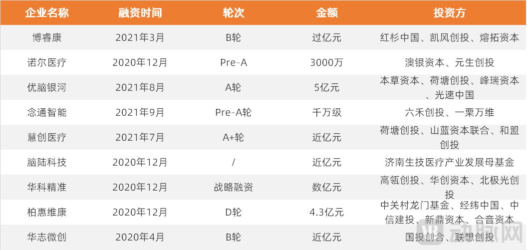
Financing Events in the Field of Functional Neurosurgery from 2020 to 2021
It is evident that, apart from surgical robots, accelerated financing in the field of functional neurosurgery is converging on a single area: brain-related data. Specifically, the acquisition, analysis, processing, and development of the three core elements of brain data—electroencephalography (EEG), neuroimaging, and brain-related clinical behavioral data—have become a popular sector for capital investment.
Among the numerous companies that have completed financing rounds, NeuroEchos stands out as a representative player in the utilization of brain science data. With a dual focus on functional neurosurgery and brain science, NeuroEchos not only develops innovative medical devices required for the precise diagnosis of various functional neurological disorders, including drug-resistant epilepsy (such as 3.0T MRI-compatible SEEG electrodes), but also advances the development of intelligent auxiliary tools to help industry-academia-research institutions process massive amounts of deep brain data. Committed to enhancing the foundational capabilities of clinical and research institutions in processing and analyzing large-scale datasets, NeuroEchos empowers scientists and clinical experts to more efficiently uncover the pathogenic mechanisms of potential neurological diseases and the fundamental operational logic of the brain. In addition to improving the efficiency and precision of epilepsy diagnosis and treatment, the company provides auxiliary diagnostic and therapeutic support for a broader range of complex brain disorders.
Currently, in terms of hardware R&D, Nuore Medical has completed the development of its first-generation 3.0T MRI-compatible stereoelectroencephalography (SEEG) electrodes and is concurrently advancing clinical trials at seven renowned clinical centers across China, including Huashan Hospital in Shanghai. The second-generation Macro-Micro radiofrequency ablation SEEG electrodes are under active development, with preclinical preparations, including animal studies, expected to commence around the end of 2021.
On the software front, Nuoer Medical is developing version 1.0 of an intelligent auxiliary detection tool based on epileptiform waves, with plans to subsequently integrate pHFO algorithms and launch version 2.0 of its intelligent auxiliary diagnostic tool based on high-frequency wavelets. Over the next one to three years, Nuoer will collaborate with numerous universities and industry-academia-research institutions, including Tsinghua University and Zhejiang University, to promote the industrialization of innovative medical devices combined with data intelligence. This effort will expand into building 3D brain functional network models and exploring combined applications such as MRI-compatible guided radiofrequency ablation and laser ablation.
Yang Huan, CEO of Nuoer Medical, stated that although Nuoer Medical entered the field of functional neurosurgery through hardware solutions, it has never positioned itself solely as a medical device company or a software intelligence firm. Instead, it is building a new organizational model driven by the dual engines of innovative medical devices and data intelligence, with the ambition to become a deep-tech company achieving breakthrough discoveries and advancements in brain science.
Deciphering the neuronal code within massive datasets is a challenge no less daunting than sending humans to Mars. Why does Nuoer Medical believe that SEEG-based deep brain data and its decoding capabilities are the core elements poised to ignite the field of neuroscience? What does deep brain data signify for breakthroughs in the brain science industry? VCBeat interviewed Yang Huan, CEO of Nuoer Medical.
The full English name of Nuor Medical is NeuroEchos. Yang Huan stated that the founding team initially named the company NeuroX, which was, to some extent, a tribute to SpaceX founded by Elon Musk. While SpaceX symbolizes the exploration of the secrets of the universe, Nuor is dedicated to exploring the three-pound universe of the human brain.
Yang Huan stated, “We do not idolize Elon Musk; rather, we revere the first-principles thinking approach, which elevates issues to the fundamental essence of the universe and addresses them from their core.”"In the field of brain science, where we are situated, the most core issue is the understanding of the fundamental mechanisms of the brain."
From its inception, Noer Medical has harbored the bold vision of not only addressing the diagnosis and treatment of functional neurological disorders but also enabling clinicians and scientists in this field to tackle core challenges in brain science. Over the past four years, Noer Medical has devised two strategic pathways to achieve this ambitious goal. For short- to medium-term commercialization, the company aims to ensure its viability by leveraging the commercial market for its innovative medical device, the SEEG electrodes. In its long-term strategy, Noer Medical uses precise diagnosis and treatment of refractory epilepsy as an entry point for brain science research, thereby accumulating essential resources and sustaining momentum for future ventures into uncharted territories aimed at decoding and deciphering the human brain.
The design of coordinated efforts across two business lines reflects Nuore Medical’s preparedness for a marathon in this field. How difficult is it really to obtain a functional map of the human brain?
In his article “The Paradigm Revolution in Brain Science: Where Will the Next One Emerge?”, Professor Gu Fanji of Fudan University pointed out that for decades, scientists have been striving to reconstruct brain functions and obtain a whole-brain mesoscale neural connectivity map. The subfield of computational neuroscience, which emerged in the late 1980s, has made progress in studying neurons, sensory information processing, and several simple circuits. However, a theoretical framework for the entire brain—particularly its higher-order functions—remains lacking, and it is still uncertain when a breakthrough in this area will be achieved.
Currently, scientists have largely elucidated the connectome of Caenorhabditis elegans (one of the simplest model organisms). Its nervous system comprises just over 300 neurons, each with a specific location and morphology; however, the mechanisms underlying their functions remain incompletely understood. This underscores the immense difficulty of mapping the human brain’s connectome—comprising 86 billion neurons and 150 trillion synaptic connections—and using it to explain human behavior.
We can divide the effort to obtain individual brain functional maps into two steps. The first step is to accurately record massive amounts of EEG signals, and the second step is to efficiently analyze and attempt to decode deep brain data such as EEG.
In this challenging field, against the backdrop of existing technologies, developing techniques capable of recording and analyzing massive amounts of data on neuronal activity has become the key to breakthroughs.
Only by effectively leveraging neuroscience data can we hope to achieve the goal of mapping brain function, and thereby develop novel therapies based on a fundamental understanding of brain mechanisms, enabling interventions for functional neurological disorders.
In the first step of EEG signal acquisition, the hardware primarily consists of various types of EEG electrodes. Currently, EEG electrodes can be mainly categorized into non-invasive scalp electrodes and invasive intracranial electrodes. Among these, invasive intracranial electrodes can be further divided into subdural cortical electrodes, which primarily capture cortical signals, and depth electrodes, which record deep intracranial EEG activity. Stereoelectroencephalography (SEEG) electrodes, used for the diagnosis and treatment of refractory epilepsy, are a type of invasive intracranial depth electrode.
In terms of scale, the development of brain electrodes has roughly gone through the centimeter level (10-2m), millimeter-level (10-3m), then to the micron level (10-3-10-5m) developmental process, with development in scale roughly corresponding to a range from 10-1EEG signal frequency band from Hz to 100 kHz.
A comparison of several types of brain electrodes reveals that non-invasive scalp electrodes, which record electroencephalogram (EEG) signals from the scalp, suffer from significant information loss and poor signal quality due to attenuation by the skull and scalp. In contrast, invasive subdural cortical electrodes can substantially improve the signal-to-noise ratio of EEG signals; however, their use is limited to short-term intraoperative monitoring because of the extensive surgical trauma involved.
More importantly, neither of these two types of electrodes can penetrate deep into the brain, and thus cannot acquire stereoelectroencephalographic (SEEG) signals that truly reflect the actual connectivity between brain networks within deep brain structures and functional brain regions. In contrast, SEEG electrodes enable long-term acquisition of deep brain electrical activity (for approximately two weeks to one month). When combined with magnetic resonance imaging (MRI) data, this approach allows for clear and precise recording and analysis of heterogeneous models of brain functional networks and epileptic networks, encompassing both neural activity and high-resolution spatial structural information.
However, SEEG electrodes are used for the diagnosis and treatment of refractory epilepsy, which imposes stricter requirements on preoperative assessment. Prior to implementation, a multidisciplinary team discussion is mandatory, involving specialists in epileptology, neurosurgery, neurophysiology, neuropsychology, and neuroradiology, to jointly determine whether SEEG electrode implantation is indicated for the patient and to formulate the implantation plan.
Overall, acquiring deep brain data via stereoelectroencephalography (SEEG) presents a high barrier to entry. Leveraging SEEG-collected deep brain data for precise diagnosis and analysis in the future will significantly test capabilities in big data architecture and massive data processing. The electroencephalogram (EEG) data from a single SEEG patient alone can reach hundreds of gigabytes or even terabytes in size, not to mention the inclusion of brain imaging data and high-definition clinical videos recorded during both interictal and ictal periods. Processing data from just 100 cases requires an architectural capacity for terabyte- or even petabyte-scale data storage and analysis. According to Li Yafeng, CTO of Nuoer Medical, the field is expected to reach petabyte-scale big data volumes with only around 1,000 cases, a scale that exceeds the capacity of conventional infrastructure.
As the critical clinical component of stereoelectroencephalography (SEEG), brain electrodes can acquire more precise and deep-seated electroencephalographic signals, which has led Nuor Medical to select MRI-compatible SEEG electrodes as the breakthrough point for its data IoT terminal and auxiliary diagnostic tools.
During the development of SEEG electrodes, Nuoer Medical’s first-generation SEEG electrodes achieved global first-in-class compatibility with 3.0T high-field MRI systems. Building on this foundation, the second-generation electrodes enable the acquisition and analysis of high signal-to-noise ratio neural discharge signals at higher frequency bands (from above 500 Hz up to 10 kHz) and in smaller spatial domains (down to single-neuron discharges). Nuoer Medical is currently advancing clinical trials and product registration for its first-generation electrodes, while having already secured multiple core patents for the second-generation products, it has initiated their R&D and industrialization.
Dr. Mo Xiaolong, Co-founder and Chief Hardware Scientist at Neuracle, offered an analogy: “In the current field of brain science research, advances in engineering technology have provided numerous tools for probing brain activity. Drawing an analogy from the history of physics, brain electrodes are akin to the astronomical telescopes crafted during humanity’s ‘Galilean era,’ offering effective tools and methods to explore the ‘three-pound universe’ and accumulate vast amounts of precise data. Research and applications in brain science still await breakthroughs in fundamental core theories. Such breakthroughs depend on gaining a deeper understanding of the underlying logic governing the operation and computational principles of the massive number of neurons in the human brain. This may well exceed the upper limits of human rational cognition. Consequently, such breakthroughs will likely rely on massive datasets and artificial intelligence methodologies—using a ‘gray box’ to understand a ‘black box.’ This approach holds greater feasibility in engineering terms and may represent the critical step for brain science to advance from the ‘Galilean era’ to the ‘Newtonian era.’”
The Nuor Medical Team believes that although "the essence of 'protecting the brain' is essentially intervention for functional neurological disorders, it is also closely related to 'understanding the brain' (recognizing the structure of brain regions and the nature of functional neural networks, attempting to elucidate how the brain works)."
“The Brain Guardian” project has accumulated extensive deep brain electrical data through interventions for functional neurological disorders, providing a robust foundation for further exploration of the mysteries of electroencephalography (EEG) and offering rich resources and effective support for the advancement of brain science. The development of deep brain electrical data represents a golden intersection between research on functional neurological disorders and brain science.
In the science fiction novel *The Three-Body Problem*, a device called the “mind-resolving camera” is described, capable of reconstructing cerebral cognitive activity within a computer. The successfully developed mind-resolving camera displayed a holographic image of brain activity: countless neural signals busily transmitting along slender synapses, resembling luminous pearls flowing through an integrated network.
Under current technological conditions, achieving a holographic image of brain activity as vast and intricate as the starry sky not only tests our ability to record EEG signals but also faces significant hurdles due to the immense computational power required.
Professor Yang Yang, Chief AI Scientist at Nuoer, stated, “Data is the prerequisite for an impending paradigm revolution at the mesoscopic level in future brain science. This provides directional clarity; however, the current key challenge lies in identifying feasible technological pathways. If we remain confined to traditional analytical methods, it will be difficult to produce effective results within a short timeframe. The emergence of artificial intelligence technology may accelerate this process, helping us to more rapidly understand functional network models of the human brain, better comprehend functional neurological disorders, and identify novel intervention strategies. Of course, this endeavor cannot proceed without the guidance and directional insight provided by clinical experts and neuroscientists, akin to the collaboration between professional Go players and AI scientists in AlphaGo, or the integration of AI researchers with geneticists and molecular biologists in AlphaFold2.”
That is to say, except for the first step of collecting EEG signals,Step 2: Data Interpretation Capability Is Equally Important. At present, domestic enterprises’ EEG analysis primarily focuses on interpreting electroencephalogram (EEG) signals to assist in disease diagnosis. By interpreting EEG signals, these companies monitor and assess brain functional disorders such as cerebral palsy, attention deficit disorders in children, epilepsy, stroke, Parkinson’s disease, cognitive impairment, Alzheimer’s disease, and psychiatric disorders.
Noer Medical is the only company in China conducting in-depth research on automated analysis algorithms for stereoelectroencephalography (SEEG). Noer’s auxiliary analysis tool leverages SEEG epileptic wave detection and pathological high-frequency oscillation (pHFO) algorithms to achieve automated recognition and annotation compatible with 3.0T MRI-integrated EEG systems. This assists electroencephalographers in improving monitoring and identification efficiency, and is expected to significantly reduce the time required for interpreting and analyzing post-SEEG implantation EEG signals in the future.
The integration of intelligent algorithms has introduced novel data mining methodologies. Intelligent EEG data analysis tools may serve as accelerators for neuroscience research. By combining AI with deep brain information, this approach could become a key to unlocking and exploring brain research, thereby helping scientists gain a better understanding of the brain.
As its industrial footprint deepens, NeuroEchos will build infrastructure capabilities to process terabyte-to-petabyte (TB–PB) scale deep-brain data, supporting leading brain science research institutions both in China and abroad. Meanwhile, Neure Medical has assembled a team of top-tier experts from China and overseas in the fields of brain science, artificial intelligence, and big data. Leveraging its technical expertise in deep brain signal processing, Neure Medical aims to provide clinical and research institutions with advanced diagnostic and therapeutic tools for stereoelectroencephalography (SEEG)-based deep brain signals, addressing complex neurological disorders.
Yang Huan, CEO of Nuoer Medical, stated, “While other ‘AI + EEG’ initiatives were still focused on conventional EEG research, we had already begun investigating high-frequency wavelets or high-frequency oscillations (HFOs) in epilepsy. High-frequency wavelets refer to EEG signals above 100 Hz. Previously, whether using standard scalp electrodes, subdural cortical electrodes, or stereoelectroencephalography (SEEG) electrodes, it was only possible to capture epileptic waves with dominant frequencies below 100 Hz. These EEG data suffered from insufficient information density due to significant noise contamination. In contrast, the global clinical consensus in this field holds that high-frequency wavelets are directly correlated with and indicative of epileptogenic zones, akin to biomarkers and therapeutic targets in oncology. Once we fully understand this ‘target’ and enable electrodes to collect, analyze, and identify high-frequency wavelets, it will bring about a qualitative leap in the diagnosis and treatment efficiency of many functional neurological disorders, including epilepsy.”
In addition to disease-specific applications, Nuor Medical also believes that the interpretation of high-frequency oscillations (HFOs) can provide fundamental driving forces for decoding functional models of the brain. Notably, basic research on human consciousness and memory circuits based on stereoelectroencephalography (SEEG) has yielded increasingly prominent results in recent years. A deeper understanding of the brain’s underlying mechanisms will further support the diagnosis and treatment of more complex neurological disorders.
Data from the life sciences database PubMed reveals that research on SEEG has experienced explosive growth in recent years, particularly since 2016.

By pursuing a path that integrates hardware and software with algorithmic breakthroughs, Nuoer Medical has achieved certain practical advancements. In September 2020, Neuralink, founded by Elon Musk, held a press conference demonstrating the implantation of electrode chips into pig brains. It showcased a coin-sized chip implantable into the brain and a surgical device capable of automated chip implantation, reigniting public interest in the concept of brain-computer interfaces (BCIs). Yang Huan stated that Nuoer Medical had already successfully conducted research on monitoring electroencephalogram (EEG) signals in pig brains in 2019. In July 2019, Nuoer Medical successfully completed the world’s first porcine epilepsy modeling. Leveraging its independently developed global-first MRI-compatible stereoelectroencephalography (SEEG) electrodes, the company achieved real-time, large-scale recording and precise identification of high-quality epileptic EEG signals and pathological high-frequency oscillations in specific brain regions. Over the past four years of development, Nuoer Medical has attracted an increasing number of top-tier experts and gained support from several leading domestic investment funds.
Currently, the core team of Nuor Medical consists of experts from renowned domestic and international institutions such as Tsinghua University, Zhejiang University, and UCLA.
This team includes Dr. Mo Xiaolong, Chief Scientist of Neurol Hardware, who has long been engaged in research on neuromodulation technologies and advanced medical device technologies at Tsinghua University; the founding initiator and Chief Clinical Expert with over 15 years of clinical experience in the field of functional neurosurgery; and Professor Yang Yang, Chief AI Scientist of Neurol, who serves as the Director of the Neurol/Zhejiang University Joint Laboratory and currently chairs the Department of Artificial Intelligence at Zhejiang University. It also includes Li Yafeng, CTO and Chief Architect of Neurol, who previously served as a Senior Research Fellow in Architecture at Shanda Games, Head of the Big Data Platform at Ctrip, and General Manager of the Shanghai R&D Center/Chief Big Data Architecture Expert at China Telecom. It is precisely the integration and drive of such a diverse yet highly aligned interdisciplinary talent pool that makes possible Neurol’s continued breakthroughs in functional neurosurgery and brain science in the future.
Faced with the Galileo era of brain science ushered in by stereoelectroencephalography (SEEG), and the widespread global recognition of SEEG technology as a key gateway to understanding the human brain, Dr. Mo Xiaolong, who has been deeply engaged in the dual fields of “SEEG + neuromodulation” for over a decade, is filled with immense excitement.
DBS and SEEG are among the few technical modalities capable of directly interfacing with deep brain structures and modulating deep neural activity.
Therapeutic deep brain stimulation (DBS) inherently prioritizes higher precision and minimal invasiveness. Taking the treatment of Parkinson’s disease as an example, DBS involves implanting only eight electrode contacts in the patient’s bilateral subthalamic nucleus (STN) or globus pallidus internus (GPi), with the implantation configuration being highly homogeneous across most patients. Consequently, when scientists seek to leverage DBS for acquiring deep brain data in neuroscience research, the resulting data are subject to significant limitations in terms of spatial coverage and spatial resolution.
In contrast, stereoelectroencephalography (SEEG) is an exploratory and localizing technique that involves implanting a greater number of electrode contacts within the patient’s brain. Taking drug-resistant epilepsy, where SEEG application is most mature, as an example, clinicians typically implant 100–200 electrode contacts into the patient’s brain to record deep brain electrical activity over an extended period (up to one month). Furthermore, given the diverse etiologies of epilepsy and the heterogeneity of epileptic networks within the brain, the placement of SEEG electrodes can cover all regions of the brain. Statistics indicate that approximately two-thirds of the implanted electrode contacts are located in normal brain areas (i.e., regions unrelated to seizure onset) during the clinical diagnosis and treatment of drug-resistant epilepsy using SEEG. These data hold significant value for neuroscience research.
Neuromodulation technologies, particularly deep brain stimulation (DBS), have achieved remarkable success in the treatment of Parkinson’s disease (PD), a degenerative functional neurological disorder. However, this success has proven difficult to replicate in other functional neurological disorders. Parkinson’s disease is characterized by a relatively standardized pathophysiology across patients; for nearly all individuals with idiopathic PD, clinicians can effectively control symptoms by implanting electrodes in the globus pallidus internus (GPi) or the subthalamic nucleus (STN) for electrical stimulation.
However, this logic of attempting to treat a neurological disorder with a universal approach may have also created significant challenges in the current efforts to expand the indications for neuromodulation technologies such as deep brain stimulation (DBS).
Unlike Parkinson’s disease (PD), many other functional neurological disorders are highly individualized, necessitating personalized techniques and approaches for their diagnosis and treatment. Epilepsy is the most typical example of such personalized functional neurological disorders. Rarely do any two epilepsy patients share identical etiologies or intracranial epileptic heterogeneous networks. The diagnosis and treatment of epilepsy must therefore rely on personalized technical methods, with stereoelectroencephalography (SEEG) being a representative personalized diagnostic and therapeutic technique. By employing SEEG, clinicians can perform personalized and targeted analyses of each patient’s epileptogenic zone and epileptic network, thereby formulating appropriate treatment plans.
Therefore, as neuromodulation techniques such as Deep Brain Stimulation (DBS) expand to encompass more indications, personalization has emerged as a critical and pivotal direction. In this context, Stereoelectroencephalography (SEEG) serves as a natural ally to neuromodulation technologies and represents an indispensable component of their developmental trajectory. Currently, there is extensive in-depth research combining SEEG with DBS for the treatment of conditions such as Treatment-Resistant Depression (MDD) and chronic pain.
Yang Huan stated, “Over the past four years of development, we have consistently adhered to our original strategy, taking every step with diligence and determination in pursuit of our ambitious vision, while maintaining a clear roadmap for future growth.”
“Although Nuore will prioritize the diagnosis and treatment of refractory epilepsy in functional neurosurgery over the next 3–5 years, with advancements in EEG data mining and decoding capabilities, it will pioneer uncharted territory. By leveraging SEEG deep brain data, particularly through high-frequency wavelet analysis, the company aims to identify key features of a broader range of functional neurological disorders and integrate intelligent SEEG technologies with wider therapeutic tools such as LITT and DBS.”
We believe that in the near future, using stereoelectroencephalography (SEEG) as a preliminary procedure to determine the optimal targets and therapeutic parameters for permanently implanted deep brain stimulation (DBS) devices, thereby delivering more precise treatment, will become the mainstream approach in neuromodulation therapy for functional neurological disorders.
As envisioned in Nuore’s slogan, “Nuore Travels the World, You See the Future,” there is a group of people in China who aspire to leverage Nuore’s technology to benefit more patients with brain disorders, enabling them to embrace a bright new future.