Ultra-Low-Cost, Ultra-Thin Japanese GI Lens Enables Affordable Disposable Endoscopes
Not long ago, in China’s endoscopy sector, Xinguangwei Medical formally submitted its prospectus to the Hong Kong Stock Exchange. The prospectus highlighted five core endoscopy technologies: single-use endoscopy technology, ultra-high-definition medical imaging technology, 3D medical imaging technology, special-light medical imaging technology, and ultra-thin endoscopic imaging technology.
Upon reviewing the entire text, we find that ultra-fine endoscopic imaging technology appears to remain shrouded in mystery among the introductions of these five major technologies. As an ultra-fine endoscope still under research and development, it lacks both detailed product specifications and an overview of competing market products.
Recently, VCBeat learned that a Japanese company is manufacturing ultra-thin GI (Graded Index) lenses for use in ultra-thin endoscopes.As one of the key components of endoscopic systems, we will delve into this through the project.
What is an “ultra-thin endoscope”?
First, we need to understand what an “ultra-thin endoscope” actually is. According to Frost & Sullivan, there is no universally accepted definition for ultra-thin endoscopes. Based on articles published by The American Society for Gastrointestinal Endoscopy, ultra-thin endoscopes primarily refer to those with an insertion tube diameter of 6 mm or less (including 6 mm), which can be inserted into the body through the patient’s nostril or mouth.
With technological advancements, many medical endoscope manufacturers on the market can already meet this standard. Therefore, the requirements for ultra-thin endoscopes have been upgraded, with an insertion section diameter of less than 1 mm becoming the new standard for “true” ultra-thin endoscopes.
Currently, many endoscope manufacturers are striving to develop such ultra-thin endoscopes. However, due to the design characteristics of ultra-thin endoscopes, achieving an insertion tube diameter of less than 1 mm typically requires their use with separate light sources and independent image processors, thereby necessitating the miniaturized integration of these two components into a single unit.
From 6 mm to 1 mm, this reduction in diameter tests the precision manufacturing capabilities of endoscope manufacturers; only a handful of companies worldwide can produce such ultra-thin endoscopes.
Although development is challenging, it is essential to develop and manufacture “ultra-thin” endoscopes.Ultra-thin endoscopes, by virtue of their slender profile, have naturally expanded their clinical applications to the diagnosis and treatment of conditions involving the lacrimal ducts, mammary ducts, root canals, and the ear. These anatomical sites are characterized by either narrow lumens or complex structures, thereby imposing stringent requirements on endoscopic technology.
For narrow body cavities, imaging was previously limited to external modalities such as ultrasound and computed tomography (CT), which suffer from low resolution and the inability to provide real-time visualization. In contrast, ultra-thin endoscopes can access bodily passages that are difficult for conventional endoscopes to reach, enabling real-time display of internal tissue images and facilitating intuitive clinical assessment by physicians.
Meanwhile, as a therapeutic modality, endoscopy enables physicians to perform interventions under direct visualization. Taking lacrimal duct endoscopy as an example, the device must integrate diagnostic illumination and imaging fibers, an irrigation system, and therapeutic laser fibers, imposing stringent requirements on its dimensions. However, the use of ultra-thin endoscopes for treatment can minimize damage to normal tissues and reduce functional impairment in other areas, while also shortening operative time and accelerating postoperative recovery.
Therefore, for departments such as ophthalmology, dentistry, otology, and otorhinolaryngology, ultra-thin endoscopes can meet the clinical demand for precision medical devices. Meanwhile, the broad application prospects of ultra-thin endoscopes are also evident.
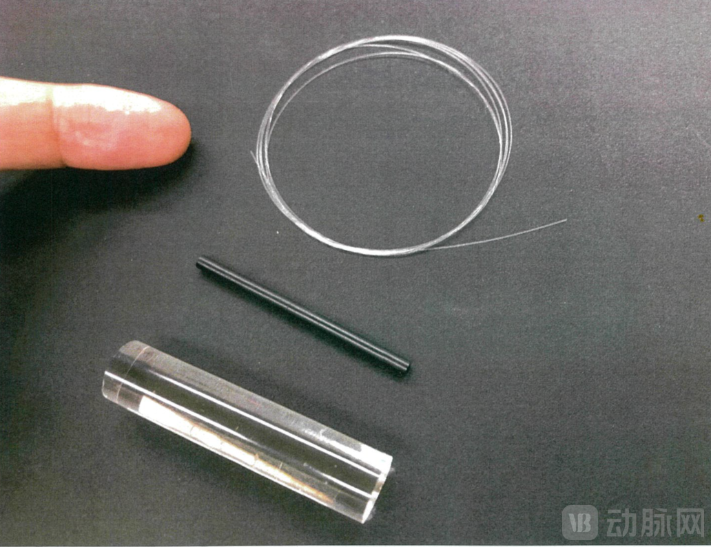
The Japanese company mentioned earlier has developed GI lenses with a diameter as small as 0.1 mm. When applied to endoscopes, these lenses enable the control of the insertion tube diameter to under 1 mm, which is half that of mainstream products currently on the market. This technology can serve as a key component in ultra-thin endoscope modules.
Source: Japanese Company
Ultra-Fine GI Endoscope: 25 Years of Meticulous Craftsmanship
What is a GI endoscope?
GI (Graded Index), or graded-index. A GI lens is a cylindrical lens with a refractive index distribution, where the refractive index decreases from the center to the periphery.
GI Endoscopes Offer Two Primary Advantages in ImagingFirst, by precisely controlling the formation of a multi-layer refractive index distribution, a local dispersion control profile can be established to manage chromatic aberration. Second, the imaging resolution currently reaches 500,000 pixels, and with further technological improvements, it is expected to achieve 2 megapixels in the future.
Japan’s optical technology is highly advanced on a global scale. Regarding GI (gastrointestinal) endoscope lenses, top-tier Japanese optical manufacturers such as Nippon Sheet Glass (NSG), Nikon, and Olympus also attempted to develop them, but ultimately discontinued their efforts due to factors including the prolonged inability to validate results and corporate operational decisions.
This Japanese company has dedicated 25 years to the research and development of GI endoscopes, overcoming numerous challenges through countless iterative trials before successfully mastering GI technology. This GI technology received funding from Japan’s Science and Technology Agency and was widely covered by major Japanese media outlets.
The Japanese company stated: “By creating a refractive index distribution in glass, Nippon Sheet Glass of Japan successfully manufactured GI lenses, which were commercialized under the brand name SELFOC and utilized in endoscopes. However, no company worldwide has yet succeeded in achieving highly precise refractive index distributions in plastics; we are currently the only enterprise to have accomplished this.”
The development of ultra-thin GI lenses originated with the plastic GI lenses developed by Professor Koike at the Faculty of Science and Technology, Keio University, Japan.
Early in the project, the research team encountered its first challenge: clear imaging was unattainable because bubbles within the lens obstructed light transmission. Through dedicated efforts, the company’s R&D team developed a novel manufacturing method that enabled the production of bubble-free GI lenses.
However, the most challenging aspect of GI lenses is controlling variations in the refractive index profile. The technique for creating a highly precise profile involves achieving the required distribution of specialized nanoparticles within polymethyl methacrylate (PMMA) material, which is only feasible under specific conditions of pressure and temperature.
Just two years ago, after several iterations, this challenge was finally overcome, and ultra-thin GI endoscopes finally achieved clear imaging quality.

Source: Japanese company
Ultra-Low-Cost GI Lenses Empowering Single-Use Endoscopes
Another prominent advantage of ultra-thin GI endoscopes is cost control.Thanks to the selection of ultra-fine GI endoscope raw materials, it not only achieves an ultra-low price but also offers excellent flexibility; it will not crack upon dropping and is difficult to break even when bent with force.
The raw material of the GI endoscope lens is PMMA (polymethyl methacrylate)., also known as acrylic or organic glass. This is a high-molecular polymer and a common alternative to glass, offering advantages such as high transparency, low cost, and ease of mechanical processing. As a result, the production cost of GI lenses is very low.
The Japanese company stated that, through design and material selection, ultra-thin GI endoscopes can reduce the price of traditional endoscopes from several million yen (approximately hundreds of thousands of RMB) to below one million yen (approximately 60,000 RMB).
Disposable endoscopes can address the critical clinical challenge of cross-infection. Currently, the market size for disposable endoscopes in China exceeds RMB 10 billion. However, to promote their widespread adoption, it is essential to resolve issues related to cost control and product performance, particularly image quality.
Leveraging its two major advantages of clear imaging and low cost, the ultra-thin gradient-index (GI) lens facilitates the low-cost mass production of disposable endoscopes. It is primarily used in rigid endoscopes and can be applied across multiple fields, including ophthalmology, digestive surgery, and thoracic surgery.
In summary, the characteristics of ultra-thin GI endoscopes are manifested in the following aspects:
The lens is extremely thin, with a diameter of 0.1 mm, and can achieve video transmission through a linear GI of only 100 micrometers;
The image resolution is high, reaching up to 500,000 pixels, and future technological improvements may enable resolutions of up to 2 million pixels;
Low cost, suitable for use as a disposable endoscope;
Good flexibility, not easy to break.
Deeply Rooted in the Field of Optical Lenses, with Products Sold Worldwide
The team involved in the ultra-fine GI endoscope project comprises organic chemistry experts from Japanese national universities, optical specialists formerly with major Japanese corporations, plastic processing engineers, and polymerization engineers, each bringing over 30 years of extensive experience in their respective fields.
The Japanese company that developed the ultra-thin GI endoscope has been deeply engaged in the optical lens vertical for 36 years, entering the medical field in 1985.Its products are purchased by renowned Japanese and global enterprises such as Apple, Google, SONY, Panasonic, Kyocera, Olympus, Omron, Toyota Motor Corporation, Honda Motor Co., Ltd., the University of Tokyo, Kyoto University, Shiseido, Kao Corporation, Kobayashi Pharmaceutical, and Taisho Pharmaceutical.
Its products have also achieved numerous world-first milestones. For instance, in 1987, the company developed the world’s first “Touch and View” video magnifier, which enables magnified observation on a monitor simply by placing it on the target object. In 1988, it pioneered the world’s first portable digital microscope; with over 350,000 units sold to date, this product has been adopted for quality control across many industries and global corporations, including NASA (National Aeronautics and Space Administration) in the United States.
Japanese companies stated:Apple’s former CEO, Steve Jobs, introduced our products in his keynote address and spoke highly of them. Our products were ultimately adopted by NASA after competing with Nikon’s offerings in Japan. They were selected by Nokia following competition with Germany’s ZEISS. Furthermore, smartphones from Japan’s NEC and Fujitsu have incorporated our company’s lenses.
Furthermore, in the healthcare sector, Japanese companies have launched new products such as psychiatric diagnostic devices for Alzheimer’s disease and depression, as well as devices for assessing pets’ emotional states. Diagnostic tests for upper respiratory tract infections, including COVID-19, SARS, and influenza, are currently undergoing medical certification. Meanwhile, therapeutic devices for Alzheimer’s disease and depression are under development.
VCBeat has learned that this ultra-thin GI endoscope has completed laboratory-stage validation and is awaiting market launch. The Japanese company stated that, to accelerate the product’s application, it seeks to transfer the technology to a capable Chinese enterprise, which would handle mass production and commercialization.
Reference: Xin Guangwei Medical's Prospectus