Home Application of DEEPQUAKE Peripheral Lithotripsy System in a Case of Severely Calcified Arteriovenous Fistula Stenosis by Professor Liu Yangdong’s Team

Application of DEEPQUAKE Peripheral Lithotripsy System in a Case of Severely Calcified Arteriovenous Fistula Stenosis by Professor Liu Yangdong’s Team

Apr 06, 2026 07:30 CST Updated 07:30
Trulive

Structural Heart Disease Device Developer

Image
Image


In the field of pan-vascular intervention, the treatment of calcified lesions is one of the key and difficult points in clinical practice. Produced by Trulive and promoted by Grand Pharmaceutical Group Limited,DEEPQUAKE Peripheral Vascular Intravascular Shockwave SystemOfficially launched (China Medical Device Registration No. 20243012335).

Stenosis of arteriovenous fistulas associated with severe calcification represents a significant challenge in the maintenance of vascular access for maintenance hemodialysis. Conventional balloon angioplasty often faces limitations such as insufficient dilation and a relatively high risk of complications. Although intravascular lithotripsy has been widely used in the treatment of calcified lesions in coronary and peripheral arteries, its application in calcified stenosis of arteriovenous fistulas remains very limited, with only a few technical reports or case studies currently available.

This issue is shared with everyoneAn Application of DEEPQUAKE Peripheral Shock Wave System in the Treatment of Severe Calcified Arteriovenous Fistula Stenosis, by South China Hospital Affiliated to Shenzhen UniversityProfessor Liu Yangdong's TeamCompleted, welcome everyone to read, study, exchange, and discuss.

【Ref:Zhou A, Zi Y, Lu Y, Hu L, Wang C, He Y, Wen L, Liu Y. Application of intravascular lithotripsy in a case of severely calcified arteriovenous fistula stenosis: A case report and technical note. J Vasc Access. 2025 Dec 14:11297298251391723. doi: 10.1177/11297298251391723. Epub ahead of print. PMID: 41392402.】


Case Summary

Patient Basic Information

Patient:54 years old, female.

Chief Complaint:"Admission due to a 20-year history of maintenance hemodialysis and decreased blood flow in the arteriovenous fistula."

History of Present Illness:The patient had undergone maintenance hemodialysis for 20 years due to chronic renal failure, with a radiocephalic arteriovenous fistula as the long-term dialysis access. Percutaneous transluminal angioplasty was performed 24 months and 7 months ago respectively due to fistula stenosis. Before this admission, the patient noticed weakened thrill of the fistula and insufficient blood flow during dialysis; ultrasound examination suggested multiple severe stenoses at the anastomosis site and outflow tract accompanied by serious circumferential calcification.

Past Medical History:Chronic renal insufficiency.

Physical Examination:The thrill at the radio-cephalic fistula site in the right forearm was weak on palpation, and auscultation revealed a low-pitched, short-duration bruit.

Preoperative Ultrasound:Multiple stenoses with severe calcification are shown, with the narrowest diameter at the anastomosis being 2.1 mm and the narrowest diameter of the outflow tract being 3.0 mm. The blood flow velocity at the anastomosis is 180 cm/s. (Figure 1)

Image

Figure 1 Preoperative imaging shows severe calcification of the radial artery-cephalic vein arteriovenous fistula and venous outflow tract, with sluggish blood flow. Local valve stenosis is visible in the distal segment of the aneurysm, with calcified plaques attached to the surface.

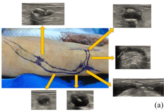
Diagnosis and Surgical Strategy
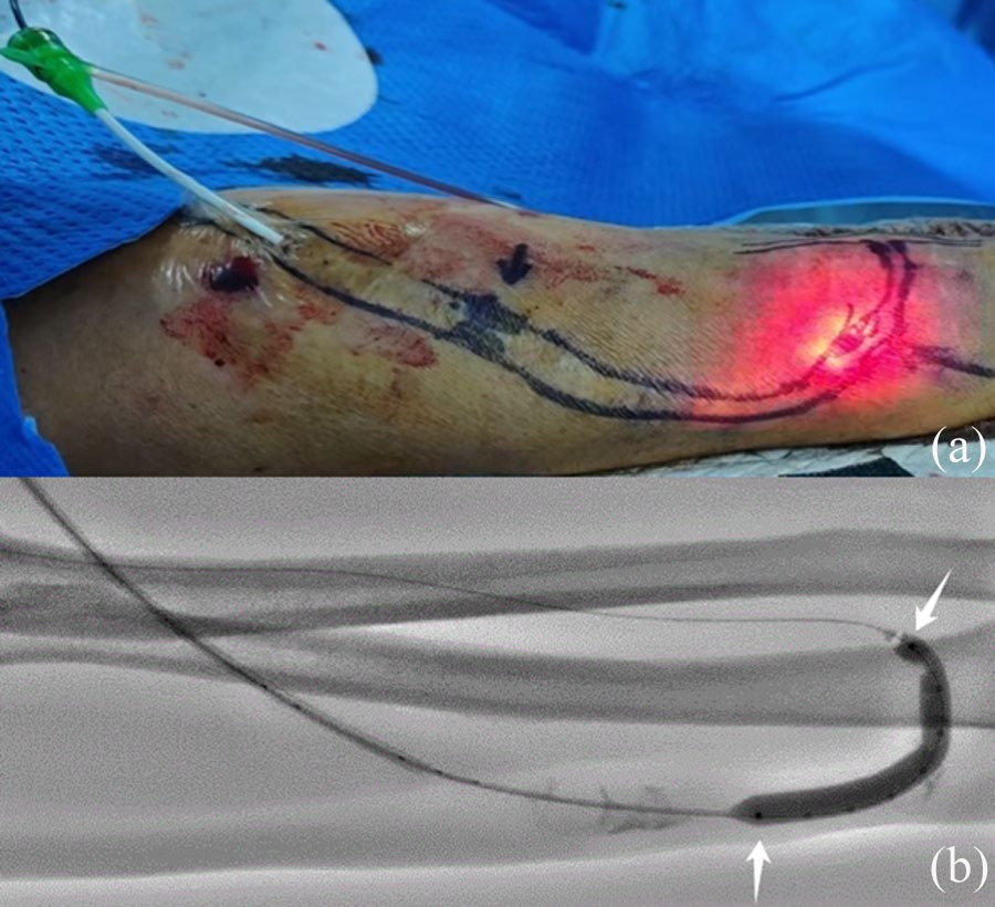
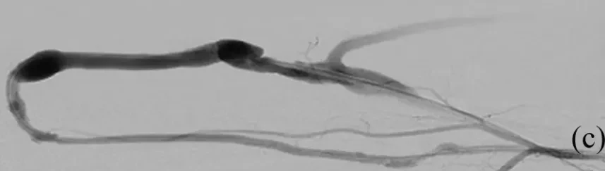
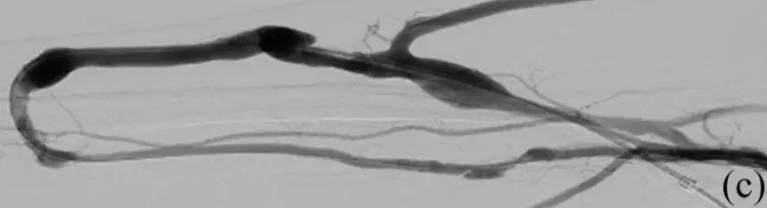
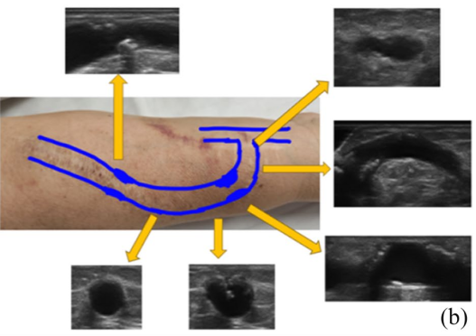
Preliminary Diagnosis:Severe Calcific Stenosis of the Radial Artery-Cephalic Vein Arteriovenous Fistula.

Proposed Surgical Strategy:Intravascular Lithotripsy (Shockwave Balloon) + Post-Dilation with High-Pressure Balloon.

Surgical Procedure

1. Puncture the right fistula vein and insert a 6F vascular sheath (Terumo). The super-selective guidewire passes through the stenotic segment, and angiography confirms the location and extent of the lesion (Figure 2). Angiography shows severe stenosis at the anastomosis and outflow tract, with significant local calcification, uneven contrast filling, and slow flow rate.

Image

Figure 2: Severe stenosis at the anastomosis of the arteriovenous fistula, with uneven filling and slow flow of contrast agent in the local calcified area.


2. A 5mm×40mm high-pressure balloon (Zenflow, China) was introduced along a 0.035-inch guidewire for pre-dilation. Subsequent angiography showed insufficient lumen expansion (Figure 3).

Image

Figure 3: After intraoperative pre-dilation with a balloon, there was no significant improvement in the local calcified lesion.


3. A 0.014-inch guidewire was used to introduce one.6mm×60mm Shock Wave Balloon (DEEPQUAKE, China)The calcified stenotic segments of the internal fistula vein and proximal arteriovenous aneurysm were dilated sequentially. After the balloon was pressurized to 4 atm, the shock wave system was first activated at level 3 energy (equivalent to an instantaneous 50 atm), applying two cycles (30 seconds per cycle) to each lesion segment. Subsequently, the energy was adjusted to level 5 (equivalent to an instantaneous 60 atm), and an additional 4-6 cycles were applied to each lesion segment (Figure 4).

Image

Figure 4 Intravascular lithotripsy treatment process (a) and fluoroscopic observation (b). The figure shows the process of using a 6 mm×60 mm DEEPQUAKE shockwave balloon to dilate and fragment calcified lesions. During the procedure, the shockwave balloon generates a beam through electrode discharge (without actual thermal effect). Under fluoroscopy, the position of the intravascular lithotripsy balloon's radiopaque marker can be observed, and based on the marker’s location (as indicated by the arrow), the lesion area can be targeted while assessing the balloon’s shape to evaluate the degree of calcification fragmentation.


4. Follow-up angiography showed significant improvement in stenosis (Figure 5).

Image

Figure 5 After treatment with the shock wave balloon, the calcified lesion in the anastomotic area decreased compared to before, and the stenotic lesion improved.


5. Post-dilation was performed using a 6mm×60mm high-pressure balloon, and the final angiography showed satisfactory lumen patency with smooth blood flow (Figure 6).

Image

Figure 6: After post-dilation balloon treatment, blood flow velocity was satisfactory, and the calcified lesion showed significant improvement compared to before.

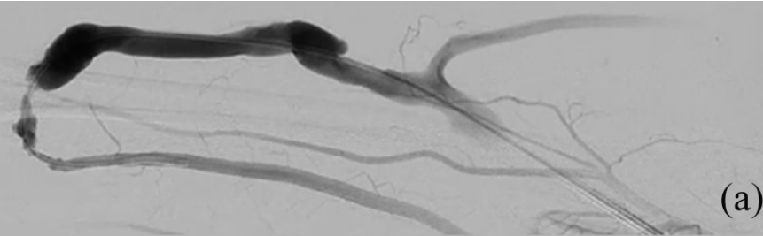
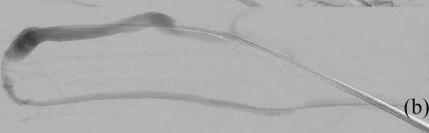
Postoperative Follow-up

During the operation, the patient only felt mild pain, and no complications such as vascular rupture, dissection, or distal embolism occurred.

Three months after the surgery, ultrasound review showed that the calcified plaque at the anastomosis stenosis had significantly decreased compared to pre-operation, and the lumen had markedly improved. The inner diameter of the narrowest segment of the original anastomosis was 4.8 mm after dilation, and the inner diameter of the narrowest segment of the original outflow tract was 5.2 mm (Figure 7). The blood flow velocity at the anastomosis increased from 180 cm/s pre-operation to 420 cm/s.

Image

Figure 7: A 3-month postoperative follow-up showed significant improvement in multiple calcified lesions compared to pre-operation, with the disappearance of calcified plaques within the local lumen and marked improvement in lumen condition.

Technical Points

1. Cautious Pre-dilationIt is the key prerequisite to ensure the efficacy of intravascular lithotripsy. It is recommended to use a standard high-pressure balloon that matches the target vessel diameter at a 1:1 ratio, rather than a cutting balloon or ultra-high-pressure balloon, to moderately trim the edges of calcified plaques with relatively low dilation pressure, establishing favorable channel conditions for subsequent shockwave treatment.

2. It is crucial to ensure the effectiveness of the shock wave energy transmission system.According to the recommendation, we added 10 mL of a 1:1 mixture of saline and contrast agent into the balloon inflation device and thoroughly evacuated the internal gas to avoid balloon damage caused by the low energy transmission efficiency of gaseous media.

3. The shock wave balloon should be gently advanced under fluoroscopic guidance, and the operational resistance should be closely monitored.If significant resistance occurs during the delivery process, it indicates that the lumen passage remains insufficient after pre-dilation. In such cases, the balloon should be withdrawn and pre-dilation repeated to establish an adequate safety pathway. Forceful delivery must be avoided to prevent balloon rupture or intimal vascular injury.

4. Treatment should adopt a stepwise, individualized energy delivery strategy.During the operation, the treatment cycle, energy level, and number of shock wave releases can be dynamically adjusted based on the fragmentation effect of the calcified lesion and the device instructions. If potential risks are encountered, the shock wave release can be paused at any time to ensure procedural safety. In this case, starting from a low energy level, the energy was gradually increased according to angiographic evaluation results, with supplementary shocks applied to areas of significant residual stenosis until satisfactory imaging results were achieved.

5. For long-segment lesions, complete coverage can be achieved by using a multi-segment overlapping approach.After each adjustment of the balloon position, alignment should be ensured via the marker bands to guarantee sufficient overlap between treatment areas, avoiding any omissions, thereby achieving comprehensive treatment of multi-segment or long, continuous calcified lesions.


Surgeon's Evaluation

1. An Effective Breakthrough for the Limitations of Traditional Balloon Dilation

Traditional Percutaneous Transluminal Angioplasty in the treatment of circumferential calcified lesions often results in balloon drift towards the less calcified side due to uneven hardness between the calcified plaque and normal vessel wall, leading to eccentric dilation and the "watermelon seed effect." This not only makes it difficult to effectively disrupt the calcified plaque but also subjects the thinner non-calcified vessel wall to greater stress, increasing the risk of vascular rupture, dissection, or perforation. In this case, an intravascular lithotripsy balloon was used, which selectively targets high-density calcified structures through acoustic shock waves, generating shear stress to fragment the calcification and avoiding the aforementioned risks. No complications such as vascular rupture or dissection occurred during the procedure.

2. Improve patient comfort during surgery
During the traditional high-pressure balloon dilation process, patients often report severe pain due to the high dilation pressure endured under local anesthesia. In this case, the patient only experienced mild pain during the procedure, significantly less than the pain felt during previous percutaneous transluminal angioplasty treatments, suggesting that the shockwave balloon has advantages in improving the patient's treatment experience.

3. Safety Considerations in the Treatment of Arteriovenous Fistula Calcification
The venous position of the arteriovenous fistula is superficial, allowing for clear assessment of the balloon's position and working status during surgery through the light spots generated by balloon discharge and the radiopaque ring under fluoroscopy, offering a high degree of visualization. Additionally, the walls of mature fistula vessels are relatively thick with no vital organs nearby, which may provide higher operational safety compared to deeper vascular anatomical locations. Considering the limited efficacy of traditional balloons on long-segment circumferential calcified lesions, intravascular lithotripsy demonstrates good applicability in treating superficial fistula calcifications. During this procedure and the three-month follow-up period post-operation, no complications such as embolism or vessel rupture were observed, consistent with the low complication rate (1.25%) and rare significant embolic events reported in the literature for intravascular lithotripsy in lower limb arteries.

4. Limitations and Prospects
This case report describes the application of intravascular lithotripsy in a patient with severe circumferential calcification of an arteriovenous fistula, preliminarily indicating that the technique is safe and effective for treating such lesions. This provides a new therapeutic approach for clinically addressing this challenge. However, its long-term efficacy and applicability in larger samples still require further research and validation.


Expert Introduction

Image

Professor Liu Yangdong

Ph.D., Postdoctoral Fellow, Chief Physician, Master's Supervisor.

Director of the Department of Interventional Vascular Surgery, South China Hospital, Shenzhen UniversityStanding Committee Member of the Endovascular Specialty Committee, Chinese Medical Doctor AssociationVice Chairman of the Hemodialysis Access Expert Committee, Endovascular Specialty Committee, Chinese Medical Doctor AssociationVice Chairman of the Vascular Access Expert Committee, Peripheral Vascular Disease Specialty Committee, Chinese Society of Integrated Traditional and Western MedicineVice Chairman of the Hemodialysis Access Expert Committee, Chinese Division of the International Union of Angiology (IUA)Vice Chairman of the Interventional Vascular Access Group, Integrated Traditional and Western Medicine Intervention Branch, China Health Information AssociationMember of the Vascular Access Group, Vascular Surgery Physician Branch, Chinese Medical Doctor AssociationMember of the Hemodialysis Access Group, Blood Purification Management Branch, Chinese Hospital AssociationStanding Committee Member of the Hemodialysis Access Group, Vascular Surgery Branch, China International Exchange and Promotion Association for Medical and HealthcareMember of the Vascular Medicine Specialty Committee, Chinese Research Hospital AssociationMember and Secretary of the Hemodialysis Vascular Access Group, Vascular Surgery Specialty Committee, Cross-Straits Medical and Health Exchange AssociationVice Chairman of the Peripheral Vascular Disease Branch, Guangdong Precision Medicine Application SocietyStanding Committee Member of the Vascular Surgery Branch, Guangdong Medical Association.


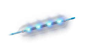
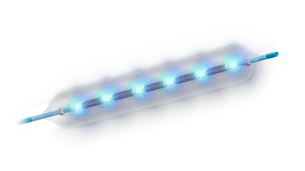
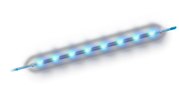
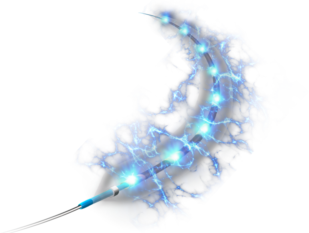
Features of DEEPQUAKE Peripheral Shock Wave System


Advantage 1LargerPulse Energy

  • Energy adjustable in five levels, with the highest energy reaching 3.2kV (fixed energy of 3.0kV for similar products).

Gear

Energy

Gear 1

2.8kV

Level 2

2.9kV

3-speed

3.0kV

4-speed

3.1kV

5 gears

3.2kV


Advantage 2MoreNumber of electrodes

  • The balloon is equipped with one pair of electrodes every 8mm, and the three lengths of 40/60/80mm have 4/6/8 pairs of electrodes respectively, ensuring uniform energy release. Treatment stitching does not require "Overlapping".

Image

40mm

4 pairs of electrodes

Image

60mm

6 pairs of electrodes

Image

80mm

8 pairs of electrodes


Advantage 3LongerBalloon Length

  • Offering an 80mm length balloon, more suitable for long lesions,Reducing operation time and IVL treatment frequency, cost-effective

Image


Previous Recommendations

【Powerful and Stable, Worry-free Endurance】Professor Zhuang Hui's Team: The First Application of DEEPQUAKE Peripheral Shock Wave System in China — A Case of Diabetic Foot Limb Salvage Treatment

【Leave Nothing Behind】Professor Jia Nan's Team: DEEPQUAKE Peripheral Shock Wave System First Application in China — Angioplasty for Dialysis Patients with Lower Limb Arteries

[Breakthrough Limits, Conquer Calcification] Professor Zhang Yan / Professor Guo You Collaboration: DEEPQUAKE Peripheral Shock Wave System First Applications in China — A Case of Long-Segment Severe Calcification Lesion in the Superficial Femoral Artery

@CAVS 2025 | Grand Pharmaceutical Group Limited's Peripheral Shock Wave Academic Symposium Successfully Convened

[More Complete Models, Comprehensive Coverage] Professor Fang Xin's Team: First Application of DEEPQUAKE Peripheral Shock Wave System in China -- A Case of Calcified Lesions in the Artery Below the Knee

[Five-level Treatment, Safe and Effective] Professor Li Xin's Team: The First Application of DEEPQUAKE Peripheral Shockwave Balloon in China — A Case of Long-segment Calcified Lesions in the External Iliac, Common Femoral, and Superficial Femoral Arteries

【Piercing the Clouds, Splitting the Stones, Achieving Excellence】Director Tian Hongyan / Professor Zhang Junbo Team: First Application of DEEPQUAKE Peripheral Shockwave Balloon in China - A Case of Severe Lower Limb Calcification

【Piercing the Clouds, Splitting the Stones; Achieving Excellence, Reaching Far】Professor Guan Sheng's Team: A Powerful Impact to Save Limbs — The Healing Journey of a Severely Calcified Patient

【Piercing the Clouds, Splitting the Stones, Achieving Excellence】Director Chen Zhong / Professor Wang Sheng Team: DEEPQUAKE Peripheral Shockwave Balloon First Applications in China — Long-Segment Calcified Lesions of the Superficial Femoral Artery

【Piercing the Clouds, Splitting the Stone, Achieving Excellence】Director Chen Zhong / Professor Yang Yaoguo's Team: First Application of DEEPQUAKE Peripheral Shockwave Balloon in China — Long-Segment Calcified Lesions in Femoropopliteal and Below-the-Knee Arteries

Grand Pharmaceutical Group Limited's DEEPQUAKE Peripheral Shock Wave System Debuts at the 4th ICCD Cardiovascular and Cerebrovascular Device Conference


Copyright Statement: This platform aims to help healthcare professionals better understand the latest developments in relevant disease areas. The information published on this platform does not imply agreement with its descriptions or viewpoints; it is only intended to provide more information. If there are any copyright issues, we kindly request the rights holders to contact us, and we will address them as soon as possible. The information is solely for healthcare professionals to stay informed and should not replace professional medical guidance in any way, nor should it be considered as medical advice. If such information is used for purposes other than staying informed, this platform and the authors assume no responsibility.Contact Email for Cooperation:vascular@edoctor.work。

Go to Mini Program

Image
Image
Image
Image